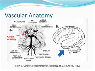
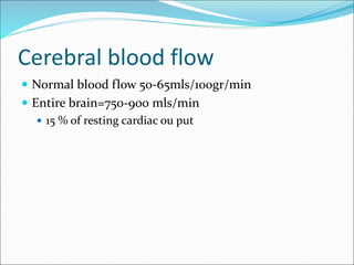
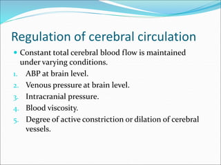
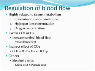
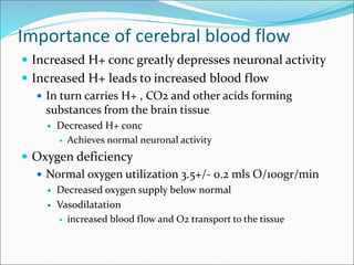
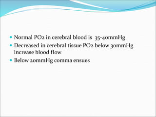
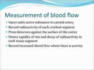
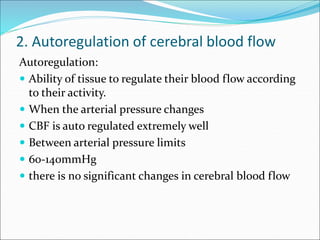
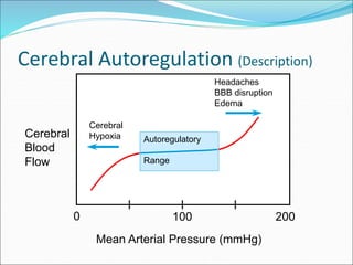
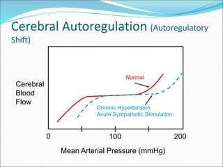
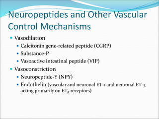
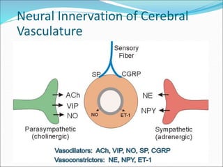
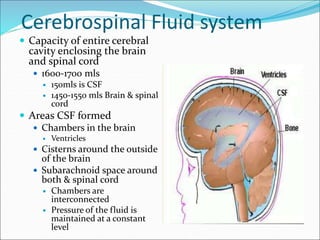
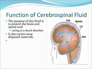
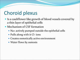
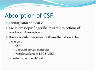
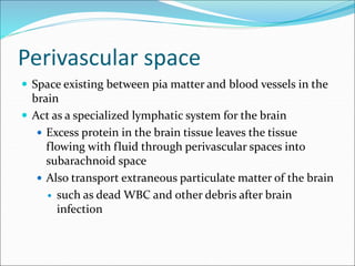
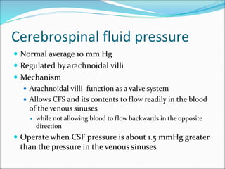
Clinical importance
 Abnormalities of
 Blood flow
 Metabolism
 Fluids
 Composition
 Pressure
 Affect brain function profoundly
 Decrease blood flow for 5-19sec
 loss of consciousness
 Increase in H+ ions
 depresses neuronal activity
 decreases brain activity
Outline
 Vascular anatomy of brain
 Control of cerebral blood flow
 Determinants of cerebral perfusion pressure
 Local regulation of cerebral blood flow
 Regulation of CBF by arterial pO2 and pCO2
 Neurohumoral regulation
 Cushing reflex
 Control by neuropeptides
 Conditions related to altered cerebral blood flow
Vascular Anatomy
(From E. Gardner, Fundamentals of Neurology. W.B. Saunders, 1963)
Circle
of Willis
Cerebral blood flow
 Normal blood flow 50-65mls/100gr/min
 Entire brain=750-900 mls/min
 15 % of resting cardiac ou put
Regulation of cerebral circulation
 Constant total cerebral blood flow is maintained
under varying conditions.
1. ABP at brain level.
2. Venous pressure at brain level.
3. Intracranial pressure.
4. Blood viscosity.
5. Degree of active constriction or dilation of cerebral
vessels.
Regulation of blood flow
 Highly related to tissue metabolism
1. Concentration of carbondioxide
2. Hydrogen ions concentration
3. Oxygen concentration
 Excess CO2 or H+
 Increase cerebral blood flow
 Vasodilator effect
 Indirect effect of CO2
 CO2 + H2O= H+ + HCO3-
 Others
 Metabolic acids
 Lactic acid & Pyruvic acid
Importance of cerebral blood flow
 Increased H+ conc greatly depresses neuronal activity
 Increased H+ leads to increased blood flow
 In turn carries H+ , CO2 and other acids forming
substances from the brain tissue
 Decreased H+ conc
 Achieves normal neuronal activity
 Oxygen deficiency
 Normal oxygen utilization 3.5+/- 0.2 mls O/100gr/min
 Decreased oxygen supply below normal
 Vasodilatation
 increased blood flow and O2 transport to the tissue
 Normal PO2 in cerebral blood is 35-40mmHg
 Decreased in cerebral tissue PO2 below 30mmHg
increase blood flow
 Below 20mmHg comma ensues
Measurement of blood flow
 Inject radio active substance in carotid artery
 Record radioactivity of each cerebral segment
 Press detectors against the surface of the cortex
 Detect rapidity of rise and decay of radioactivity in
each tissue segment
 Record increased blood flow where there is activity
2. Autoregulation of cerebral blood flow
Autoregulation:
 Ability of tissue to regulate their blood flow according
to their activity.
 When the arterial pressure changes
 CBF is auto regulated extremely well
 Between arterial pressure limits
 60-140mmHg
 there is no significant changes in cerebral blood flow
Cerebral Autoregulation (Description)
Cerebral
Blood
Flow
Mean Arterial Pressure (mmHg)
0 200
100
Autoregulatory
Range
Cerebral
Hypoxia
Headaches
BBB disruption
Edema
Cerebral Autoregulation (Autoregulatory
Shift)
Cerebral
Blood
Flow
Mean Arterial Pressure (mmHg)
0 200
100
Normal
Chronic Hypertension
Acute Sympathetic Stimulation
Cerebral Autoregulation
(Possible Mechanisms)
 Metabolic
 Decreased perfusion pressure leads to:
  pO2 (decreased O2 delivery)
  pCO2 (decreased CO2 washout)
  H+ (decreased H+ washout plus lactic acid)
  adenosine (hypoxia resulting in net loss of ATP)
 Each of the above changes produces vasodilation
 Myogenic
 Decreased perfusion pressure decreases stretching of
arteriolar smooth muscle which causes relaxation
3. Autonomic Control
 Sympathetic
 Innervation from superior cervical ganglion primarily
to larger cerebral arteries on brain surface
 Very weak sympathetic vascular tone
 Sympathetic blockade has little effect on flow
 Maximal sympathetic stimulation increases resistance
by 20-30% (cp >500% in muscle)
 Shifts autoregulatory curve to right
 Parasympathetic
 Innervation from facial nerve (VII)
 Weak dilator effect on pial vessels
 Baroreceptor reflexes
 Very weak
Sympathetic Control
Cerebral
Blood
Flow
(ml/min•100g)
Level of Sympathetic Activity
0
50
100
None Maximal
(From Lassen, N.A., Brain. In: Peripheral Circulation, P.C. Johnson, ed. Wiley, 1978)
Role of sympathetic NS in controlling CBF
 There is strong sympathetic innervations in the brain
 Inhibition or mild to moderate sympathetic
stimulation has mild or no effect in CBF changes
 Auto regulation overrides the nervous effect
 Important in preventing stroke
 Cerebral vascular accidents
 Incase of acute increase of Arterial pressure
 Strenuous exercise
 Excessive circulatory activity
 Constrict large and intermediate sized brain arteries
 High pressure is prevented reaching smaller brain vessels
 Prevent vascular hemorrhages
4. Effects of Intracranial Pressure
(CNS Ischemic Reflex)
 Increased intracranial pressure leads to
mechanical compression of cerebral vasculature
and decreased flow
 Increased intracranial pressure elicits arterial
hypertension (“Cushing reflex”)
 May be caused by bulbar ischemia, which in turn
stimulates medullary cardiovascular centers and
increases sympathetic outflow to systemic
vasculature
 Bradycardia often accompanies the hypertension
because of baroreceptor activation of vagal efferents
to the heart
5. Humoral Control
 Catecholamines
 Weak alpha-adrenergic vasoconstriction is masked by
autoregulation although very high doses of
epinephrine can decrease flow
 Beta-adrenoceptors cause vasodilation; however, this
is masked by autoregulation
 Angiotensin II
 Very little or no effect
Neuropeptides and Other Vascular
Control Mechanisms
 Vasodilation
 Calcitonin gene-related peptide (CGRP)
 Substance-P
 Vasoactive intestinal peptide (VIP)
 Vasoconstriction
 Neuropeptide-Y (NPY)
 Endothelin (vascular and neuronal ET-1 and neuronal ET-3
acting primarily on ETA receptors)
Neural Innervation of Cerebral
Vasculature
Cerebrospinal Fluid system
 Capacity of entire cerebral
cavity enclosing the brain
and spinal cord
 1600-1700 mls
 150mls is CSF
 1450-1550 mls Brain & spinal
cord
 Areas CSF formed
 Chambers in the brain
 Ventricles
 Cisterns around the outside
of the brain
 Subarachnoid space around
both & spinal cord
 Chambers are
interconnected
 Pressure of the fluid is
maintained at a constant
level
Function of Cerebrospinal Fluid
 The purpose of this fluid is
to protect the brain and
spinal cord
 acting as a shock absorber.
 It also carries away
disposed materials.

Functions of CSF, continued,…
2. Facilitation of pulsatile cerebral blood flow,
3. Distribution of peptides, hormones, neuroendocrine
factors and other nutrients and essential substances to
cells of the body,
4. Wash away waste products.
Formation, flow and absorption
 Rate per day
 500mls/day
 3-4 times its volume
 2/3rds secreted by choroid
plexus in ventricles
 Mainly 2 lateral ventricles
 Ependymal surface of all
ventricles
 Arachnoid membranes
 Brain itself
Composition of the CSF
 The composition of CSF is essentially the same as
brain ECF
Substance CSF Plasma
Na+ 147 150
K+ 2.9 4.6
HCO3- 25 24.8
PCO2 50 39.5
pH 7.33 7.4
Osmolality
Glucose
289
64
289
100
flow
1. From lateral ventricles
2. to 3rd ventricles
3. to Aqueduct of sylvius
4. to 4th ventricle
5. then passes through the three small openings
1. Two lateral foramina of lushka
2. Foramen magandie on the middle
3. And then enters cisterna magna
6. Then upwards through the subarachnoid space
surrounding the cerebrum
7. Finally to large sagital venous sinuses
Choroid plexus
 Is a cauliflower like growth of blood vessels covered by
a thin layer of epithelial cells
 Mechanism of CSF formation
 Na+ actively pumped outside the epithelial cells
 Pulls along with it Cl- ions
 Creates osmotically active environment
 Water flows by osmosis
Absorption of CSF
 Through arachnoidal villi
 Are microscopic fingerlike inward projections of
arachnoidal membrane
 Have vesicular passages in them that allows the
passage of
 CSF
 Dissolved protein molecules
 Particles as large as RBC & WBC
 into the venous blood
Perivascular space
 Space existing between pia matter and blood vessels in the
brain
 Act as a specialized lymphatic system for the brain
 Excess protein in the brain tissue leaves the tissue
flowing with fluid through perivascular spaces into
subarachnoid space
 Also transport extraneous particulate matter of the brain
 such as dead WBC and other debris after brain
infection
Cerebrospinal fluid pressure
 Normal average 10 mm Hg
 Regulated by arachnoidal villi
 Mechanism
 Arachnoidal villi function as a valve system
 Allows CFS and its contents to flow readily in the blood
of the venous sinuses
 while not allowing blood to flow backwards in the opposite
direction
 Operate when CSF pressure is about 1.5 mmHg greater
than the pressure in the venous sinuses
Disorders of CSF circulation
 Disease states
 Blocks the system
 Increase CSF pressure
1. Large brain tumors-
 decrease reabsorbption of CSF into the blood
 Increase 4x above normal
2. Haemorhage
3. Infection
 In cranial vault
 Release Large number of RBC+/- WBC in CSF
 block the system
Disorders of CSF circulation
4. Abnormal villi system
 Few arachnoid villi
 Or abnormal absorptive properties
 Hydrocephallus
 Excess water in the cranial vault
 Cause
 Obstruction of CSF flow
 Types
 Communicating
 Non communicating
Disorders of CSF circulation
 Communicating
 Fluid flow normal from ventricular system to arachnoid
spaces
 Non communicating
 There is blockage in fluid flow from one or two
ventricles
 Blockage of aqueduct of sylvius secondary top atresia
(closure) before birth
 Brain tumor at any age
 Communicating
 Blockage of arachnoid villi or subarachnoid spaces
 Fluids collects outside the brain
Brain barriers
1. Blood cerebral spinal fluid barrier
 Barrier formed between blood and cerebral spinal fluid
2. Blood brain barrier
 Barrier formed between blood and brain fluid
 Permeability Characteristics
 Highly permeable to H2O,CO2,O2, and most lipid
soluble substances e.g alcoholic & anaesthetics
 Slightly permeable to electrolytes Na+, Cl- and K+
 Glucose : its passive penetration is slow, but is
transported across brain capillaries by GLUT1
 totally impermeable to placenta proteins and non lipid
soluble large organic molecules
Cause of barrier
 Manner in which the endothelial cells of the brain
tissue capillaries are joined to one another
 Joined by tightly junctions
 Membranes of adjacent endothelial cells are tightly
fused rather than having large slit spores between
them
 Unlike other capillaries which have large pores
Functions of BBB
 Maintanins the constancy of the environment of the
neurons in the CNS.
 Protection of the brain from endogenous and
exogenous toxins.
 Prevent escape of the neurotransmitters into the
general circulation.
Development of BBB
 Premature infants with hyperbilirubinemia, free
bilirubin pass BBB, and may stain basal ganglia
causing damage (Kernicterus).
Clinical implications
 Some drugs penetrate BBB with difficulty e.g.
antibiotics and dopamine.
 BBB breaks down in areas of infection, injury, tumors,
sudden increase in blood pressure, and I.V injection of
hypertonic fluids.
 Injection of radiolabeled materials help diagnose
tumors as BBB is broken down at tumor site because of
increased vascularity by abnormal vessels.
Circumventricular organs
• Posterior pituitary.
• Area postrema.
• Organum vasculosum of the lamina terminalis (OVLT).
• Subfornical organ (SFO).
These areas are outside the blood brain barrier. They have
fenestrated capillaries .
Functions:
- Chemoreceptor trigger zone. As area postrema that trigger
vomiting & cardiovascular control.
- Ang II acts on SFO and OVLT to increase H2O intake.
- IL2 induce fever by (+) circumventricular organs.

CSF FLOW.ppt

  • 2.
    Clinical importance  Abnormalitiesof  Blood flow  Metabolism  Fluids  Composition  Pressure  Affect brain function profoundly  Decrease blood flow for 5-19sec  loss of consciousness  Increase in H+ ions  depresses neuronal activity  decreases brain activity
  • 3.
    Outline  Vascular anatomyof brain  Control of cerebral blood flow  Determinants of cerebral perfusion pressure  Local regulation of cerebral blood flow  Regulation of CBF by arterial pO2 and pCO2  Neurohumoral regulation  Cushing reflex  Control by neuropeptides  Conditions related to altered cerebral blood flow
  • 4.
    Vascular Anatomy (From E.Gardner, Fundamentals of Neurology. W.B. Saunders, 1963) Circle of Willis
  • 5.
    Cerebral blood flow Normal blood flow 50-65mls/100gr/min  Entire brain=750-900 mls/min  15 % of resting cardiac ou put
  • 6.
    Regulation of cerebralcirculation  Constant total cerebral blood flow is maintained under varying conditions. 1. ABP at brain level. 2. Venous pressure at brain level. 3. Intracranial pressure. 4. Blood viscosity. 5. Degree of active constriction or dilation of cerebral vessels.
  • 7.
    Regulation of bloodflow  Highly related to tissue metabolism 1. Concentration of carbondioxide 2. Hydrogen ions concentration 3. Oxygen concentration  Excess CO2 or H+  Increase cerebral blood flow  Vasodilator effect  Indirect effect of CO2  CO2 + H2O= H+ + HCO3-  Others  Metabolic acids  Lactic acid & Pyruvic acid
  • 8.
    Importance of cerebralblood flow  Increased H+ conc greatly depresses neuronal activity  Increased H+ leads to increased blood flow  In turn carries H+ , CO2 and other acids forming substances from the brain tissue  Decreased H+ conc  Achieves normal neuronal activity  Oxygen deficiency  Normal oxygen utilization 3.5+/- 0.2 mls O/100gr/min  Decreased oxygen supply below normal  Vasodilatation  increased blood flow and O2 transport to the tissue
  • 9.
     Normal PO2in cerebral blood is 35-40mmHg  Decreased in cerebral tissue PO2 below 30mmHg increase blood flow  Below 20mmHg comma ensues
  • 10.
    Measurement of bloodflow  Inject radio active substance in carotid artery  Record radioactivity of each cerebral segment  Press detectors against the surface of the cortex  Detect rapidity of rise and decay of radioactivity in each tissue segment  Record increased blood flow where there is activity
  • 11.
    2. Autoregulation ofcerebral blood flow Autoregulation:  Ability of tissue to regulate their blood flow according to their activity.  When the arterial pressure changes  CBF is auto regulated extremely well  Between arterial pressure limits  60-140mmHg  there is no significant changes in cerebral blood flow
  • 12.
    Cerebral Autoregulation (Description) Cerebral Blood Flow MeanArterial Pressure (mmHg) 0 200 100 Autoregulatory Range Cerebral Hypoxia Headaches BBB disruption Edema
  • 13.
    Cerebral Autoregulation (Autoregulatory Shift) Cerebral Blood Flow MeanArterial Pressure (mmHg) 0 200 100 Normal Chronic Hypertension Acute Sympathetic Stimulation
  • 14.
    Cerebral Autoregulation (Possible Mechanisms) Metabolic  Decreased perfusion pressure leads to:   pO2 (decreased O2 delivery)   pCO2 (decreased CO2 washout)   H+ (decreased H+ washout plus lactic acid)   adenosine (hypoxia resulting in net loss of ATP)  Each of the above changes produces vasodilation  Myogenic  Decreased perfusion pressure decreases stretching of arteriolar smooth muscle which causes relaxation
  • 15.
    3. Autonomic Control Sympathetic  Innervation from superior cervical ganglion primarily to larger cerebral arteries on brain surface  Very weak sympathetic vascular tone  Sympathetic blockade has little effect on flow  Maximal sympathetic stimulation increases resistance by 20-30% (cp >500% in muscle)  Shifts autoregulatory curve to right  Parasympathetic  Innervation from facial nerve (VII)  Weak dilator effect on pial vessels  Baroreceptor reflexes  Very weak
  • 16.
    Sympathetic Control Cerebral Blood Flow (ml/min•100g) Level ofSympathetic Activity 0 50 100 None Maximal (From Lassen, N.A., Brain. In: Peripheral Circulation, P.C. Johnson, ed. Wiley, 1978)
  • 17.
    Role of sympatheticNS in controlling CBF  There is strong sympathetic innervations in the brain  Inhibition or mild to moderate sympathetic stimulation has mild or no effect in CBF changes  Auto regulation overrides the nervous effect  Important in preventing stroke  Cerebral vascular accidents  Incase of acute increase of Arterial pressure  Strenuous exercise  Excessive circulatory activity  Constrict large and intermediate sized brain arteries  High pressure is prevented reaching smaller brain vessels  Prevent vascular hemorrhages
  • 18.
    4. Effects ofIntracranial Pressure (CNS Ischemic Reflex)  Increased intracranial pressure leads to mechanical compression of cerebral vasculature and decreased flow  Increased intracranial pressure elicits arterial hypertension (“Cushing reflex”)  May be caused by bulbar ischemia, which in turn stimulates medullary cardiovascular centers and increases sympathetic outflow to systemic vasculature  Bradycardia often accompanies the hypertension because of baroreceptor activation of vagal efferents to the heart
  • 19.
    5. Humoral Control Catecholamines  Weak alpha-adrenergic vasoconstriction is masked by autoregulation although very high doses of epinephrine can decrease flow  Beta-adrenoceptors cause vasodilation; however, this is masked by autoregulation  Angiotensin II  Very little or no effect
  • 20.
    Neuropeptides and OtherVascular Control Mechanisms  Vasodilation  Calcitonin gene-related peptide (CGRP)  Substance-P  Vasoactive intestinal peptide (VIP)  Vasoconstriction  Neuropeptide-Y (NPY)  Endothelin (vascular and neuronal ET-1 and neuronal ET-3 acting primarily on ETA receptors)
  • 21.
    Neural Innervation ofCerebral Vasculature
  • 22.
    Cerebrospinal Fluid system Capacity of entire cerebral cavity enclosing the brain and spinal cord  1600-1700 mls  150mls is CSF  1450-1550 mls Brain & spinal cord  Areas CSF formed  Chambers in the brain  Ventricles  Cisterns around the outside of the brain  Subarachnoid space around both & spinal cord  Chambers are interconnected  Pressure of the fluid is maintained at a constant level
  • 23.
    Function of CerebrospinalFluid  The purpose of this fluid is to protect the brain and spinal cord  acting as a shock absorber.  It also carries away disposed materials. 
  • 24.
    Functions of CSF,continued,… 2. Facilitation of pulsatile cerebral blood flow, 3. Distribution of peptides, hormones, neuroendocrine factors and other nutrients and essential substances to cells of the body, 4. Wash away waste products.
  • 25.
    Formation, flow andabsorption  Rate per day  500mls/day  3-4 times its volume  2/3rds secreted by choroid plexus in ventricles  Mainly 2 lateral ventricles  Ependymal surface of all ventricles  Arachnoid membranes  Brain itself
  • 26.
    Composition of theCSF  The composition of CSF is essentially the same as brain ECF Substance CSF Plasma Na+ 147 150 K+ 2.9 4.6 HCO3- 25 24.8 PCO2 50 39.5 pH 7.33 7.4 Osmolality Glucose 289 64 289 100
  • 27.
    flow 1. From lateralventricles 2. to 3rd ventricles 3. to Aqueduct of sylvius 4. to 4th ventricle 5. then passes through the three small openings 1. Two lateral foramina of lushka 2. Foramen magandie on the middle 3. And then enters cisterna magna 6. Then upwards through the subarachnoid space surrounding the cerebrum 7. Finally to large sagital venous sinuses
  • 29.
    Choroid plexus  Isa cauliflower like growth of blood vessels covered by a thin layer of epithelial cells  Mechanism of CSF formation  Na+ actively pumped outside the epithelial cells  Pulls along with it Cl- ions  Creates osmotically active environment  Water flows by osmosis
  • 30.
    Absorption of CSF Through arachnoidal villi  Are microscopic fingerlike inward projections of arachnoidal membrane  Have vesicular passages in them that allows the passage of  CSF  Dissolved protein molecules  Particles as large as RBC & WBC  into the venous blood
  • 31.
    Perivascular space  Spaceexisting between pia matter and blood vessels in the brain  Act as a specialized lymphatic system for the brain  Excess protein in the brain tissue leaves the tissue flowing with fluid through perivascular spaces into subarachnoid space  Also transport extraneous particulate matter of the brain  such as dead WBC and other debris after brain infection
  • 32.
    Cerebrospinal fluid pressure Normal average 10 mm Hg  Regulated by arachnoidal villi  Mechanism  Arachnoidal villi function as a valve system  Allows CFS and its contents to flow readily in the blood of the venous sinuses  while not allowing blood to flow backwards in the opposite direction  Operate when CSF pressure is about 1.5 mmHg greater than the pressure in the venous sinuses
  • 33.
    Disorders of CSFcirculation  Disease states  Blocks the system  Increase CSF pressure 1. Large brain tumors-  decrease reabsorbption of CSF into the blood  Increase 4x above normal 2. Haemorhage 3. Infection  In cranial vault  Release Large number of RBC+/- WBC in CSF  block the system
  • 34.
    Disorders of CSFcirculation 4. Abnormal villi system  Few arachnoid villi  Or abnormal absorptive properties  Hydrocephallus  Excess water in the cranial vault  Cause  Obstruction of CSF flow  Types  Communicating  Non communicating
  • 35.
    Disorders of CSFcirculation  Communicating  Fluid flow normal from ventricular system to arachnoid spaces  Non communicating  There is blockage in fluid flow from one or two ventricles  Blockage of aqueduct of sylvius secondary top atresia (closure) before birth  Brain tumor at any age  Communicating  Blockage of arachnoid villi or subarachnoid spaces  Fluids collects outside the brain
  • 36.
    Brain barriers 1. Bloodcerebral spinal fluid barrier  Barrier formed between blood and cerebral spinal fluid 2. Blood brain barrier  Barrier formed between blood and brain fluid  Permeability Characteristics  Highly permeable to H2O,CO2,O2, and most lipid soluble substances e.g alcoholic & anaesthetics  Slightly permeable to electrolytes Na+, Cl- and K+  Glucose : its passive penetration is slow, but is transported across brain capillaries by GLUT1  totally impermeable to placenta proteins and non lipid soluble large organic molecules
  • 37.
    Cause of barrier Manner in which the endothelial cells of the brain tissue capillaries are joined to one another  Joined by tightly junctions  Membranes of adjacent endothelial cells are tightly fused rather than having large slit spores between them  Unlike other capillaries which have large pores
  • 38.
    Functions of BBB Maintanins the constancy of the environment of the neurons in the CNS.  Protection of the brain from endogenous and exogenous toxins.  Prevent escape of the neurotransmitters into the general circulation.
  • 39.
    Development of BBB Premature infants with hyperbilirubinemia, free bilirubin pass BBB, and may stain basal ganglia causing damage (Kernicterus).
  • 40.
    Clinical implications  Somedrugs penetrate BBB with difficulty e.g. antibiotics and dopamine.  BBB breaks down in areas of infection, injury, tumors, sudden increase in blood pressure, and I.V injection of hypertonic fluids.  Injection of radiolabeled materials help diagnose tumors as BBB is broken down at tumor site because of increased vascularity by abnormal vessels.
  • 41.
    Circumventricular organs • Posteriorpituitary. • Area postrema. • Organum vasculosum of the lamina terminalis (OVLT). • Subfornical organ (SFO). These areas are outside the blood brain barrier. They have fenestrated capillaries . Functions: - Chemoreceptor trigger zone. As area postrema that trigger vomiting & cardiovascular control. - Ang II acts on SFO and OVLT to increase H2O intake. - IL2 induce fever by (+) circumventricular organs.