
Recommended
Recommended
More Related Content
Similar to CT Head (Cerebral) Angiography.pptx, internal carotid artery. 2- vertebral artery. Aneurysm Narrowing of artery in the brain, Abnormal blood vessels
Similar to CT Head (Cerebral) Angiography.pptx, internal carotid artery. 2- vertebral artery. Aneurysm Narrowing of artery in the brain, Abnormal blood vessels (20)
More from RukamaneeYadav
More from RukamaneeYadav (10)
Recently uploaded
Recently uploaded (20)
CT Head (Cerebral) Angiography.pptx, internal carotid artery. 2- vertebral artery. Aneurysm Narrowing of artery in the brain, Abnormal blood vessels
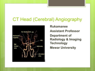
- 1. CT Head (Cerebral) Angiography Rukamanee Assistant Professor Department of Radiology & Imaging Technology Mewar University
- 2. What is Cerebral Angiography Study of blood vessels in the body by injecting a contrast media through particular arteries and veins that is called the angiography. Cerebral angiography is a procedure that use the contrast media and CT equipment to see the blood flow of brain. And diagnosis the brain pathology. This is minimally invasive procedure.
- 3. Anatomy of Head Arterial supply of the brain. Major blood supply to the brain by the two artery- 1- internal carotid artery. 2- vertebral artery. Internal carotid artery arise from the point of neck. These arteries are in pairs and supply the blood in both side of brain. The major blood supply by internal carotid artery to cerebrum. 80 percent. It is the part of front brain.
- 4. Blood supply of brain
- 5. Indications Aneurysm Narrowing of artery in the brain Abnormal blood vessels Investigating source of haemorrhage. Blockage of carotid artery Stenosis
- 6. Conti.. To determine future risk of stroke Severe headaches Slurred speech Dizziness Blurred or double vision weakness or numbness Loss of coordination or balance.
- 7. Contraindication Pregnancy Unstable vital sign Allergic patient Kidney problems Severe diabetes Hepatic failure Patient declined consent
- 8. Equipments CT SCAN equipment with control console Contrast media Needle (18 gauge) Surgical blade Saline Heparin Local anesthesia Elastoplast Gloves Gown Anti-allergic drugs Pressure injector
- 9. Patient Preparation Take previous history and clinical examination. Informed consent. Patient should be well hydrated. Fasting 4 hours prior to procedure. Shave and clean the arterial puncture site. Xylocaine sensitivity test.
- 10. Con…. Following investigations to be done : Hb% and and platelet count ESR PT, (prothrombin time) BT and CT HBsAg (hepatitis B surface Antigen) Pulse chart Any history of drug intake. History of diabetes .
- 11. Patient Position Patient position on CT scan table in supine position. Head first and arm beside the trunk patient head will place using a strap, tape or a foam head holder so patient cannot move it during the procedure. Placed the IV cannula .(18 gauge) To ensure that cannula is not block. Scan orientation –caudo cranial
- 12. INJECTION OF CONTRAST WITH NEEDLE IN SITU Feel the inguinal ligament. Feel the artery and fix it with three fingers of left hand below the inguinal ligament. Inject the local anaesthetic agent at the site of the puncture . Make a hole in skin using a thick needle 2½ cm (1 inch) below the inguinal ligament or give a stab incision. Then inject the contrast media. Volume of contrast media- Then take the image.
- 13. Complications 1.Due to local anaesthesia (a) Allergic (b) Toxic-due to injection in the vein, e.g.cardiac arrhythmias 2. Due to contrast media Mild Moderate Severe contrast reactions Pain may follow injection of contrast material into the vessel.
- 14. TOPOGRAM IMAGE
- 15. Conti…..
- 16. Post processing technique Commonly used technique- MIP (Maximum Intensity Projection) MPR (Multiplanar Reconstruction) VRT (Volume Rendering Technique)
- 17. Advantages CT angiography may give more accurate anatomical details compare to MRI . Minimal invasive and minimum risk . Low cost examination. Aneurysms can be best diagnosed by angiography. Many patient can undergo CT angiography instead of catheter angiography to daignose the blood vessels.
- 18. Disadvantages Radiation exposure Risk of contrast reactions.
- 19. After care Bed rest Keep the puncture part without moving for atleast 6-8 hrs. Watch for any recurrence of bleeding. Peripheral pulses should be monitored/Vitals monitored. Hydrate the patient well. Deterioration of renal function should be watched .
- 20. THANK YOU