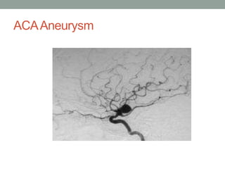
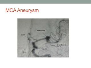
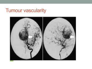
SlideShare a Scribd company logo
CEREBRAL
ANGIOGRAPHY(DSA)-
TECHNIQUEAND
NORMALANATOMY
NORMALANATOMY
• ARTERIAL SUPPLY
• 2 Internal Carotid Artery
• 2 Vertebral Arteries
CIRCLE OF WILLIS
• Grand Vascular Station of the Brain
• Classical –18% to 20%
• Majority circles shows anomaly-52%
• Most frequent anomaly is hypoplasia of ACA-24%
• Accesory vessels in the form of duplication/triplicationsof
ACOM (2 M.C.) -12%
Fetal posterior cerebral artery-10%
CIRCLE OF WILLIS
COMPONENTS:
• Internal carotid arteries
• Horizontal segments of Anterior cerebrals(A1)
• Anterior communicating artery
• Proximal segments of posterior cerebrals(P1)
• Posterior communicating arteries
• Basilar artery
Branches of ACA
• A1- medial lenticulostriate artery
• ACoA- Perforating branches
• A2- Recurrent artery of Heubner (RAH)
• Orbitofrontal artery
• Frontopolar artery
• A3- Pericallosal and Callosomarginal a.
• A4 & A5- Cortical branches
ACA
MIDDLE CEREBRALARTERY
• Larger terminal branch of ICA
• Run laterally in stem of lateral sulcus
• Curves on superolateral surface
• Runs backwards in depth of posterior ramus of lateral
sulcus
• M1 segment =horizontal segment from origin to its
bifurcation (it is in sylvian fissure)
• M2 segment =lacunar segment -in the insula loops over
insula—laterally to exit from sylvian fissure
• M3 segment = opercular branch-from sylvian fissure &
ramify over cerebral cortex
• Anomalies of MCA are uncommon
POSTERIOR CEREBRALARTERY
 P1-Peduncular/Precommunicating
 P2-Ambient segment
 P3-Quadrigeminal segment
 P4-Cortical branches
• 2 major terminal br of PCA—
•
• parieto occipital art & calcarine art
POSTERIOR FOSSA
• Vertebral artery
• Basilar artrery
• Vertebral arteries
• Originate from the subclavian arteries
• Left VA is dominant in 60% cases
Branches of vertebral artery
• Extracranial -numerous branches to the meninges,spinal
cord & muscles
• -Posterior meningeal artery
• Intracranial-
-Anterior spinal artery
• -Posterior inferior cerebellar Art.
• Anterior medullary
• Lateral medullary
• Tonsillomedullary
• Telovelotonsillar
• Cortical branches
BASILAR ARTERY
• Right and left VA unite to form basilar artery
• Courses infront of pons (Prepontine cistern)
terminates in the interpeduncular cistern
• 3cm in length,1.5 to 4mm in width
Branches of basilar artery
• Anterior inferior cerebellar artery (AICA)
• Superior cerebellar artery
• Pontine artery
• Labyrinthine artery
Normal VARIANTS
• Fenestrations and duplications
• Variants of the circle of Willis
• Persistent carotid-basilar anastomoses
• Anomalies identified in the skull base
Normal Variants of the Circle of Willis
CEREBRALANGIOGRAPHY
• Angiography –
• Radiological study of blood vessel in the body
after the introduction of iodinated contrast
media.
Imaging Techniques
• Vascular structures of brain can be imaged by 4
means:
• 1. DSA: gold standard
• Invasive and risk of nephrotoxic contrast,ionising
radiation
• 2. Vascular ultrasound: least invasive, can be
done bedside, cost effective.
• Best choice for imaging vessels close to skin
surface.
• Drawback: limited anatomic coverage, deep
vessels cant be imaged, operator dependent,
requires skill.
• 3 CT angio: main drawbacks are contrast use and
radiation exposure,
• calcifications are overestimated.
• Preferred for aorta and coronaries
• 4 MRA : non invasive, no radiation exposure.
• Preferred for carotids and intracranial vessels as MRI
brain can also be obtained
• widely used in neurological disorders
• ANGIO?
• ‘ANGIO means blood vessel’
• SUBTRACTION?
• It is simply a technique by which bone
structures images are subtracted or canceled
out from a film of bones plus opacified vessels,
“leaving an unobscured image of the vessels”
DSA
• Acquisition of digital fluoroscopic images
combined with injection of contrast material
and real time subtraction of pre- and post
contrast images to perform angiography is
referred to as digital subtraction angiography
HISTORY
Portuguese neurologist
Prize winner 1949), in
Egas Moniz,( Nobel
1927 developed the
technique of contrast x-ray cerebral angiography
to diagnose diseases,
such as tumors and arteriovenous
malformations.
• Idea of subtraction images was first proposed
by the Dutch radiologist Ziedses des Plantes in
the 1935, when he was able to produce
subtracted images using plain films.
HISTORICAL DEVELOPMENT
• CONVENTIONAL SUBTRACTION TECHNIQUE:
• Photographic method used to eliminate
unwanted images.
• No addition of information; only purpose to
make diagnostically important information
• Developed in 1970s, University of Wisconsin,
University of Arizona, University of Kiel.
• USA
• Commercial systems introduced in 1980.
• 3 conditions:
• SCOUT FILM
• ANGIOGRAM FILM-CONTRAST
• NO MOTION OF HEAD
PRINCIPAL
• Principles of subtraction
following:
are based on the
• Scout film shows the structural details of the
skull and the adjacent soft tissue.
• Angiogram film shows exactly the same
anatomic details, if the patient does not move,
plus the opacified blood vessels.
• If all the information in the scout film could be
subtracted from the angiogram film, only the
opacified vessel pattern would remain visible.
DIGITAL IMAGE PROCESSOR
WHY DSA?
• Digital subtraction angiography (DSA) was developed
to improve vessel contrast.
• Technique that uses a computer to subtract two
images, obtained before and after contrast media is
injected into the vessels of interest.
• Anatomical structures that are the same in the two
images can be removed and the resulting image
shows the vessels only
• Modern DSA systems are based on digital
fluoroscopy/fluorography systems, which are
equipped with special software and display
facilities
• Image before the contrast agent is
administered is called the mask image.
•
• Once the contrast is administered, a sequence
of images are taken by a television camera in
analog form, which is then digitised by
computer
• DSA processor has two separate image
memories, one for the mask and the other for
the images with contrast medium.
• These two image memories are subtracted
from one another arithmetically, and the result
goes to an image processing and display unit.
Indications
•Diagnostic:
traumatic
• Non
(SAH)
• Arterial dissection
• laceration
• Aneurysm
Subarachnoid Hemorrhage
Indications
• Pseudoaneurysm
• Thrombosis (Extracranial & Intracranial)
• Arterio-venous malformation (AVM).
• Arterio-venous fistula (AVF).
• Tumor vascularity
•Therapeutic:
• Embolisation
• Stenting
• Thrombolysis
• Thrombectomy
Contra indications
• No absolute contraindication.
• Poor renal reserve.
• Deranged coagulogram.
• Allergic to contrast media
Contrast Media
• Blood vessels are not normally seen in an x-ray
image, because of low tissue contrast.
• To increase image contrast, contrast agents,
which are dense fluids with elements of high
atomic numbers, such as iodine, are injected into
a blood vessel during angiography. Because of its
higher density and high atomic number, iodine
absorbs photons more than blood and tissue.
Creates detailed images of the blood vessels in
real time.
• First contrast media used for intravascular
injection were called high-osmolar contrast
media (HOCM).
• High osmolarity caused adverse effects such as
pain, endothelial damage, thrombosis, and
increased pressure in the pulmonary circulation.
• Low-osmolar contrast media (LOCM) were first
developed in the 1970's reduce side effects.
• Major risks of modern iodine contrast media is an
allergic reaction to iodine
• Non ionic Iso-osmolar contrast media.
• 30-40% dilution with normal saline.
• 50 ml of diluted contrast media is enough to do
a standard cerebral angiogram with total 8
projections.
• Approx. 5-8 ml diluted contrast / injection.
Materials used
• Catheters
• Arterial sheath
• Medicut
• Guidewires
• Contrast
• Connector/100
cm.tubing
• Surgical blade
• Saline
• Disposable syringes
• Local anesthesia
• Heparin
• Surgical gloves
• Elastoplast
Picard Pigtail
Sim -1
•
Guide Wires Medicut
PREPARATION
• Nil orally 4-6 hrs.
• On trolley
• In hospital gown
• Groin shave
• Records
• Should be well
hydrated.
• Should void before
procedure.
• Peripheral pulses
marked.
• I.V line in place.
• Informed consent
PROCEDURE
• Gaining arterial access.
• Selective arterial catheterization.
• Image acquisition.
• Closure of arterial access.
• Post processing
• Hard copy
• Patients may be sedated to reduce anxiety.
• Monitor of vitals
• Local anesthetic is usually used in the area
where the catheter is to be inserted,
• Most common femoral artery
PROCEDURE
• Small incision given, medicut is inserted into the artery,
• Fluoroscopy is used to guide the needle to the proper
position .
• Needle is then removed after placing guide wire in the
artery and vascular sheath is inserted over the guide wire
.
• Catheter is then inserted along the guide wire through the
sheath
• When the catheter is in the correct position, the wire is
pulled out and dye is injected through the catheter.
• Images are acquired during contrast injection.
• Injections can be made directly into the artery of interest
(selective arteriography)
COMPLICATIONS
• 0.16% major complication rate.
vessel
• Local complications: hematoma,
dissection, pseudoaneurysm ,AVfistula.
• Systemic complications: contrast
sepsis, dehydration, death.
• CNS complication: aggravation
complaints, neurological deficit.
laceration,
reactions, fever,
of preexisting
POSTPROCEDURAL CARE
• After the catheter is removed compression is applied to
the puncture site
• Bed rest for a minimum of 4 hours
• During rest patient is monitored and vital sign like
peripheral pulse like distal to Puncture are regularly
• Extremity is also checked for warmth, color, numbness to
ensure circulation has not been disrupted.
Cerebro-vascular anatomy as
appreciated in DSA
Diagnostic usage
• Acom aneurysm:
ACAAneurysm
MCAAneurysm
Pcom Aneurysm
AV Malformation
Tumour vascularity
Digital subtraction angiography based
advances
• Better visualization and less radiation exposure
are key tenets in the development of digital
subtraction angiography
• Complexity of disease types increases and
more technically challenging, cutting-edge
procedures are performed
• Digital subtraction angiography systems with flat
panel : ADVANTAGES-
• High spatial resolution, wide dynamic range,
square field of view, and real-time imaging
capabilities with no geometric distortion—all of
which may be used for improve image quality,
reduced patient exposure to radiation
• Hatakeyama et al have shown that two-dimensional (2D)
and three-dimensional (3D)digital subtraction angiography
using the flat panel detector system of the direct
conversion type, with low radiation dose, is superior in
image quality for visualizing small intracranial vessels with
significantly decreased radiation exposure compared with
digital subtraction angiography with the conventional
image intensifier television system
• Hatakeyama Y
,Kakeda S, Korogi Y
,et al. Intracranial 2D and 3DDSA with flat panel detector of the
direct conversion type: initial experience. Eur Radiol 2006;16:2594 –2602.
Digital subtraction techniques
• Mask subtraction
• Dual energy subtraction
• Time interval differencing
• Temporal filtering
• Allura Xper FD20/10 biplane flat detector
system with integrated 3D for intricate
neurovascular procedures.
• 3D- reconstructions
• Xper CT
• SPECTRA BEAM
• 3D Roadmapping
• multi-modality information integration
• Redefines image clarity and captures
information at a resolution four times greater
than conventional X-ray systems.
Xper CT
• With XperCT clinicians can access CT-like imaging right
on the angio system so can assess soft tissue, bone
structure and other body structures before, during or after
an interventional procedure.
• XperCT reconstruction is created from rotational
acquisition performed on the Allura Xper system.
• This reconstruction can be overlaid with the 3D vascular
image.
• 3D soft tissue imaging supports diagnosis
planning, interventions and treatment follow-up
• XperCT can be combined with Allura 3D-RA
images to visualize soft tissue and vascular
anatomy on one image.
Spectra Beam
• Basically a selectable copper beam filtration
• Combination of Spectra Beam with the MRC-
tube allows increased X-ray output with better
filtration of soft radiation.
• Reduces patient X-ray dose for cardiac and
vascular applications, while maintaining the same
image quality.
Disadvantage of DSA
• Limited spatial resolution
• Artifacts
• Small visual field
• Not a good technique for neoplasm
Conclusion
• Despite recent advances in CT angiography and
MR angiography, DSA remains the standard
imaging technique for evaluation of the
cerebral vasculature .
• 3D reconstruction
during rotational
of the dataset acquired
DSA represents the latest
development in the neurovascular imaging .
has taken a prominent role in
• 3D-DSA
treatment planning by enabling better
appreciation of the morphology of complex
vascular lesions before endovascular or surgical
management.
• Superior in the performance of sophisticated
tasks such as aneurysm volume measurement
• On the other hand, inability of 3D-DSA to
simultaneously image osseous and vascular
structures is noted as a weakness of this
technique compared with CT angiography .
• THANK YOU

More Related Content

Similar to cerebralangiography.pptx

Carotid endarterectomy
Carotid endarterectomyCarotid endarterectomy
Carotid endarterectomy
Dheeraj Sharma
 
Cardiovascular testing 1
Cardiovascular testing 1Cardiovascular testing 1
Cardiovascular testing 1
Dr Madhuri Rudraraju
 
CAROTID DOPPLER STUDY
CAROTID DOPPLER STUDYCAROTID DOPPLER STUDY
CAROTID DOPPLER STUDY
Dr I Gurubharath .
 
Carotid doppler Ultrasound
Carotid doppler UltrasoundCarotid doppler Ultrasound
Carotid doppler Ultrasound
Syed Yousaf Gilani
 
Intravenous Ultrasound
Intravenous UltrasoundIntravenous Ultrasound
Intravenous Ultrasound
Dhileeban Maharajan
 
Orbital imaging (X-RAY,CT SCAN,AND MRI)
Orbital imaging (X-RAY,CT SCAN,AND MRI)Orbital imaging (X-RAY,CT SCAN,AND MRI)
Orbital imaging (X-RAY,CT SCAN,AND MRI)
Prashant Patel
 
Vascular Interventions and their Clinical Applications
Vascular Interventions and their Clinical ApplicationsVascular Interventions and their Clinical Applications
Vascular Interventions and their Clinical Applications
Dr.Anmol Uberoi
 
7th round seminar
7th round seminar7th round seminar
7th round seminar
farzana khantoon
 
Peripheral angiography
Peripheral angiographyPeripheral angiography
Peripheral angiography
InosRagan
 
CAROTID DOPPLER BY DR NITIN WADHWANI
CAROTID DOPPLER BY DR NITIN WADHWANICAROTID DOPPLER BY DR NITIN WADHWANI
CAROTID DOPPLER BY DR NITIN WADHWANI
dypradio
 
ANATOMY EMBRYOLOGY AND IMAGING OF SVC.pptx
ANATOMY EMBRYOLOGY AND IMAGING OF SVC.pptxANATOMY EMBRYOLOGY AND IMAGING OF SVC.pptx
ANATOMY EMBRYOLOGY AND IMAGING OF SVC.pptx
GajananWattamwar1
 
ANATOMY EMBRYOLOGY AND IMAGING OF SVC.pptx
ANATOMY EMBRYOLOGY AND IMAGING OF SVC.pptxANATOMY EMBRYOLOGY AND IMAGING OF SVC.pptx
ANATOMY EMBRYOLOGY AND IMAGING OF SVC.pptx
GajananWattamwar1
 
Qi carotid study 5 10-12 -cme
Qi carotid study 5 10-12 -cmeQi carotid study 5 10-12 -cme
Qi carotid study 5 10-12 -cme
inertia182
 
Cerebral aneurysm
Cerebral aneurysm Cerebral aneurysm
Cerebral aneurysm
Milan Silwal
 
cerebral aneurysm mohammad abu sad (1).pptx
cerebral aneurysm mohammad abu sad (1).pptxcerebral aneurysm mohammad abu sad (1).pptx
cerebral aneurysm mohammad abu sad (1).pptx
MohamadAbusaad
 
Carotid Cavernous Fistulas
Carotid Cavernous FistulasCarotid Cavernous Fistulas
Carotid Cavernous Fistulas
RejoyceAnto
 
Venography
VenographyVenography
Venography
SUJAN KARKI
 
PEDIATRIC Cardiac ct
PEDIATRIC Cardiac ctPEDIATRIC Cardiac ct
PEDIATRIC Cardiac ct
Indhu Reddy
 
Basics srikanths angio
Basics srikanths angioBasics srikanths angio
Basics srikanths angio
Srikanth Yadav
 
Arteriography and interventional radiology
Arteriography and interventional radiologyArteriography and interventional radiology
Arteriography and interventional radiology
Milan Silwal
 

Similar to cerebralangiography.pptx (20)

Carotid endarterectomy
Carotid endarterectomyCarotid endarterectomy
Carotid endarterectomy
 
Cardiovascular testing 1
Cardiovascular testing 1Cardiovascular testing 1
Cardiovascular testing 1
 
CAROTID DOPPLER STUDY
CAROTID DOPPLER STUDYCAROTID DOPPLER STUDY
CAROTID DOPPLER STUDY
 
Carotid doppler Ultrasound
Carotid doppler UltrasoundCarotid doppler Ultrasound
Carotid doppler Ultrasound
 
Intravenous Ultrasound
Intravenous UltrasoundIntravenous Ultrasound
Intravenous Ultrasound
 
Orbital imaging (X-RAY,CT SCAN,AND MRI)
Orbital imaging (X-RAY,CT SCAN,AND MRI)Orbital imaging (X-RAY,CT SCAN,AND MRI)
Orbital imaging (X-RAY,CT SCAN,AND MRI)
 
Vascular Interventions and their Clinical Applications
Vascular Interventions and their Clinical ApplicationsVascular Interventions and their Clinical Applications
Vascular Interventions and their Clinical Applications
 
7th round seminar
7th round seminar7th round seminar
7th round seminar
 
Peripheral angiography
Peripheral angiographyPeripheral angiography
Peripheral angiography
 
CAROTID DOPPLER BY DR NITIN WADHWANI
CAROTID DOPPLER BY DR NITIN WADHWANICAROTID DOPPLER BY DR NITIN WADHWANI
CAROTID DOPPLER BY DR NITIN WADHWANI
 
ANATOMY EMBRYOLOGY AND IMAGING OF SVC.pptx
ANATOMY EMBRYOLOGY AND IMAGING OF SVC.pptxANATOMY EMBRYOLOGY AND IMAGING OF SVC.pptx
ANATOMY EMBRYOLOGY AND IMAGING OF SVC.pptx
 
ANATOMY EMBRYOLOGY AND IMAGING OF SVC.pptx
ANATOMY EMBRYOLOGY AND IMAGING OF SVC.pptxANATOMY EMBRYOLOGY AND IMAGING OF SVC.pptx
ANATOMY EMBRYOLOGY AND IMAGING OF SVC.pptx
 
Qi carotid study 5 10-12 -cme
Qi carotid study 5 10-12 -cmeQi carotid study 5 10-12 -cme
Qi carotid study 5 10-12 -cme
 
Cerebral aneurysm
Cerebral aneurysm Cerebral aneurysm
Cerebral aneurysm
 
cerebral aneurysm mohammad abu sad (1).pptx
cerebral aneurysm mohammad abu sad (1).pptxcerebral aneurysm mohammad abu sad (1).pptx
cerebral aneurysm mohammad abu sad (1).pptx
 
Carotid Cavernous Fistulas
Carotid Cavernous FistulasCarotid Cavernous Fistulas
Carotid Cavernous Fistulas
 
Venography
VenographyVenography
Venography
 
PEDIATRIC Cardiac ct
PEDIATRIC Cardiac ctPEDIATRIC Cardiac ct
PEDIATRIC Cardiac ct
 
Basics srikanths angio
Basics srikanths angioBasics srikanths angio
Basics srikanths angio
 
Arteriography and interventional radiology
Arteriography and interventional radiologyArteriography and interventional radiology
Arteriography and interventional radiology
 

Recently uploaded

Aortic Association CBL Pilot April 19 – 20 Bern
Aortic Association CBL Pilot April 19 – 20 BernAortic Association CBL Pilot April 19 – 20 Bern
Aortic Association CBL Pilot April 19 – 20 Bern
suvadeepdas911
 
THERAPEUTIC ANTISENSE MOLECULES .pptx
THERAPEUTIC ANTISENSE MOLECULES    .pptxTHERAPEUTIC ANTISENSE MOLECULES    .pptx
THERAPEUTIC ANTISENSE MOLECULES .pptx
70KRISHPATEL
 
The Electrocardiogram - Physiologic Principles
The Electrocardiogram - Physiologic PrinciplesThe Electrocardiogram - Physiologic Principles
The Electrocardiogram - Physiologic Principles
MedicoseAcademics
 
CBL Seminar 2024_Preliminary Program.pdf
CBL Seminar 2024_Preliminary Program.pdfCBL Seminar 2024_Preliminary Program.pdf
CBL Seminar 2024_Preliminary Program.pdf
suvadeepdas911
 
Hemodialysis: Chapter 4, Dialysate Circuit - Dr.Gawad
Hemodialysis: Chapter 4, Dialysate Circuit - Dr.GawadHemodialysis: Chapter 4, Dialysate Circuit - Dr.Gawad
Hemodialysis: Chapter 4, Dialysate Circuit - Dr.Gawad
NephroTube - Dr.Gawad
 
Promoting Wellbeing - Applied Social Psychology - Psychology SuperNotes
Promoting Wellbeing - Applied Social Psychology - Psychology SuperNotesPromoting Wellbeing - Applied Social Psychology - Psychology SuperNotes
Promoting Wellbeing - Applied Social Psychology - Psychology SuperNotes
PsychoTech Services
 
8 Surprising Reasons To Meditate 40 Minutes A Day That Can Change Your Life.pptx
8 Surprising Reasons To Meditate 40 Minutes A Day That Can Change Your Life.pptx8 Surprising Reasons To Meditate 40 Minutes A Day That Can Change Your Life.pptx
8 Surprising Reasons To Meditate 40 Minutes A Day That Can Change Your Life.pptx
Holistified Wellness
 
The Best Ayurvedic Antacid Tablets in India
The Best Ayurvedic Antacid Tablets in IndiaThe Best Ayurvedic Antacid Tablets in India
The Best Ayurvedic Antacid Tablets in India
Swastik Ayurveda
 
Chapter 11 Nutrition and Chronic Diseases.pptx
Chapter 11 Nutrition and Chronic Diseases.pptxChapter 11 Nutrition and Chronic Diseases.pptx
Chapter 11 Nutrition and Chronic Diseases.pptx
Earlene McNair
 
REGULATION FOR COMBINATION PRODUCTS AND MEDICAL DEVICES.pptx
REGULATION FOR COMBINATION PRODUCTS AND MEDICAL DEVICES.pptxREGULATION FOR COMBINATION PRODUCTS AND MEDICAL DEVICES.pptx
REGULATION FOR COMBINATION PRODUCTS AND MEDICAL DEVICES.pptx
LaniyaNasrink
 
TEST BANK For Basic and Clinical Pharmacology, 14th Edition by Bertram G. Kat...
TEST BANK For Basic and Clinical Pharmacology, 14th Edition by Bertram G. Kat...TEST BANK For Basic and Clinical Pharmacology, 14th Edition by Bertram G. Kat...
TEST BANK For Basic and Clinical Pharmacology, 14th Edition by Bertram G. Kat...
rightmanforbloodline
 
Abortion PG Seminar Power point presentation
Abortion PG Seminar Power point presentationAbortion PG Seminar Power point presentation
Abortion PG Seminar Power point presentation
AksshayaRajanbabu
 
TEST BANK For An Introduction to Brain and Behavior, 7th Edition by Bryan Kol...
TEST BANK For An Introduction to Brain and Behavior, 7th Edition by Bryan Kol...TEST BANK For An Introduction to Brain and Behavior, 7th Edition by Bryan Kol...
TEST BANK For An Introduction to Brain and Behavior, 7th Edition by Bryan Kol...
rightmanforbloodline
 
Clinic ^%[+27633867063*Abortion Pills For Sale In Tembisa Central
Clinic ^%[+27633867063*Abortion Pills For Sale In Tembisa CentralClinic ^%[+27633867063*Abortion Pills For Sale In Tembisa Central
Clinic ^%[+27633867063*Abortion Pills For Sale In Tembisa Central
19various
 
Part II - Body Grief: Losing parts of ourselves and our identity before, duri...
Part II - Body Grief: Losing parts of ourselves and our identity before, duri...Part II - Body Grief: Losing parts of ourselves and our identity before, duri...
Part II - Body Grief: Losing parts of ourselves and our identity before, duri...
bkling
 
Diabetic nephropathy diagnosis treatment
Diabetic nephropathy diagnosis treatmentDiabetic nephropathy diagnosis treatment
Diabetic nephropathy diagnosis treatment
arahmanzai5
 
Artificial Intelligence Symposium (THAIS)
Artificial Intelligence Symposium (THAIS)Artificial Intelligence Symposium (THAIS)
Artificial Intelligence Symposium (THAIS)
Josep Vidal-Alaball
 
A Classical Text Review on Basavarajeeyam
A Classical Text Review on BasavarajeeyamA Classical Text Review on Basavarajeeyam
A Classical Text Review on Basavarajeeyam
Dr. Jyothirmai Paindla
 
Best Ayurvedic medicine for Gas and Indigestion
Best Ayurvedic medicine for Gas and IndigestionBest Ayurvedic medicine for Gas and Indigestion
Best Ayurvedic medicine for Gas and Indigestion
Swastik Ayurveda
 
Ketone bodies and metabolism-biochemistry
Ketone bodies and metabolism-biochemistryKetone bodies and metabolism-biochemistry
Ketone bodies and metabolism-biochemistry
Dhayanithi C
 

Recently uploaded (20)

Aortic Association CBL Pilot April 19 – 20 Bern
Aortic Association CBL Pilot April 19 – 20 BernAortic Association CBL Pilot April 19 – 20 Bern
Aortic Association CBL Pilot April 19 – 20 Bern
 
THERAPEUTIC ANTISENSE MOLECULES .pptx
THERAPEUTIC ANTISENSE MOLECULES    .pptxTHERAPEUTIC ANTISENSE MOLECULES    .pptx
THERAPEUTIC ANTISENSE MOLECULES .pptx
 
The Electrocardiogram - Physiologic Principles
The Electrocardiogram - Physiologic PrinciplesThe Electrocardiogram - Physiologic Principles
The Electrocardiogram - Physiologic Principles
 
CBL Seminar 2024_Preliminary Program.pdf
CBL Seminar 2024_Preliminary Program.pdfCBL Seminar 2024_Preliminary Program.pdf
CBL Seminar 2024_Preliminary Program.pdf
 
Hemodialysis: Chapter 4, Dialysate Circuit - Dr.Gawad
Hemodialysis: Chapter 4, Dialysate Circuit - Dr.GawadHemodialysis: Chapter 4, Dialysate Circuit - Dr.Gawad
Hemodialysis: Chapter 4, Dialysate Circuit - Dr.Gawad
 
Promoting Wellbeing - Applied Social Psychology - Psychology SuperNotes
Promoting Wellbeing - Applied Social Psychology - Psychology SuperNotesPromoting Wellbeing - Applied Social Psychology - Psychology SuperNotes
Promoting Wellbeing - Applied Social Psychology - Psychology SuperNotes
 
8 Surprising Reasons To Meditate 40 Minutes A Day That Can Change Your Life.pptx
8 Surprising Reasons To Meditate 40 Minutes A Day That Can Change Your Life.pptx8 Surprising Reasons To Meditate 40 Minutes A Day That Can Change Your Life.pptx
8 Surprising Reasons To Meditate 40 Minutes A Day That Can Change Your Life.pptx
 
The Best Ayurvedic Antacid Tablets in India
The Best Ayurvedic Antacid Tablets in IndiaThe Best Ayurvedic Antacid Tablets in India
The Best Ayurvedic Antacid Tablets in India
 
Chapter 11 Nutrition and Chronic Diseases.pptx
Chapter 11 Nutrition and Chronic Diseases.pptxChapter 11 Nutrition and Chronic Diseases.pptx
Chapter 11 Nutrition and Chronic Diseases.pptx
 
REGULATION FOR COMBINATION PRODUCTS AND MEDICAL DEVICES.pptx
REGULATION FOR COMBINATION PRODUCTS AND MEDICAL DEVICES.pptxREGULATION FOR COMBINATION PRODUCTS AND MEDICAL DEVICES.pptx
REGULATION FOR COMBINATION PRODUCTS AND MEDICAL DEVICES.pptx
 
TEST BANK For Basic and Clinical Pharmacology, 14th Edition by Bertram G. Kat...
TEST BANK For Basic and Clinical Pharmacology, 14th Edition by Bertram G. Kat...TEST BANK For Basic and Clinical Pharmacology, 14th Edition by Bertram G. Kat...
TEST BANK For Basic and Clinical Pharmacology, 14th Edition by Bertram G. Kat...
 
Abortion PG Seminar Power point presentation
Abortion PG Seminar Power point presentationAbortion PG Seminar Power point presentation
Abortion PG Seminar Power point presentation
 
TEST BANK For An Introduction to Brain and Behavior, 7th Edition by Bryan Kol...
TEST BANK For An Introduction to Brain and Behavior, 7th Edition by Bryan Kol...TEST BANK For An Introduction to Brain and Behavior, 7th Edition by Bryan Kol...
TEST BANK For An Introduction to Brain and Behavior, 7th Edition by Bryan Kol...
 
Clinic ^%[+27633867063*Abortion Pills For Sale In Tembisa Central
Clinic ^%[+27633867063*Abortion Pills For Sale In Tembisa CentralClinic ^%[+27633867063*Abortion Pills For Sale In Tembisa Central
Clinic ^%[+27633867063*Abortion Pills For Sale In Tembisa Central
 
Part II - Body Grief: Losing parts of ourselves and our identity before, duri...
Part II - Body Grief: Losing parts of ourselves and our identity before, duri...Part II - Body Grief: Losing parts of ourselves and our identity before, duri...
Part II - Body Grief: Losing parts of ourselves and our identity before, duri...
 
Diabetic nephropathy diagnosis treatment
Diabetic nephropathy diagnosis treatmentDiabetic nephropathy diagnosis treatment
Diabetic nephropathy diagnosis treatment
 
Artificial Intelligence Symposium (THAIS)
Artificial Intelligence Symposium (THAIS)Artificial Intelligence Symposium (THAIS)
Artificial Intelligence Symposium (THAIS)
 
A Classical Text Review on Basavarajeeyam
A Classical Text Review on BasavarajeeyamA Classical Text Review on Basavarajeeyam
A Classical Text Review on Basavarajeeyam
 
Best Ayurvedic medicine for Gas and Indigestion
Best Ayurvedic medicine for Gas and IndigestionBest Ayurvedic medicine for Gas and Indigestion
Best Ayurvedic medicine for Gas and Indigestion
 
Ketone bodies and metabolism-biochemistry
Ketone bodies and metabolism-biochemistryKetone bodies and metabolism-biochemistry
Ketone bodies and metabolism-biochemistry
 

cerebralangiography.pptx

  • 2. NORMALANATOMY • ARTERIAL SUPPLY • 2 Internal Carotid Artery • 2 Vertebral Arteries
  • 3.
  • 4. CIRCLE OF WILLIS • Grand Vascular Station of the Brain • Classical –18% to 20% • Majority circles shows anomaly-52% • Most frequent anomaly is hypoplasia of ACA-24% • Accesory vessels in the form of duplication/triplicationsof ACOM (2 M.C.) -12% Fetal posterior cerebral artery-10%
  • 6. COMPONENTS: • Internal carotid arteries • Horizontal segments of Anterior cerebrals(A1) • Anterior communicating artery • Proximal segments of posterior cerebrals(P1) • Posterior communicating arteries • Basilar artery
  • 7. Branches of ACA • A1- medial lenticulostriate artery • ACoA- Perforating branches • A2- Recurrent artery of Heubner (RAH) • Orbitofrontal artery • Frontopolar artery • A3- Pericallosal and Callosomarginal a. • A4 & A5- Cortical branches
  • 8. ACA
  • 9. MIDDLE CEREBRALARTERY • Larger terminal branch of ICA • Run laterally in stem of lateral sulcus • Curves on superolateral surface • Runs backwards in depth of posterior ramus of lateral sulcus
  • 10. • M1 segment =horizontal segment from origin to its bifurcation (it is in sylvian fissure) • M2 segment =lacunar segment -in the insula loops over insula—laterally to exit from sylvian fissure • M3 segment = opercular branch-from sylvian fissure & ramify over cerebral cortex • Anomalies of MCA are uncommon
  • 11.
  • 12. POSTERIOR CEREBRALARTERY  P1-Peduncular/Precommunicating  P2-Ambient segment  P3-Quadrigeminal segment  P4-Cortical branches
  • 13. • 2 major terminal br of PCA— • • parieto occipital art & calcarine art
  • 14. POSTERIOR FOSSA • Vertebral artery • Basilar artrery • Vertebral arteries • Originate from the subclavian arteries • Left VA is dominant in 60% cases
  • 15. Branches of vertebral artery • Extracranial -numerous branches to the meninges,spinal cord & muscles • -Posterior meningeal artery • Intracranial- -Anterior spinal artery • -Posterior inferior cerebellar Art. • Anterior medullary • Lateral medullary • Tonsillomedullary • Telovelotonsillar • Cortical branches
  • 16. BASILAR ARTERY • Right and left VA unite to form basilar artery • Courses infront of pons (Prepontine cistern) terminates in the interpeduncular cistern • 3cm in length,1.5 to 4mm in width
  • 17. Branches of basilar artery • Anterior inferior cerebellar artery (AICA) • Superior cerebellar artery • Pontine artery • Labyrinthine artery
  • 18. Normal VARIANTS • Fenestrations and duplications • Variants of the circle of Willis • Persistent carotid-basilar anastomoses • Anomalies identified in the skull base
  • 19. Normal Variants of the Circle of Willis
  • 20.
  • 21.
  • 22. CEREBRALANGIOGRAPHY • Angiography – • Radiological study of blood vessel in the body after the introduction of iodinated contrast media.
  • 23. Imaging Techniques • Vascular structures of brain can be imaged by 4 means: • 1. DSA: gold standard • Invasive and risk of nephrotoxic contrast,ionising radiation • 2. Vascular ultrasound: least invasive, can be done bedside, cost effective. • Best choice for imaging vessels close to skin surface.
  • 24. • Drawback: limited anatomic coverage, deep vessels cant be imaged, operator dependent, requires skill.
  • 25. • 3 CT angio: main drawbacks are contrast use and radiation exposure, • calcifications are overestimated. • Preferred for aorta and coronaries • 4 MRA : non invasive, no radiation exposure. • Preferred for carotids and intracranial vessels as MRI brain can also be obtained • widely used in neurological disorders
  • 26. • ANGIO? • ‘ANGIO means blood vessel’ • SUBTRACTION? • It is simply a technique by which bone structures images are subtracted or canceled out from a film of bones plus opacified vessels, “leaving an unobscured image of the vessels”
  • 27. DSA • Acquisition of digital fluoroscopic images combined with injection of contrast material and real time subtraction of pre- and post contrast images to perform angiography is referred to as digital subtraction angiography
  • 28. HISTORY Portuguese neurologist Prize winner 1949), in Egas Moniz,( Nobel 1927 developed the technique of contrast x-ray cerebral angiography to diagnose diseases, such as tumors and arteriovenous malformations.
  • 29. • Idea of subtraction images was first proposed by the Dutch radiologist Ziedses des Plantes in the 1935, when he was able to produce subtracted images using plain films.
  • 30. HISTORICAL DEVELOPMENT • CONVENTIONAL SUBTRACTION TECHNIQUE: • Photographic method used to eliminate unwanted images. • No addition of information; only purpose to make diagnostically important information
  • 31. • Developed in 1970s, University of Wisconsin, University of Arizona, University of Kiel. • USA • Commercial systems introduced in 1980.
  • 32. • 3 conditions: • SCOUT FILM • ANGIOGRAM FILM-CONTRAST • NO MOTION OF HEAD
  • 33. PRINCIPAL • Principles of subtraction following: are based on the • Scout film shows the structural details of the skull and the adjacent soft tissue. • Angiogram film shows exactly the same anatomic details, if the patient does not move, plus the opacified blood vessels.
  • 34. • If all the information in the scout film could be subtracted from the angiogram film, only the opacified vessel pattern would remain visible.
  • 36.
  • 37. WHY DSA? • Digital subtraction angiography (DSA) was developed to improve vessel contrast. • Technique that uses a computer to subtract two images, obtained before and after contrast media is injected into the vessels of interest. • Anatomical structures that are the same in the two images can be removed and the resulting image shows the vessels only
  • 38. • Modern DSA systems are based on digital fluoroscopy/fluorography systems, which are equipped with special software and display facilities
  • 39. • Image before the contrast agent is administered is called the mask image. • • Once the contrast is administered, a sequence of images are taken by a television camera in analog form, which is then digitised by computer
  • 40. • DSA processor has two separate image memories, one for the mask and the other for the images with contrast medium. • These two image memories are subtracted from one another arithmetically, and the result goes to an image processing and display unit.
  • 41. Indications •Diagnostic: traumatic • Non (SAH) • Arterial dissection • laceration • Aneurysm Subarachnoid Hemorrhage
  • 42. Indications • Pseudoaneurysm • Thrombosis (Extracranial & Intracranial) • Arterio-venous malformation (AVM). • Arterio-venous fistula (AVF). • Tumor vascularity
  • 43. •Therapeutic: • Embolisation • Stenting • Thrombolysis • Thrombectomy
  • 44. Contra indications • No absolute contraindication. • Poor renal reserve. • Deranged coagulogram. • Allergic to contrast media
  • 45. Contrast Media • Blood vessels are not normally seen in an x-ray image, because of low tissue contrast. • To increase image contrast, contrast agents, which are dense fluids with elements of high atomic numbers, such as iodine, are injected into a blood vessel during angiography. Because of its higher density and high atomic number, iodine absorbs photons more than blood and tissue.
  • 46. Creates detailed images of the blood vessels in real time. • First contrast media used for intravascular injection were called high-osmolar contrast media (HOCM).
  • 47. • High osmolarity caused adverse effects such as pain, endothelial damage, thrombosis, and increased pressure in the pulmonary circulation. • Low-osmolar contrast media (LOCM) were first developed in the 1970's reduce side effects. • Major risks of modern iodine contrast media is an allergic reaction to iodine
  • 48. • Non ionic Iso-osmolar contrast media. • 30-40% dilution with normal saline. • 50 ml of diluted contrast media is enough to do a standard cerebral angiogram with total 8 projections. • Approx. 5-8 ml diluted contrast / injection.
  • 49. Materials used • Catheters • Arterial sheath • Medicut • Guidewires • Contrast • Connector/100 cm.tubing • Surgical blade • Saline • Disposable syringes • Local anesthesia • Heparin • Surgical gloves • Elastoplast
  • 51.
  • 53. PREPARATION • Nil orally 4-6 hrs. • On trolley • In hospital gown • Groin shave • Records • Should be well hydrated. • Should void before procedure. • Peripheral pulses marked. • I.V line in place. • Informed consent
  • 54. PROCEDURE • Gaining arterial access. • Selective arterial catheterization. • Image acquisition. • Closure of arterial access. • Post processing • Hard copy
  • 55. • Patients may be sedated to reduce anxiety. • Monitor of vitals • Local anesthetic is usually used in the area where the catheter is to be inserted, • Most common femoral artery
  • 56. PROCEDURE • Small incision given, medicut is inserted into the artery, • Fluoroscopy is used to guide the needle to the proper position . • Needle is then removed after placing guide wire in the artery and vascular sheath is inserted over the guide wire . • Catheter is then inserted along the guide wire through the sheath
  • 57. • When the catheter is in the correct position, the wire is pulled out and dye is injected through the catheter. • Images are acquired during contrast injection. • Injections can be made directly into the artery of interest (selective arteriography)
  • 58. COMPLICATIONS • 0.16% major complication rate. vessel • Local complications: hematoma, dissection, pseudoaneurysm ,AVfistula. • Systemic complications: contrast sepsis, dehydration, death. • CNS complication: aggravation complaints, neurological deficit. laceration, reactions, fever, of preexisting
  • 59. POSTPROCEDURAL CARE • After the catheter is removed compression is applied to the puncture site • Bed rest for a minimum of 4 hours • During rest patient is monitored and vital sign like peripheral pulse like distal to Puncture are regularly • Extremity is also checked for warmth, color, numbness to ensure circulation has not been disrupted.
  • 61.
  • 62.
  • 63.
  • 64.
  • 71. Digital subtraction angiography based advances • Better visualization and less radiation exposure are key tenets in the development of digital subtraction angiography • Complexity of disease types increases and more technically challenging, cutting-edge procedures are performed
  • 72. • Digital subtraction angiography systems with flat panel : ADVANTAGES- • High spatial resolution, wide dynamic range, square field of view, and real-time imaging capabilities with no geometric distortion—all of which may be used for improve image quality, reduced patient exposure to radiation
  • 73. • Hatakeyama et al have shown that two-dimensional (2D) and three-dimensional (3D)digital subtraction angiography using the flat panel detector system of the direct conversion type, with low radiation dose, is superior in image quality for visualizing small intracranial vessels with significantly decreased radiation exposure compared with digital subtraction angiography with the conventional image intensifier television system • Hatakeyama Y ,Kakeda S, Korogi Y ,et al. Intracranial 2D and 3DDSA with flat panel detector of the direct conversion type: initial experience. Eur Radiol 2006;16:2594 –2602.
  • 74.
  • 75. Digital subtraction techniques • Mask subtraction • Dual energy subtraction • Time interval differencing • Temporal filtering
  • 76. • Allura Xper FD20/10 biplane flat detector system with integrated 3D for intricate neurovascular procedures. • 3D- reconstructions • Xper CT • SPECTRA BEAM • 3D Roadmapping • multi-modality information integration
  • 77.
  • 78. • Redefines image clarity and captures information at a resolution four times greater than conventional X-ray systems.
  • 79. Xper CT • With XperCT clinicians can access CT-like imaging right on the angio system so can assess soft tissue, bone structure and other body structures before, during or after an interventional procedure. • XperCT reconstruction is created from rotational acquisition performed on the Allura Xper system. • This reconstruction can be overlaid with the 3D vascular image.
  • 80. • 3D soft tissue imaging supports diagnosis planning, interventions and treatment follow-up • XperCT can be combined with Allura 3D-RA images to visualize soft tissue and vascular anatomy on one image.
  • 81. Spectra Beam • Basically a selectable copper beam filtration • Combination of Spectra Beam with the MRC- tube allows increased X-ray output with better filtration of soft radiation. • Reduces patient X-ray dose for cardiac and vascular applications, while maintaining the same image quality.
  • 82. Disadvantage of DSA • Limited spatial resolution • Artifacts • Small visual field • Not a good technique for neoplasm
  • 83. Conclusion • Despite recent advances in CT angiography and MR angiography, DSA remains the standard imaging technique for evaluation of the cerebral vasculature . • 3D reconstruction during rotational of the dataset acquired DSA represents the latest development in the neurovascular imaging .
  • 84. has taken a prominent role in • 3D-DSA treatment planning by enabling better appreciation of the morphology of complex vascular lesions before endovascular or surgical management. • Superior in the performance of sophisticated tasks such as aneurysm volume measurement
  • 85. • On the other hand, inability of 3D-DSA to simultaneously image osseous and vascular structures is noted as a weakness of this technique compared with CT angiography .