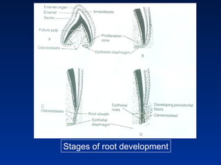
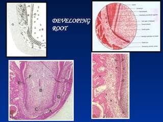
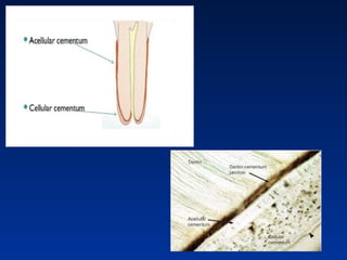
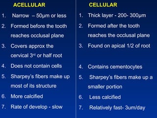
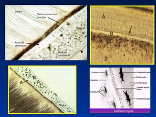
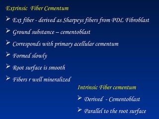
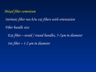
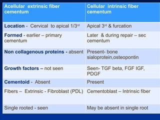
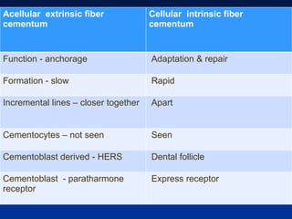
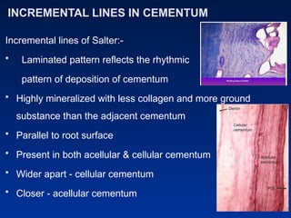
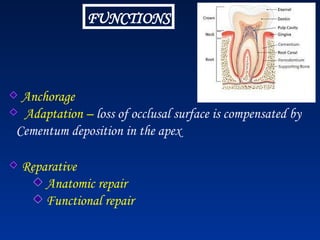
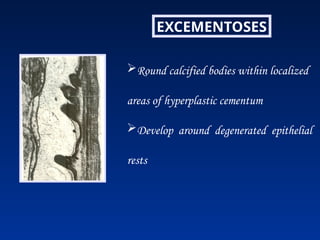
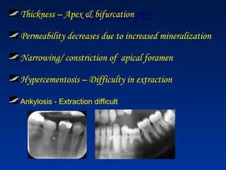
Stages of root development
DEVELOPING
ROOT
At late bell stage – Amelogenesis, Dentinogenesis are
well advanced
EEE & IEE at the cervical loop  double layered epithelial
root sheath
Growth of the Hertwig's epithelial sheath occur to enclose
Dental Papilla, except at the base- the pri apical foramen
Epithelium appeared angled to form root diaphragm
After the diaphragm is formed the location is stationary &
growth of HERS occurs coronally , proliferation occurs at the
cervical end & it lengthens
HERS proliferate to map out shape, no & location
of root & induces the DP cells  odontoblasts
At the cervical portion inner layer of epithelial sheath induce
the peripheral cells of DP to differentiate into Odontoblasts
Following the onset dentinogenesis in the root, epithelial cells
of the root sheath loose their continuity, becoming separated
from the surface of the developing root dentine to form
Epithelial rests of Malassez in the PDL
 Mesenchymal cells diff  Cementoblast  Cementum
Cementogenesis
• Deposition of dentin along the inner aspect of HERS
• HERS allowing the newly formed dentin to come in
direct contact with CT of the dental follicle
Cells derived
Cementum
• HERS – degeneration or loss of its basal lamina on the
cementum side
• Sheath cells- epithelial rests of malassez  migrates to
PDL
Stages of cementum formation
• Phase I : Laying down of cementoid tissue (matrix formation)
 Collagen fibrils
 Ground substance – Glycoproteins & proteoglycans.
• Phase II : Mineralization
 Apatite crystals r deposited along the fibrils.
 Cementum formation -rhythmically.
 Thin layer of cementoid  on the surface, lined by
cementoblasts
 Ca & PO4 ions present in tissue fluids  deposit into matrix
 in the form of hydroxyapatite
• Cementum is laid down slowly - tooth is erupting.
- Acellular or primary.
- Mineral content first seen as thin plates or lamellae
• Tooth comes in occlusion, more cementum forms around
the apical two-thirds of the root
• Which has greater proportion of collagen.
Cementoblasts become trapped in lacunae within this matrix.
- cellular ( secondary) cementum.
- minerals – globules,
- scattered throughout the matrix & b/w cementoblasts
- spherules increase in size and engulf the cementoblasts.
 Rate of formation of cellular cementum is much more rapid
than that of acellular cementum.
Physical Characteristics
 Calcified str – hardness & calcification is less than dentin
 More permeable than dentin
 Colour – light yellow
 Softer & lighter than dentin
 Lacks luster & is dark -differentiate from enamel
 Less readily resorbed than bone
Chemical composition
Inorganic- 45-50%
Organic & water – 50-55%
Inorganic:
Ca & Phosphate – hydroxyapatite crystal
Numerous traces of elements – varying amounts
Highest fluoride content
Organic :
Type I collagen & protein polysaccharides (proteoglycans)
Other types of collagen fibers –type III, V, VI & XII
Noncollagenous proteins:
imp role in matrix deposition, initiation, control of mineralization
& matrix remodeling
Bone sialoprotein & osteopontin – major
- fill up large interfibrillar spaces
Osteopontin – regulates the mineralization
Cementum derived attachment protein- adhesion molecule
- helps in the attachment of mesenchymal cells to extracellular
matrix
Bone & cementum has similar types of proteoglycans
Chondroitin sulfate, heparan sulfate, hyaluronate,
keratin sulfates- fibromodulin & lumican, versican, biglycan &
osteoadherin.
Matrix – growth factors – control the activities of many types of cells
TGF beta, various BMPs, FGF, IGF, EGF & platelet derived GF
Classification
• According to three factors –
1.Time of formation
• Primary and Secondary Cementum
2.Presence and absence of cells within its matrix
• Acellular and cellular Cementum
3. Origin of the collagenous fibers & organic matrix
• Intrinsic, Extrinsic and mixed fibers
According to fibers
• Acellular extrinsic fiber Cementum
• Cellular intrinsic fiber Cementum
BASIC STRUCTURE OF CEMENTUM
1) Cells :
a. Cementoblasts
b. Cementocytes
c. Cementoclasts
2) Fibers :
a. Intrinsic
b. Extrinsic
3) Ground Substance
Cells – cuboidal with large nucleus
 Origin – Undifferentiated
mesenchymal cells adjacent CT differentiate 
cementoblasts
 Secretes – Collagen and Protein polysaccharide
- Organic matrix
Cementoblasts
 Function – Organic matrix of cementum
 EM –
Numerous mitochondria
Well formed golgi apparatus
Large amount of granular
endoplasmic reticulum
 outlined by microvilli
Cementocytes
• Cementoblast  incorporated in cementum
cementocyte – cellular cementum
• Lie in lacunae space
• Has numerous cell processes or Canaliculi
• Cytoplasm - few cell organelles
• Canaliculi are radiating from its cell body
• Processes may branched and frequently anastomosis
with neighboring cells
• Processes are directed towards the periodontal
ligament – source of nutrition
Differences between cementocytes & osteocytes
Cementocytes Osteocytes
Lacunae Ovoid to tubular Invariably oval
Canaliculi Less complicated & sparse Radiating, more
dense
Arrangement Facing towards PDL Complex network
IHC Fibromodulin & Lumican +Ve -Ve
Cementoclasts
Odontoclast
• Resemble osteoclasts
• Areas of resorption they are located in Howships
lacunae
Cementoid tissue
• Cementum – Rhythmic process
• New layer of Cementoid will formed and old one calcified
• Cementoid tissue - lined by Cementoblasts cells
• CT fibers from the PDL pass between the cementoblasts
into the cementum
• H & E – eosinophilic & non mineralized
• Collagen fibers & ground substance
• Average width – 8μm
Cervical – thin
Apex – maximum width
• Provide resistance cementoclasia
PRIMARY CEMENTUM
Formed before tooth reaches
occlusion
Formed after tooth reaches occlusion
Acellular cementum Cellular cementum
Forms slowly Forms rapidly
More mineralized Less mineralized
2/3 of the root Apical third & inter radicular
Collagen fibers –
perpendicular to the root surface parallel to the root surface
Incremental lines are closely
spaced, thin, and even
Incremental lines are wide apart,
thick and uneven
SECONDARY CEMENTUM
Pre cementum layer narrow Pre cementum layer wider
ACELLULAR
1. Narrow – 50μm or less
2. Formed before the tooth
reaches occlusal plane
3. Covers approx the
cervical 3rd
or half root
4. Does not contain cells
5. Sharpey’s fibers make up
most of its structure
6. More calcified
7. Rate of develop - slow
CELLULAR
1. Thick layer - 200- 300μm
2. Formed after the tooth
reaches the occlusal plane
3. Found on apical 1/2 of root
4. Contains cementocytes
5. Sharpey’s fibers make up a
smaller portion
6. Less calcified
7. Relatively fast- 3um/day
Extrinsic Fiber Cementum
 Ext fiber - derived as Sharpeys fibers from PDL Fibroblast
 Ground substance – cementoblast
 Corresponds with primary acellular cementum
 Formed slowly
 Root surface is smooth
 Fibers r well mineralized
Intrinsic Fiber cementum
 Derived - Cementoblast
 Parallel to the root surface
Mixed fiber cementum
Intrinsic fiber run b/w ext fibers with orientation
Fiber bundle size
Ext fiber – ovoid / round bundles, 5-7μm in diameter
Int fiber – 1-2 μm in diameter
Acellular extrinsic fiber
cementum
Cellular intrinsic fiber
cementum
Location - Cervical to apical 1/3rd
Apical 3rd
& furcation
Formed - earlier – primary
cementum
Later & during repair – sec
cementum
Non collagenous proteins - absent Present- bone
sialoprotein,osteopontin
Growth factors – not seen Seen- TGF beta, FGF IGF,
PDGF
Cementoid - Absent Present
Fibers – Extrinsic - Fibroblast (PDL) Cementoblast – Intrinsic fiber
Single rooted - seen May be absent in single root
Acellular extrinsic fiber
cementum
Cellular intrinsic fiber
cementum
Function - anchorage Adaptation & repair
Formation - slow Rapid
Incremental lines – closer together Apart
Cementocytes – not seen Seen
Cementoblast derived - HERS Dental follicle
Cementoblast - paratharmone
receptor
Express receptor
INCREMENTAL LINES IN CEMENTUM
Incremental lines of Salter:-
• Laminated pattern reflects the rhythmic
pattern of deposition of cementum
• Highly mineralized with less collagen and more ground
substance than the adjacent cementum
• Parallel to root surface
• Present in both acellular & cellular cementum
• Wider apart - cellular cementum
• Closer - acellular cementum
CLINICAL SIGNIFICANCE
One cementum ring is laid down each year
It has been suggested that it could also occur in humans
Sharpey’s fibers
• Principal collagen bundles of the PDL - embedded in
the cementum or the bone
• Sharpey’s fibers in the primary acellular cementum
are fully mineralized
• Sharpey’s fibers in cellular cementum and bone are
generally only partially mineralized
Cementodentinal junction
• Dentine surface upon which Cementum is deposited
• CDJ – Deciduous – Scalloped
• Decalcification section – CDJ clearly seen – LM
• EM – a narrow interface zone between Cementum
and dentin
• Cementum is more electron dense than dentin
• Collagen fibers in cementum – distinct bundles
• In dentin – Haphazardly
• Some times dentin is separated from cementum by a
zone – Intermediate cementum
INTERMEDIATE CEMENTUM
• Layer of Hopewell Smith or Hyaline layer
• Highly calcified amorphous layer found at the CDJ
- structureless hyaline layer – 10 μ thick
• Located - adj to Tome’s granular layer & internal layer of
acellular cementum
• Formed by deposition of enamel matrix proteins.
• It is neither dentin or cementum
A. Acellular cementum
B. Hope well smith
C. Tomes layer
D. Dentin
• Predominantly seen – Apical 2/3rd
of roots of molars & PMs
• Continuous layer or isolated areas
• Product of HERS
- Enamel matrix proteins
Amelogenin, PTH receptor & certain basement memb
constituents
• Cementum related proteins like BSP, Osteopontin & fibrillar
collagen – controversial role of HERS in cementum formation
• Noncollagenous hypercalcified matrix - glycosylated
tryptophan, proline and histidine
Cementoenamel junction
• Junction between cementum and enamel at the
cervical region
• 30% - will meet cementum with enamel – Butt joint
• 10% - enamel and cementum will not meet – Gap
junction
• 60% - cementum overlaps the cervical end of the
enamel for short distance – Overlap junction
60% - Over lapping, 30% - Butt joint, 10% - gap junction
Cemento enamel junction
• Over lap Jun – HERS degenerates – cervical 3rd

connective tissue  direct contact with the enamel surface
- Afibrillar cementum
• Gap Jun – Enamel epithelium in the cervical portion of
the root  delayed in its separation from dentin
Deciduous teeth – Edge type overlap type
FUNCTIONS
Anchorage
Adaptation – loss of occlusal surface is compensated by
Cementum deposition in the apex
Reparative
Anatomic repair
Functional repair
REPAIR OF CEMENTUM
Trauma / excessive occlusal forces 
resorption
Resorption ceased  repaired by cellular
or acellular C  reestablish the former
Outline of the root surface
-- ANATOMIC REPAIR
FUCTIONAL REPAIR
 Thin layer  deposited deep
resorption bay like concave area
 Bony projection  periodontal space
width restored functional relationship
Hypercementosis
• Abnormal thickening of cementum-
localized & Generalized
• Diffuse or circumscribed
• May involve all teeth / single tooth / part of a single tooth
Etiology
Periapical inflamation
Mechanical stimulation Orthodontic forces
Non functional / unerupted teeth
Paget’s disease
Accelerated elongation of tooth
Tooth repairs
Idiopathic
Cementum Hypertrophy: If the overgrowth occurs in functional teeth
& improves the functional qualities of the Cementum
Cementum Hyperplasia: Overgrowth occurs in non-functional teeth
Clinical consideration
• Orthodontic tooth movement
• Anatomic repair
• Functional repair
• Transverse fracture
• Fragments of root in the socket after extraction
• Surface changes of pathologically exposed cementum
• Hypercementosis
• Ankylosis
Orthodontic tooth movement
♣ Cementum is resistant resorption
♣ Bone is resorbed - pressure
♣ New bone - tension
♣ Diff in the resistance to pressure - bone is richly vascularized
- cementum is avascular
♣ Degeneration process - affected by interference On
♣ Cementum – slow metabolism , is not damaged by a pressure
equal exerted on bone
Cementicles
Round or ovoid calcified bodies found free in PDL
Exhibit dystrophic calcification of the degenerated pdtal tissue or
the epithelial cell rests of Malassez
Cementicles may be
1. Free in the PDL
2. Attached to Cementum - Excementosis
3. Embedded in the Cementum during its growth by age
EXCEMENTOSES
Round calcified bodies within localized
areas of hyperplastic cementum
Develop around degenerated epithelial
rests
AGE CHANGES
Thickness – Apex & bifurcation
Permeability decreases due to increased mineralization
Narrowing/ constriction of apical foramen
Hypercementosis – Difficulty in extraction
Ankylosis - Extraction difficult
Age Changes
Thickness of Cementum increases with age – apex
- because of passive eruption
Cementum exposed – gingival recession thickness decreases
Cementocytes have lowest proliferative capacity – gradually die –
because of lack of nutrition as the width of Cementum increases.
Any
Questions?
??????????

Cementum 10 (1) presentation from dadh.ppt

  • 1.
    Stages of rootdevelopment
  • 2.
  • 3.
    At late bellstage – Amelogenesis, Dentinogenesis are well advanced EEE & IEE at the cervical loop  double layered epithelial root sheath Growth of the Hertwig's epithelial sheath occur to enclose Dental Papilla, except at the base- the pri apical foramen Epithelium appeared angled to form root diaphragm
  • 4.
    After the diaphragmis formed the location is stationary & growth of HERS occurs coronally , proliferation occurs at the cervical end & it lengthens HERS proliferate to map out shape, no & location of root & induces the DP cells  odontoblasts
  • 5.
    At the cervicalportion inner layer of epithelial sheath induce the peripheral cells of DP to differentiate into Odontoblasts Following the onset dentinogenesis in the root, epithelial cells of the root sheath loose their continuity, becoming separated from the surface of the developing root dentine to form Epithelial rests of Malassez in the PDL  Mesenchymal cells diff  Cementoblast  Cementum
  • 6.
    Cementogenesis • Deposition ofdentin along the inner aspect of HERS • HERS allowing the newly formed dentin to come in direct contact with CT of the dental follicle Cells derived Cementum
  • 7.
    • HERS –degeneration or loss of its basal lamina on the cementum side • Sheath cells- epithelial rests of malassez  migrates to PDL
  • 8.
    Stages of cementumformation • Phase I : Laying down of cementoid tissue (matrix formation)  Collagen fibrils  Ground substance – Glycoproteins & proteoglycans. • Phase II : Mineralization  Apatite crystals r deposited along the fibrils.  Cementum formation -rhythmically.  Thin layer of cementoid  on the surface, lined by cementoblasts  Ca & PO4 ions present in tissue fluids  deposit into matrix  in the form of hydroxyapatite
  • 9.
    • Cementum islaid down slowly - tooth is erupting. - Acellular or primary. - Mineral content first seen as thin plates or lamellae • Tooth comes in occlusion, more cementum forms around the apical two-thirds of the root • Which has greater proportion of collagen.
  • 10.
    Cementoblasts become trappedin lacunae within this matrix. - cellular ( secondary) cementum. - minerals – globules, - scattered throughout the matrix & b/w cementoblasts - spherules increase in size and engulf the cementoblasts.  Rate of formation of cellular cementum is much more rapid than that of acellular cementum.
  • 11.
    Physical Characteristics  Calcifiedstr – hardness & calcification is less than dentin  More permeable than dentin  Colour – light yellow  Softer & lighter than dentin  Lacks luster & is dark -differentiate from enamel  Less readily resorbed than bone
  • 12.
    Chemical composition Inorganic- 45-50% Organic& water – 50-55% Inorganic: Ca & Phosphate – hydroxyapatite crystal Numerous traces of elements – varying amounts Highest fluoride content
  • 13.
    Organic : Type Icollagen & protein polysaccharides (proteoglycans) Other types of collagen fibers –type III, V, VI & XII Noncollagenous proteins: imp role in matrix deposition, initiation, control of mineralization & matrix remodeling
  • 14.
    Bone sialoprotein &osteopontin – major - fill up large interfibrillar spaces Osteopontin – regulates the mineralization Cementum derived attachment protein- adhesion molecule - helps in the attachment of mesenchymal cells to extracellular matrix
  • 15.
    Bone & cementumhas similar types of proteoglycans Chondroitin sulfate, heparan sulfate, hyaluronate, keratin sulfates- fibromodulin & lumican, versican, biglycan & osteoadherin. Matrix – growth factors – control the activities of many types of cells TGF beta, various BMPs, FGF, IGF, EGF & platelet derived GF
  • 16.
    Classification • According tothree factors – 1.Time of formation • Primary and Secondary Cementum 2.Presence and absence of cells within its matrix • Acellular and cellular Cementum 3. Origin of the collagenous fibers & organic matrix • Intrinsic, Extrinsic and mixed fibers
  • 17.
    According to fibers •Acellular extrinsic fiber Cementum • Cellular intrinsic fiber Cementum
  • 18.
    BASIC STRUCTURE OFCEMENTUM 1) Cells : a. Cementoblasts b. Cementocytes c. Cementoclasts 2) Fibers : a. Intrinsic b. Extrinsic 3) Ground Substance
  • 19.
    Cells – cuboidalwith large nucleus  Origin – Undifferentiated mesenchymal cells adjacent CT differentiate  cementoblasts  Secretes – Collagen and Protein polysaccharide - Organic matrix Cementoblasts
  • 20.
     Function –Organic matrix of cementum  EM – Numerous mitochondria Well formed golgi apparatus Large amount of granular endoplasmic reticulum  outlined by microvilli
  • 21.
    Cementocytes • Cementoblast incorporated in cementum cementocyte – cellular cementum • Lie in lacunae space • Has numerous cell processes or Canaliculi • Cytoplasm - few cell organelles
  • 22.
    • Canaliculi areradiating from its cell body • Processes may branched and frequently anastomosis with neighboring cells • Processes are directed towards the periodontal ligament – source of nutrition
  • 23.
    Differences between cementocytes& osteocytes Cementocytes Osteocytes Lacunae Ovoid to tubular Invariably oval Canaliculi Less complicated & sparse Radiating, more dense Arrangement Facing towards PDL Complex network IHC Fibromodulin & Lumican +Ve -Ve
  • 24.
    Cementoclasts Odontoclast • Resemble osteoclasts •Areas of resorption they are located in Howships lacunae
  • 27.
    Cementoid tissue • Cementum– Rhythmic process • New layer of Cementoid will formed and old one calcified • Cementoid tissue - lined by Cementoblasts cells • CT fibers from the PDL pass between the cementoblasts into the cementum
  • 28.
    • H &E – eosinophilic & non mineralized • Collagen fibers & ground substance • Average width – 8μm Cervical – thin Apex – maximum width • Provide resistance cementoclasia
  • 29.
    PRIMARY CEMENTUM Formed beforetooth reaches occlusion Formed after tooth reaches occlusion Acellular cementum Cellular cementum Forms slowly Forms rapidly More mineralized Less mineralized 2/3 of the root Apical third & inter radicular Collagen fibers – perpendicular to the root surface parallel to the root surface Incremental lines are closely spaced, thin, and even Incremental lines are wide apart, thick and uneven SECONDARY CEMENTUM Pre cementum layer narrow Pre cementum layer wider
  • 31.
    ACELLULAR 1. Narrow –50μm or less 2. Formed before the tooth reaches occlusal plane 3. Covers approx the cervical 3rd or half root 4. Does not contain cells 5. Sharpey’s fibers make up most of its structure 6. More calcified 7. Rate of develop - slow CELLULAR 1. Thick layer - 200- 300μm 2. Formed after the tooth reaches the occlusal plane 3. Found on apical 1/2 of root 4. Contains cementocytes 5. Sharpey’s fibers make up a smaller portion 6. Less calcified 7. Relatively fast- 3um/day
  • 33.
    Extrinsic Fiber Cementum Ext fiber - derived as Sharpeys fibers from PDL Fibroblast  Ground substance – cementoblast  Corresponds with primary acellular cementum  Formed slowly  Root surface is smooth  Fibers r well mineralized Intrinsic Fiber cementum  Derived - Cementoblast  Parallel to the root surface
  • 34.
    Mixed fiber cementum Intrinsicfiber run b/w ext fibers with orientation Fiber bundle size Ext fiber – ovoid / round bundles, 5-7μm in diameter Int fiber – 1-2 μm in diameter
  • 35.
    Acellular extrinsic fiber cementum Cellularintrinsic fiber cementum Location - Cervical to apical 1/3rd Apical 3rd & furcation Formed - earlier – primary cementum Later & during repair – sec cementum Non collagenous proteins - absent Present- bone sialoprotein,osteopontin Growth factors – not seen Seen- TGF beta, FGF IGF, PDGF Cementoid - Absent Present Fibers – Extrinsic - Fibroblast (PDL) Cementoblast – Intrinsic fiber Single rooted - seen May be absent in single root
  • 36.
    Acellular extrinsic fiber cementum Cellularintrinsic fiber cementum Function - anchorage Adaptation & repair Formation - slow Rapid Incremental lines – closer together Apart Cementocytes – not seen Seen Cementoblast derived - HERS Dental follicle Cementoblast - paratharmone receptor Express receptor
  • 37.
    INCREMENTAL LINES INCEMENTUM Incremental lines of Salter:- • Laminated pattern reflects the rhythmic pattern of deposition of cementum • Highly mineralized with less collagen and more ground substance than the adjacent cementum • Parallel to root surface • Present in both acellular & cellular cementum • Wider apart - cellular cementum • Closer - acellular cementum
  • 38.
    CLINICAL SIGNIFICANCE One cementumring is laid down each year It has been suggested that it could also occur in humans
  • 39.
    Sharpey’s fibers • Principalcollagen bundles of the PDL - embedded in the cementum or the bone • Sharpey’s fibers in the primary acellular cementum are fully mineralized • Sharpey’s fibers in cellular cementum and bone are generally only partially mineralized
  • 40.
    Cementodentinal junction • Dentinesurface upon which Cementum is deposited • CDJ – Deciduous – Scalloped • Decalcification section – CDJ clearly seen – LM • EM – a narrow interface zone between Cementum and dentin • Cementum is more electron dense than dentin
  • 41.
    • Collagen fibersin cementum – distinct bundles • In dentin – Haphazardly • Some times dentin is separated from cementum by a zone – Intermediate cementum
  • 42.
    INTERMEDIATE CEMENTUM • Layerof Hopewell Smith or Hyaline layer • Highly calcified amorphous layer found at the CDJ - structureless hyaline layer – 10 μ thick • Located - adj to Tome’s granular layer & internal layer of acellular cementum • Formed by deposition of enamel matrix proteins. • It is neither dentin or cementum A. Acellular cementum B. Hope well smith C. Tomes layer D. Dentin
  • 43.
    • Predominantly seen– Apical 2/3rd of roots of molars & PMs • Continuous layer or isolated areas • Product of HERS - Enamel matrix proteins Amelogenin, PTH receptor & certain basement memb constituents • Cementum related proteins like BSP, Osteopontin & fibrillar collagen – controversial role of HERS in cementum formation • Noncollagenous hypercalcified matrix - glycosylated tryptophan, proline and histidine
  • 44.
    Cementoenamel junction • Junctionbetween cementum and enamel at the cervical region • 30% - will meet cementum with enamel – Butt joint • 10% - enamel and cementum will not meet – Gap junction • 60% - cementum overlaps the cervical end of the enamel for short distance – Overlap junction
  • 45.
    60% - Overlapping, 30% - Butt joint, 10% - gap junction Cemento enamel junction
  • 46.
    • Over lapJun – HERS degenerates – cervical 3rd  connective tissue  direct contact with the enamel surface - Afibrillar cementum • Gap Jun – Enamel epithelium in the cervical portion of the root  delayed in its separation from dentin Deciduous teeth – Edge type overlap type
  • 47.
    FUNCTIONS Anchorage Adaptation – lossof occlusal surface is compensated by Cementum deposition in the apex Reparative Anatomic repair Functional repair
  • 48.
    REPAIR OF CEMENTUM Trauma/ excessive occlusal forces  resorption Resorption ceased  repaired by cellular or acellular C  reestablish the former Outline of the root surface -- ANATOMIC REPAIR
  • 49.
    FUCTIONAL REPAIR  Thinlayer  deposited deep resorption bay like concave area  Bony projection  periodontal space width restored functional relationship
  • 50.
    Hypercementosis • Abnormal thickeningof cementum- localized & Generalized • Diffuse or circumscribed • May involve all teeth / single tooth / part of a single tooth
  • 51.
    Etiology Periapical inflamation Mechanical stimulationOrthodontic forces Non functional / unerupted teeth Paget’s disease Accelerated elongation of tooth Tooth repairs Idiopathic Cementum Hypertrophy: If the overgrowth occurs in functional teeth & improves the functional qualities of the Cementum Cementum Hyperplasia: Overgrowth occurs in non-functional teeth
  • 52.
    Clinical consideration • Orthodontictooth movement • Anatomic repair • Functional repair • Transverse fracture • Fragments of root in the socket after extraction • Surface changes of pathologically exposed cementum • Hypercementosis • Ankylosis
  • 53.
    Orthodontic tooth movement ♣Cementum is resistant resorption ♣ Bone is resorbed - pressure ♣ New bone - tension ♣ Diff in the resistance to pressure - bone is richly vascularized - cementum is avascular ♣ Degeneration process - affected by interference On ♣ Cementum – slow metabolism , is not damaged by a pressure equal exerted on bone
  • 54.
    Cementicles Round or ovoidcalcified bodies found free in PDL Exhibit dystrophic calcification of the degenerated pdtal tissue or the epithelial cell rests of Malassez Cementicles may be 1. Free in the PDL 2. Attached to Cementum - Excementosis 3. Embedded in the Cementum during its growth by age
  • 55.
    EXCEMENTOSES Round calcified bodieswithin localized areas of hyperplastic cementum Develop around degenerated epithelial rests
  • 56.
    AGE CHANGES Thickness –Apex & bifurcation Permeability decreases due to increased mineralization Narrowing/ constriction of apical foramen Hypercementosis – Difficulty in extraction Ankylosis - Extraction difficult
  • 57.
    Age Changes Thickness ofCementum increases with age – apex - because of passive eruption Cementum exposed – gingival recession thickness decreases Cementocytes have lowest proliferative capacity – gradually die – because of lack of nutrition as the width of Cementum increases.
  • 58.

Editor's Notes

  • #25 Macrophage CSF
  • #47 Hypophosphatasia, Normal condition Cementum doesn’t resorb unlike bone- remodelling