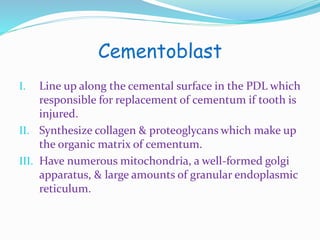
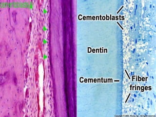
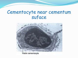
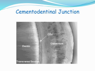
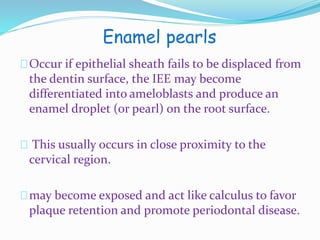
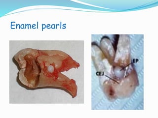
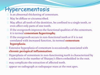
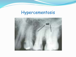
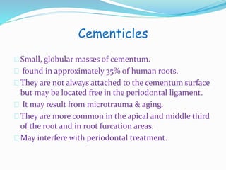
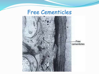
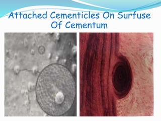
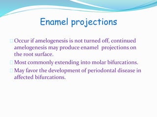
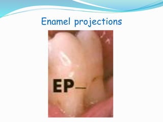
CEMENTUM
CEMENTUM: is a specialized hard layer of calcified 
mesenchymal tissues which forms the outer covering of the 
anatomical root. 
Begins at cervical portion of the tooth at the cementoenamel 
junction & continues to the apex.
Function 
• Furnishes a medium for the attachment of 
collagen fibers that bind the tooth to 
surrounding structures. 
• Serves as major reparative tissue for root 
surface.
Distrbution 
I. It varies in thickness at different levels of the root. It is 
thickest at the root apex and in the interradicular areas 
of multirooted teeth, and thinnest cervically. 
II. The thickness cervically is 10-50μm, and apically, 50- 
200μm. It is contiguous with the periodontal ligament on 
its outer surface and is firmly adherent to dentine on its 
deep surface. 
III. As cementum is slowly formed throughout life, this 
allows for continual reattachment of the periodontal 
ligament fibers. 
IV. It is deposited throughout life and there is always a thin 
layer of uncalcified matrix on its surface.
Physical Characteristics 
I. Hardness is less than that of dentin and bone. 
II. Light yellow in color, It’s lighter in color than 
dentin but more darker than enamel. 
III. Can be distinguished from enamel by its lack of 
luster & its darker hue. 
IV. Permeable to a variety of materials, Its 
permeability decreases with age. 
V. Avascular receive nutrient from the surrounding 
periodontal space and not innervated.
Chemical Composion 
I. Contains 45% to 50% inorganic substances 
mainly calicum, hydroxyapatite, phosphate & 
fluride. 
II. 50% to 55% organic material & water. 
III. Organic portion consists primarily of type I 
collagen & proteoglycans. 
IV. Cementum has the highest fluoride content of 
all the mineralized tissues.
Cementogenesis 
Cementum develops from the activity of 
esenchymal cells of dental follicle after 
fragmentation of the epithelial root sheath. 
After formation of dentin, loss of continuity 
occurs in the epithelial root sheath. 
This allows adjacent cells of the investing layer of 
the dental follicle to come to lie on surface of root 
dentin & these are induced to differentiate into 
cementoblasts.
Structure 
I. Cells 
II. Matrix 
III. Mineralized fibers
Cellular components of cementum 
Cementocytes 
I. Soon after Hertwig’s sheath breaks up, undifferentiated 
mesenchymal cells from adjacent connective tissue of the 
dental sac differentiate into cementoblasts. 
II. The spaces that the cementocytes accupy in the tissue are 
called lacunae, and the channels that their processes 
extend along are the canaliculi, adjacent canaliculi are 
often connected. 
III. They are preferentially orientated towards the 
periodontal ligament to get their chief source of 
nutrition. 
IV. Some unmineralized matrix may be seen in perilacuna 
space.
Cementoblast 
I. Line up along the cemental surface in the PDL which 
responsible for replacement of cementum if tooth is 
injured. 
II. Synthesize collagen & proteoglycans which make up 
the organic matrix of cementum. 
III. Have numerous mitochondria, a well-formed golgi 
apparatus, & large amounts of granular endoplasmic 
reticulum.
Cementocyte near cementum 
suface
Fibers 
The fibrous matrix consist both: 
 Sharpey's fibres(Extrinsic fibers ): are the terminal 
ends of principal fibres of the periodontal 
ligament, that insert into the cementum and 
into the periosteumof the alveolar bone,it’s 
perpendicular or oblique to the root surface. 
 Intrinsic fibers: It’s non periodontal collagen 
fibers made up by cementoblast and all of these 
fibers run parallel to the DCJ.
Sharpey's fibres
Classification of cementum 
Acellular cementum Cellular cementum 
I. Covers the apical third and 
interradicular. 
II. Formed after the tooth reaches 
the occlusal plane. 
III. It contains embeded 
cementocytes. 
IV. Thickness is in the range of 100- 
1000 μm 
V. Lesser number of Sharpy’s 
fibers. 
VI. Main function is adaptation. 
VII. Formed at faster rate. 
VIII.Deposited over the acellular 
cementum. 
I. Covers the cervical third of the 
root. 
II. Formed before the tooth reaches 
the occlusal plane. 
III. Does not contain embedded 
cementocytes. 
IV. Thickness is in the range of 30- 
230 μm 
V. Abundance of sharpey’s fibers. 
VI. Main function is anchorage. 
VII.Formed at slow rate. 
VIII.First layer of cementum.
Acellular cementum
Cellular cementum
Cementodentinal Junction 
Smooth in permanent teeth, But Scalloped in 
deciduous teeth. 
Dentin is separated from cementum by a zone 
known as the intermediate cementum layer. 
This layer is predominantly seen in apical two-thirds 
of roots of molars & premolars.
Cementodentinal Junction
Cementoenamel Junction 
In 60% of the teeth, cementumoverlaps the cervical 
end of enamel for a short distance. 
In 30% of all teeth, cementum meets the cervical end 
of enamel in a relatively sharp line. 
In 10% of the teeth, enamel & cementum do not meet.
Relationship between cementum & 
enamel at the CEJ
Developmental 
Anomalies 
& 
Clinical 
Considerations
Enamel pearls 
Occur if epithelial sheath fails to be displaced from 
the dentin surface, the IEE may become 
differentiated into ameloblasts and produce an 
enamel droplet (or pearl) on the root surface. 
This usually occurs in close proximity to the 
cervical region. 
may become exposed and act like calculus to favor 
plaque retention and promote periodontal disease.
Enamel pearls
Hypercementosis 
Is an abnormal thickening of cementum. 
May be diffuse or circumscribed. 
May affect all teeth of the dentition, be confined to a single tooth, or 
even affect only parts of one tooth. 
If the overgrowth improves the functional qualities of the cementum, 
it is termed cementum hypertrophy. 
If the overgrowth occurs in non-functional teeth or if it is not 
correlated with increased function, its termed cementum 
hyperplasia. 
Extensive hyperplasia of cementum is occasionally associated with 
chronic periapical inflammation. 
Hyperplasia of cementum in non-functioning teeth is characterized by 
a reduction in the number of Sharpey’s fibers embedded in the root. 
may complicate the extraction of affected teeth. 
appear on radiograph as radiopaque mass at the root apex.
Hypercementosis
Cementicles 
Small, globular masses of cementum. 
found in approximately 35% of human roots. 
They are not always attached to the cementumsurface 
but may be located free in the periodontal ligament. 
It may result from microtrauma & aging. 
They are more common in the apical and middle third 
of the root and in root furcation areas. 
May interfere with periodontal treatment.
Free Cementicles
Attached Cementicles On Surfuse 
Of Cementum
Enamel projections 
Occur if amelogenesis is not turned off, continued 
amelogenesis may produce enamel projections on 
the root surface. 
Most commonly extending into molar bifurcations. 
May favor the development of periodontal disease in 
affected bifurcations.
Enamel projections
Cementum exposure 
Gingival recession 
Gum recession is not directly linked with age but 
generally it is a more common condition in adults over 
the age of 40. 
Causes: poor plaque control, abrasion due to “hard” 
tooth brushing habits, mouth trauma and occlusion. 
problems associated: tooth sensitivity, cemental caries 
risk, periodontal disease, esthetic problems. 
Bone loss also occurs with gingival recession, giving less 
support to the teeth.
Gingival recession
Clinical Concederation 
1. Anchoring function: it mediates the attachment 
of the tooth to the gingival connective tissue, as 
well as to the periodontal ligament and, hence, the 
alveolar bone. 
2. Protective function: as it is less susceptible 
to resorption than bone. 
This allows pressure induced movement of the tooth 
through bone, as in orthodontics, while 
minimizing resorptive damage to the tooth.
Causes Of Cemental Resorption 
Trauma from Occlusion . 
Deficiency of Ca . 
Cyst & Tumors Deficiency of Vit. A & D. 
Periapical Pathology . 
Hypothyroidism . 
Excessive orthodontic forces .
3. Reparative function: New cementum formation 
is a key process during therapeutic procedures 
aimed at gaining new attachment, as it mediates 
new attachment of the tooth to the periodontal 
ligament and bone. 
While it is possible for bone to fuse directly with 
the dentin and cementum of the tooth through 
ankylosis, this is considered an undesirable 
process, as it results in progressive resorption of 
the tooth structure because of ongoing 
osteoclastic (odontoclastic) activity. 
4. Regular Cementum deposition at the root apex, 
helps to replenish the lost tooth height due to 
occlusal wear.
Aging Of Cementum 
Surface become rough and irrigular. 
Cemental Resorption. 
Permeability Decreases. 
More Cemental deposition is greater in the apical 
zone, which may lead to closure of the apical foramen. 
The lacunae of cellular cementumappear empty (loss 
of cementocytes).

Cementum ^_^ Emad

  • 1.
  • 2.
    CEMENTUM: is aspecialized hard layer of calcified mesenchymal tissues which forms the outer covering of the anatomical root. Begins at cervical portion of the tooth at the cementoenamel junction & continues to the apex.
  • 3.
    Function • Furnishesa medium for the attachment of collagen fibers that bind the tooth to surrounding structures. • Serves as major reparative tissue for root surface.
  • 4.
    Distrbution I. Itvaries in thickness at different levels of the root. It is thickest at the root apex and in the interradicular areas of multirooted teeth, and thinnest cervically. II. The thickness cervically is 10-50μm, and apically, 50- 200μm. It is contiguous with the periodontal ligament on its outer surface and is firmly adherent to dentine on its deep surface. III. As cementum is slowly formed throughout life, this allows for continual reattachment of the periodontal ligament fibers. IV. It is deposited throughout life and there is always a thin layer of uncalcified matrix on its surface.
  • 5.
    Physical Characteristics I.Hardness is less than that of dentin and bone. II. Light yellow in color, It’s lighter in color than dentin but more darker than enamel. III. Can be distinguished from enamel by its lack of luster & its darker hue. IV. Permeable to a variety of materials, Its permeability decreases with age. V. Avascular receive nutrient from the surrounding periodontal space and not innervated.
  • 6.
    Chemical Composion I.Contains 45% to 50% inorganic substances mainly calicum, hydroxyapatite, phosphate & fluride. II. 50% to 55% organic material & water. III. Organic portion consists primarily of type I collagen & proteoglycans. IV. Cementum has the highest fluoride content of all the mineralized tissues.
  • 7.
    Cementogenesis Cementum developsfrom the activity of esenchymal cells of dental follicle after fragmentation of the epithelial root sheath. After formation of dentin, loss of continuity occurs in the epithelial root sheath. This allows adjacent cells of the investing layer of the dental follicle to come to lie on surface of root dentin & these are induced to differentiate into cementoblasts.
  • 8.
    Structure I. Cells II. Matrix III. Mineralized fibers
  • 9.
    Cellular components ofcementum Cementocytes I. Soon after Hertwig’s sheath breaks up, undifferentiated mesenchymal cells from adjacent connective tissue of the dental sac differentiate into cementoblasts. II. The spaces that the cementocytes accupy in the tissue are called lacunae, and the channels that their processes extend along are the canaliculi, adjacent canaliculi are often connected. III. They are preferentially orientated towards the periodontal ligament to get their chief source of nutrition. IV. Some unmineralized matrix may be seen in perilacuna space.
  • 10.
    Cementoblast I. Lineup along the cemental surface in the PDL which responsible for replacement of cementum if tooth is injured. II. Synthesize collagen & proteoglycans which make up the organic matrix of cementum. III. Have numerous mitochondria, a well-formed golgi apparatus, & large amounts of granular endoplasmic reticulum.
  • 13.
  • 14.
    Fibers The fibrousmatrix consist both:  Sharpey's fibres(Extrinsic fibers ): are the terminal ends of principal fibres of the periodontal ligament, that insert into the cementum and into the periosteumof the alveolar bone,it’s perpendicular or oblique to the root surface.  Intrinsic fibers: It’s non periodontal collagen fibers made up by cementoblast and all of these fibers run parallel to the DCJ.
  • 15.
  • 16.
    Classification of cementum Acellular cementum Cellular cementum I. Covers the apical third and interradicular. II. Formed after the tooth reaches the occlusal plane. III. It contains embeded cementocytes. IV. Thickness is in the range of 100- 1000 μm V. Lesser number of Sharpy’s fibers. VI. Main function is adaptation. VII. Formed at faster rate. VIII.Deposited over the acellular cementum. I. Covers the cervical third of the root. II. Formed before the tooth reaches the occlusal plane. III. Does not contain embedded cementocytes. IV. Thickness is in the range of 30- 230 μm V. Abundance of sharpey’s fibers. VI. Main function is anchorage. VII.Formed at slow rate. VIII.First layer of cementum.
  • 17.
  • 18.
  • 19.
    Cementodentinal Junction Smoothin permanent teeth, But Scalloped in deciduous teeth. Dentin is separated from cementum by a zone known as the intermediate cementum layer. This layer is predominantly seen in apical two-thirds of roots of molars & premolars.
  • 20.
  • 21.
    Cementoenamel Junction In60% of the teeth, cementumoverlaps the cervical end of enamel for a short distance. In 30% of all teeth, cementum meets the cervical end of enamel in a relatively sharp line. In 10% of the teeth, enamel & cementum do not meet.
  • 22.
    Relationship between cementum& enamel at the CEJ
  • 23.
    Developmental Anomalies & Clinical Considerations
  • 24.
    Enamel pearls Occurif epithelial sheath fails to be displaced from the dentin surface, the IEE may become differentiated into ameloblasts and produce an enamel droplet (or pearl) on the root surface. This usually occurs in close proximity to the cervical region. may become exposed and act like calculus to favor plaque retention and promote periodontal disease.
  • 25.
  • 26.
    Hypercementosis Is anabnormal thickening of cementum. May be diffuse or circumscribed. May affect all teeth of the dentition, be confined to a single tooth, or even affect only parts of one tooth. If the overgrowth improves the functional qualities of the cementum, it is termed cementum hypertrophy. If the overgrowth occurs in non-functional teeth or if it is not correlated with increased function, its termed cementum hyperplasia. Extensive hyperplasia of cementum is occasionally associated with chronic periapical inflammation. Hyperplasia of cementum in non-functioning teeth is characterized by a reduction in the number of Sharpey’s fibers embedded in the root. may complicate the extraction of affected teeth. appear on radiograph as radiopaque mass at the root apex.
  • 27.
  • 28.
    Cementicles Small, globularmasses of cementum. found in approximately 35% of human roots. They are not always attached to the cementumsurface but may be located free in the periodontal ligament. It may result from microtrauma & aging. They are more common in the apical and middle third of the root and in root furcation areas. May interfere with periodontal treatment.
  • 29.
  • 30.
    Attached Cementicles OnSurfuse Of Cementum
  • 31.
    Enamel projections Occurif amelogenesis is not turned off, continued amelogenesis may produce enamel projections on the root surface. Most commonly extending into molar bifurcations. May favor the development of periodontal disease in affected bifurcations.
  • 32.
  • 33.
    Cementum exposure Gingivalrecession Gum recession is not directly linked with age but generally it is a more common condition in adults over the age of 40. Causes: poor plaque control, abrasion due to “hard” tooth brushing habits, mouth trauma and occlusion. problems associated: tooth sensitivity, cemental caries risk, periodontal disease, esthetic problems. Bone loss also occurs with gingival recession, giving less support to the teeth.
  • 34.
  • 35.
    Clinical Concederation 1.Anchoring function: it mediates the attachment of the tooth to the gingival connective tissue, as well as to the periodontal ligament and, hence, the alveolar bone. 2. Protective function: as it is less susceptible to resorption than bone. This allows pressure induced movement of the tooth through bone, as in orthodontics, while minimizing resorptive damage to the tooth.
  • 36.
    Causes Of CementalResorption Trauma from Occlusion . Deficiency of Ca . Cyst & Tumors Deficiency of Vit. A & D. Periapical Pathology . Hypothyroidism . Excessive orthodontic forces .
  • 37.
    3. Reparative function:New cementum formation is a key process during therapeutic procedures aimed at gaining new attachment, as it mediates new attachment of the tooth to the periodontal ligament and bone. While it is possible for bone to fuse directly with the dentin and cementum of the tooth through ankylosis, this is considered an undesirable process, as it results in progressive resorption of the tooth structure because of ongoing osteoclastic (odontoclastic) activity. 4. Regular Cementum deposition at the root apex, helps to replenish the lost tooth height due to occlusal wear.
  • 38.
    Aging Of Cementum Surface become rough and irrigular. Cemental Resorption. Permeability Decreases. More Cemental deposition is greater in the apical zone, which may lead to closure of the apical foramen. The lacunae of cellular cementumappear empty (loss of cementocytes).

Editor's Notes