This document provides an overview of the skeletal system including:
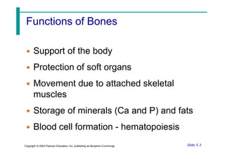
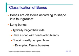
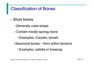
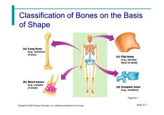
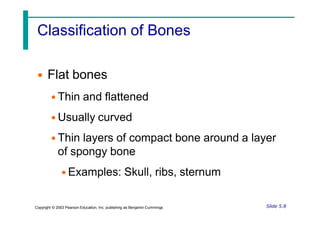
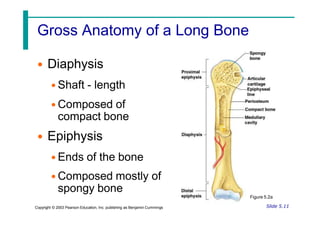
- The 206 bones of the adult human body which are classified based on shape into long, short, flat, and irregular bones.
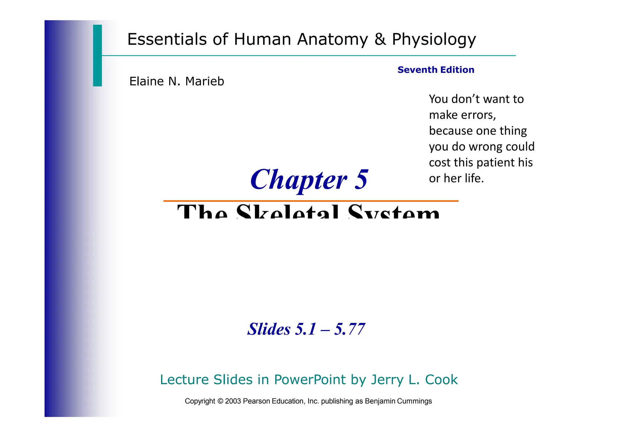
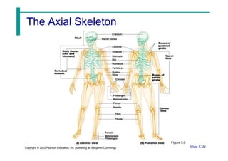
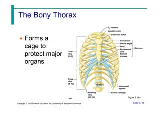
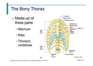
- The axial skeleton which forms the longitudinal axis of the body and includes the skull, vertebral column, and thoracic cage.
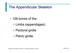
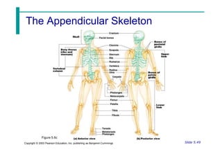
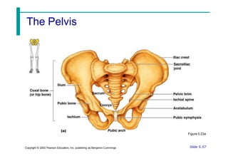
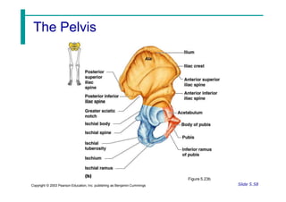
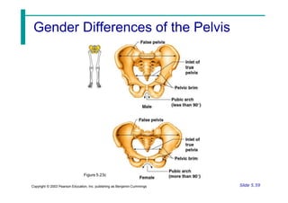
- The appendicular skeleton which includes the 126 bones of the upper and lower limbs attached to the axial skeleton via the shoulder and pelvic girdles.
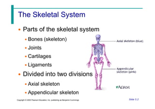
- Key functions of the skeletal system including support, protection, movement, mineral storage, and blood cell formation.


![ONFH[AVN HIP] -TRIPLE REGIME -A NOVAL SURGICAL CONCEPT .pptx](https://cdn.slidesharecdn.com/ss_thumbnails/onfhavnhip2026koaconcalicutdrgokuldevdrmashraf-260210064517-213ec005-thumbnail.jpg?width=640&height=640&fit=bounds)


