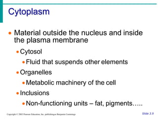
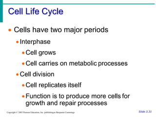
Essentials of Human Anatomy & Physiology
For. Romualdo B. De Guzman, Jr.,
MSA, MBio
Associate Professor II
Chapter 2
Plant Cell and Cell
Physiology
Cell Dicovery
Robert Brown (1831) a British botanist.
❑identify the central part of the cell and called it the
nucleus and also described the cytoplasm as the
grayish fluid that suspend inside the cell.
Dujardin (1835) a French biologist.
❑observed that cells are composed of fluid filled with
thick, jelly-like fluids the surround the cells and
called them protoplasm.
Cell Dicovery
Robert Hooked (1665)a British scientist.
❑He discovered tiny cubical shape mass from the
slice cork on his self-constructed microscope and
named it “cells”.
Anton Van Leeuwenhoek (1674) Dutch scientist.
❑made discoveries concerning protozoa, red blood
cells, capillary systems and the life cycle of the
insects out of his modified compound microscope.
Cell Dicovery
Matthias Schleiden (1838) and Theodore Schwann
(1839) a botanist and zoologist.
❑concluded that plants and animals are composed of
cells and developed the theory about cell.
Rudolf Vichow1858 by a German pathologist.
❑theory was basis of statement that, “all cells must
come only from pre-existing cells which become the
interest of other scientific studies about cell.”
Cell Dicovery
German Engineer Max Knoll and Ernst Ruska in 1932.
❑ built and developed the first transmission electron
microscope that gives more detail in discovering the
different organelles inside the cell.
James Watson, American biochemist and Francis Crick,
British biophysicist in 1953.
❑ significant discovery about cells is the DNA structure.
❑ gave way to the science of molecular biology and it is used
in advance study like, genetics, molecular systematic,
organic chemistry, biochemistry and others.
Cell Defined as:
❑it is the basic fundamental unit of all living
organisms capable of duplication and
multiplication.
Types of Cells
1. Prokaryotic cells (Greek pro, before, and
Karyon, nucleus).
❑type of cell has no nucleus common characteristic
of bacteria with a size ranging from (1 to 10
microns).
❑outer part is composed of cell wall & plasma
membrane.
❑The cytoplasm contains ribosome, thylakoids and
innumerable enzyme.
❑nucleoid contain single chromosome consist of the
DNA.
Types of Cells
2. Eukaryotic cells (Greek eu, true and karyon,nucleus).
❑a true nucleus common characteristic of protists
(unicellular cell), fungi, plants andanimals.
❑consist plasma membrane, cytoplasm and a central
nucleus.
❑nucleus is membrane bounded containing multiple
chromatin and DNA inside it.
❑plasma membrane made of phospholipids bi-layer .
❑Cytoplasm contains organelles for control, synthesisand
metabolic processes inside the cell.
Cells and Tissues – C, O, H, N
• Carry out all chemical activities needed
to sustain life.
• Cells are the building blocks of all living
things
• Tissues are groups of cells that are
similar in structure and function
• Structure reflects function
Slide 3.1
Copyright © 2003 Pearson Education, Inc. publishingas Benjamin Cummings
Anatomy of the Generalized Cell
• Cells are not all the same
• All cells share general structures
• Cells are organized into three main
regions
•Nucleus
•Cytoplasm
•Plasma membrane
Figure 3.1a
Slide 3.2
Copyright © 2003 Pearson Education, Inc. publishingas Benjamin Cummings
The Nucleus
• Control center
of the cell
• Contains genetic
material (DNA)
• Three regions
• Nuclear
membrane
• Nucleolus
• Chromatin Figure 3.1b
Slide 3.3
Copyright © 2003 Pearson Education, Inc. publishingas Benjamin Cummings
Nuclear Membrane – double
membrane or envelope
• Barrier of nucleus
• Consists of a double phospholipid
membrane
• Contain nuclear pores that allow for
exchange of material with the rest of the
cell – selectively permeable
Slide 3.4
Copyright © 2003 Pearson Education, Inc. publishingas Benjamin Cummings
Nucleoli
• Nucleus contains one or more nucleoli
• Sites of ribosome production
• Ribosomes then migrate to the
cytoplasm through nuclear pores
Slide 3.5
Copyright © 2003 Pearson Education, Inc. publishingas Benjamin Cummings
Chromatin (when not dividing)
• Composed of DNA and protein
• Scattered throughout the nucleus
• Chromatin condenses to form
chromosomes when the cell divides
Slide 3.6
Copyright © 2003 Pearson Education, Inc. publishingas Benjamin Cummings
Plasma Membrane
Slide 3.7a
Copyright © 2003 Pearson Education, Inc. publishingas Benjamin Cummings
• Barrier for cell contents
• Double phospholipid layer (fat – water)
•Hydrophilic heads
•Hydrophobic tails
• Other materials in plasma membrane
•Protein
•Cholesterol
•Glycoproteins
Plasma Membrane
3.7b
Copyright © 2003 Pearson Education, Inc. publishing as Benjamin Cummings Slide
Figure 3.2
Cytoplasm
Slide 3.9
Copyright © 2003 Pearson Education, Inc. publishingas Benjamin Cummings
• Material outside the nucleus and inside
the plasma membrane
•Cytosol
•Fluid that suspends other elements
•Organelles
•Metabolic machinery of the cell
•Inclusions
•Non-functioning units – fat, pigments…..
Cytoplasmic Organelles
10
Copyright © 2003 Pearson Education, Inc. publishing as Benjamin Cummings Slide 3.
Figure 3.4
Cytoplasmic Organelles
• Ribosomes
•Made of protein and RNA
•Sites of protein synthesis
•Found at two locations
•Free in the cytoplasm
•Attached to rough endoplasmic reticulum
Slide 3.11
Copyright © 2003 Pearson Education, Inc. publishingas Benjamin Cummings
Cytoplasmic Organelles
• Endoplasmic reticulum (ER)
• Fluid-filled tubules for carrying substances
• Two types of ER
• Rough Endoplasmic Reticulum
• Studded with ribosomes
• Site where building materials of cellular
membrane are formed
• Smooth Endoplasmic Reticulum
• Functions in cholesterol synthesis and
breakdown, fat metabolism, and detoxification
of drugs
Slide 3.12
Copyright © 2003 Pearson Education, Inc. publishingas Benjamin Cummings
Cytoplasmic Organelles
Slide 3.13a
Copyright © 2003 Pearson Education, Inc. publishingas Benjamin Cummings
• Golgi apparatus
•Modifies and packages proteins
Cytoplasmic Organelles
Slide 3.13b
Copyright © 2003 Pearson Education, Inc. publishingas Benjamin Cummings
Figure 3.5
Cytoplasmic Organelles
Slide 3.15
Copyright © 2003 Pearson Education, Inc. publishingas Benjamin Cummings
• Mitochondria
• “Powerhouses” of the cell
• Change shape continuously
• Carry out reactions where oxygen is used
to break down food
• Provides ATP for cellular energy
Cytoplasmic Organelles
Slide 3.16a
Copyright © 2003 Pearson Education, Inc. publishingas Benjamin Cummings
• Cytoskeleton
•Network of protein structures that extend
throughout the cytoplasm
•Provides the cell with an internal framework
Cytoplasmic Organelles
Slide 3.16b
Copyright © 2003 Pearson Education, Inc. publishingas Benjamin Cummings
• Cytoskeleton
•Three different types
• Microfilaments
• Intermediate
filaments
• Microtubules
Figure 3.6
Cytoplasmic Organelles
• Centrioles
•Rod-shaped bodies made of microtubules
•Direct formation of mitotic spindle during
cell division
Slide 3.17
Copyright © 2003 Pearson Education, Inc. publishingas Benjamin Cummings
Cellular Physiology:
Membrane Transport
• Membrane Transport – movement of
substance into and out of the cell
• Transport is by two basic methods
•Passive transport
•No energy is required
•Active transport
•The cell must provide metabolic energy
Slide 3.20
Copyright © 2003 Pearson Education, Inc. publishingas Benjamin Cummings
Selective Permeability
• The plasma membrane allows some
materials to pass while excluding others
• This permeability includes movement
into and out of the cell
Slide 3.22
Copyright © 2003 Pearson Education, Inc. publishingas Benjamin Cummings
Passive Transport Processes
• Diffusion
• Particles tend to distribute themselves evenly
within a solution
• Movement is
from high
concentration
to low
concentration,
or down a
concentration
gradient
Figure 3.8
Slide 3.23
Copyright © 2003 Pearson Education, Inc. publishingas Benjamin Cummings
Passive Transport Processes
Slide 3.24b
Copyright © 2003 Pearson Education, Inc. publishingas Benjamin Cummings
• Types of diffusion
•Osmosis – simple diffusion of water
•Highly polar water easily crosses the
plasma membrane
•Facilitated diffusion (glucose)
•Substances require a protein carrier for
passive transport
Diffusion through the Plasma
Membrane
Figure 3.9
Slide 3.25
Copyright © 2003 Pearson Education, Inc. publishingas Benjamin Cummings
Passive Transport Processes
• Filtration
•Water and solutes are forced through a
membrane by fluid, or hydrostatic pressure
•A pressure gradient must exist
•Solute-containing fluid is pushed from a
high pressure area to a lower pressure
area
Slide 3.26
Copyright © 2003 Pearson Education, Inc. publishingas Benjamin Cummings
Active Transport Processes (ATP)
• Transport substances that are unable to pass
by diffusion
• They may be too large
• They may not be able to dissolve in the fat core
of the membrane
• They may have to move against a concentration
gradient
• Two common forms of active transport
• Solute pumping
• Bulk transport
Slide 3.27
Copyright © 2003 Pearson Education, Inc. publishingas Benjamin Cummings
Active Transport Processes
Slide 3.28a
Copyright © 2003 Pearson Education, Inc. publishingas Benjamin Cummings
• Solute pumping
•Amino acids, some sugars and ions are
transported by solute pumps
•ATP energizes protein carriers, and in most
cases, moves substances against
concentration gradients
Active Transport Processes
Slide 3.28b
Figure 3.10
Copyright © 2003 Pearson Education, Inc. publishingas Benjamin Cummings
Active Transport Processes
Slide 3.29a
Copyright © 2003 Pearson Education, Inc. publishingas Benjamin Cummings
• Bulk transport
•Exocytosis
• Moves materials out of the cell
• Material is carried in a membranous vesicle
• Vesicle migrates to plasma membrane
• Vesicle combines with plasma membrane
• Material is emptied to the outside
Active Transport Processes
Slide 3.29b
Copyright © 2003 Pearson Education, Inc. publishingas Benjamin Cummings
Figure 3.11
Active Transport Processes
Slide 3.30a
Copyright © 2003 Pearson Education, Inc. publishingas Benjamin Cummings
• Bulk transport
•Endocytosis
•Extracellular substances are engulfed by
being enclosed in a membranous
vescicle
•Types of endocytosis
•Phagocytosis – cell eating
•Pinocytosis – cell drinking
Active Transport Processes
Slide 3.30b
opyright © 2003 Pearson Education, Inc. publishingas Benjamin Cummings
Figure 3.12
Cell Life Cycle
• Cells have two major periods
•Interphase
•Cell grows
•Cell carries on metabolic processes
•Cell division
•Cell replicates itself
•Function is to produce more cells for
growth and repair processes
Slide 3.31
Copyright © 2003 Pearson Education, Inc. publishingas Benjamin Cummings
DNA Replication
• Genetic material
duplicated and
readies a cell for
division into two cells
• Occurs toward the
end of interphase
• DNA uncoils and
each side serves
as a template
Slide 3.32
Copyright © 2003 Pearson Education, Inc. publishingas Benjamin Cummings
Figure 3.13
Events of Cell Division
• Mitosis
•Division of the nucleus
•Results in the formation of two daughter
nuclei
• Cytokinesis
•Division of the cytoplasm
•Begins when mitosis is near completion
•Results in the formation of two daughter
cells
Slide 3.33
Copyright © 2003 Pearson Education, Inc. publishingas Benjamin Cummings
Stages of Mitosis
Slide 3.36a
Copyright © 2003 Pearson Education, Inc. publishingas Benjamin Cummings
Figure 3.14; 1
Stages of Mitosis
Slide 3.36b
Copyright © 2003 Pearson Education, Inc. publishingas Benjamin Cummings
Figure 3.14; 2
Protein Synthesis
• Gene – DNA segment that carries a
blueprint for building one protein
• Proteins have many functions
•Building materials for cells
•Act as enzymes (biological catalysts)
• RNA is essential for protein synthesis
Slide 3.37
Copyright © 2003 Pearson Education, Inc. publishingas Benjamin Cummings
Role of RNA
• Transfer RNA(tRNA)
•Transfers appropriate amino acids to the
ribosome for building the protein
• Ribosomal RNA(rRNA)
•Helps form the ribosomes where proteins
are built
• Messenger RNA
•Carries the instructions for building a
protein from the nucleus to the ribosome
Slide 3.38
Copyright © 2003 Pearson Education, Inc. publishingas Benjamin Cummings
Transcription and Translation
• Transcription
•Transfer of information from DNA’s base
sequence to the complimentary base
sequence of mRNA
• Translation
•Base sequence of nucleic acid is translated
to an amino acid sequence
•Amino acids are the building blocks of
proteins
Slide 3.39
Copyright © 2003 Pearson Education, Inc. publishingas Benjamin Cummings
Protein Synthesis
Slide 3.40
Inc. publishing as Benjamin Cummings
Figure 3.15
opyright © 2003 Pearson Education,
Chapter 2 Cellular Structure & Physiology.pdf

Chapter 2 Cellular Structure & Physiology.pdf

  • 1.
    Essentials of HumanAnatomy & Physiology For. Romualdo B. De Guzman, Jr., MSA, MBio Associate Professor II Chapter 2 Plant Cell and Cell Physiology
  • 2.
    Cell Dicovery Robert Brown(1831) a British botanist. ❑identify the central part of the cell and called it the nucleus and also described the cytoplasm as the grayish fluid that suspend inside the cell. Dujardin (1835) a French biologist. ❑observed that cells are composed of fluid filled with thick, jelly-like fluids the surround the cells and called them protoplasm.
  • 3.
    Cell Dicovery Robert Hooked(1665)a British scientist. ❑He discovered tiny cubical shape mass from the slice cork on his self-constructed microscope and named it “cells”. Anton Van Leeuwenhoek (1674) Dutch scientist. ❑made discoveries concerning protozoa, red blood cells, capillary systems and the life cycle of the insects out of his modified compound microscope.
  • 4.
    Cell Dicovery Matthias Schleiden(1838) and Theodore Schwann (1839) a botanist and zoologist. ❑concluded that plants and animals are composed of cells and developed the theory about cell. Rudolf Vichow1858 by a German pathologist. ❑theory was basis of statement that, “all cells must come only from pre-existing cells which become the interest of other scientific studies about cell.”
  • 5.
    Cell Dicovery German EngineerMax Knoll and Ernst Ruska in 1932. ❑ built and developed the first transmission electron microscope that gives more detail in discovering the different organelles inside the cell. James Watson, American biochemist and Francis Crick, British biophysicist in 1953. ❑ significant discovery about cells is the DNA structure. ❑ gave way to the science of molecular biology and it is used in advance study like, genetics, molecular systematic, organic chemistry, biochemistry and others.
  • 6.
    Cell Defined as: ❑itis the basic fundamental unit of all living organisms capable of duplication and multiplication.
  • 7.
    Types of Cells 1.Prokaryotic cells (Greek pro, before, and Karyon, nucleus). ❑type of cell has no nucleus common characteristic of bacteria with a size ranging from (1 to 10 microns). ❑outer part is composed of cell wall & plasma membrane. ❑The cytoplasm contains ribosome, thylakoids and innumerable enzyme. ❑nucleoid contain single chromosome consist of the DNA.
  • 8.
    Types of Cells 2.Eukaryotic cells (Greek eu, true and karyon,nucleus). ❑a true nucleus common characteristic of protists (unicellular cell), fungi, plants andanimals. ❑consist plasma membrane, cytoplasm and a central nucleus. ❑nucleus is membrane bounded containing multiple chromatin and DNA inside it. ❑plasma membrane made of phospholipids bi-layer . ❑Cytoplasm contains organelles for control, synthesisand metabolic processes inside the cell.
  • 9.
    Cells and Tissues– C, O, H, N • Carry out all chemical activities needed to sustain life. • Cells are the building blocks of all living things • Tissues are groups of cells that are similar in structure and function • Structure reflects function Slide 3.1 Copyright © 2003 Pearson Education, Inc. publishingas Benjamin Cummings
  • 10.
    Anatomy of theGeneralized Cell • Cells are not all the same • All cells share general structures • Cells are organized into three main regions •Nucleus •Cytoplasm •Plasma membrane Figure 3.1a Slide 3.2 Copyright © 2003 Pearson Education, Inc. publishingas Benjamin Cummings
  • 11.
    The Nucleus • Controlcenter of the cell • Contains genetic material (DNA) • Three regions • Nuclear membrane • Nucleolus • Chromatin Figure 3.1b Slide 3.3 Copyright © 2003 Pearson Education, Inc. publishingas Benjamin Cummings
  • 12.
    Nuclear Membrane –double membrane or envelope • Barrier of nucleus • Consists of a double phospholipid membrane • Contain nuclear pores that allow for exchange of material with the rest of the cell – selectively permeable Slide 3.4 Copyright © 2003 Pearson Education, Inc. publishingas Benjamin Cummings
  • 13.
    Nucleoli • Nucleus containsone or more nucleoli • Sites of ribosome production • Ribosomes then migrate to the cytoplasm through nuclear pores Slide 3.5 Copyright © 2003 Pearson Education, Inc. publishingas Benjamin Cummings
  • 14.
    Chromatin (when notdividing) • Composed of DNA and protein • Scattered throughout the nucleus • Chromatin condenses to form chromosomes when the cell divides Slide 3.6 Copyright © 2003 Pearson Education, Inc. publishingas Benjamin Cummings
  • 15.
    Plasma Membrane Slide 3.7a Copyright© 2003 Pearson Education, Inc. publishingas Benjamin Cummings • Barrier for cell contents • Double phospholipid layer (fat – water) •Hydrophilic heads •Hydrophobic tails • Other materials in plasma membrane •Protein •Cholesterol •Glycoproteins
  • 16.
    Plasma Membrane 3.7b Copyright ©2003 Pearson Education, Inc. publishing as Benjamin Cummings Slide Figure 3.2
  • 17.
    Cytoplasm Slide 3.9 Copyright ©2003 Pearson Education, Inc. publishingas Benjamin Cummings • Material outside the nucleus and inside the plasma membrane •Cytosol •Fluid that suspends other elements •Organelles •Metabolic machinery of the cell •Inclusions •Non-functioning units – fat, pigments…..
  • 18.
    Cytoplasmic Organelles 10 Copyright ©2003 Pearson Education, Inc. publishing as Benjamin Cummings Slide 3. Figure 3.4
  • 19.
    Cytoplasmic Organelles • Ribosomes •Madeof protein and RNA •Sites of protein synthesis •Found at two locations •Free in the cytoplasm •Attached to rough endoplasmic reticulum Slide 3.11 Copyright © 2003 Pearson Education, Inc. publishingas Benjamin Cummings
  • 20.
    Cytoplasmic Organelles • Endoplasmicreticulum (ER) • Fluid-filled tubules for carrying substances • Two types of ER • Rough Endoplasmic Reticulum • Studded with ribosomes • Site where building materials of cellular membrane are formed • Smooth Endoplasmic Reticulum • Functions in cholesterol synthesis and breakdown, fat metabolism, and detoxification of drugs Slide 3.12 Copyright © 2003 Pearson Education, Inc. publishingas Benjamin Cummings
  • 21.
    Cytoplasmic Organelles Slide 3.13a Copyright© 2003 Pearson Education, Inc. publishingas Benjamin Cummings • Golgi apparatus •Modifies and packages proteins
  • 22.
    Cytoplasmic Organelles Slide 3.13b Copyright© 2003 Pearson Education, Inc. publishingas Benjamin Cummings Figure 3.5
  • 23.
    Cytoplasmic Organelles Slide 3.15 Copyright© 2003 Pearson Education, Inc. publishingas Benjamin Cummings • Mitochondria • “Powerhouses” of the cell • Change shape continuously • Carry out reactions where oxygen is used to break down food • Provides ATP for cellular energy
  • 24.
    Cytoplasmic Organelles Slide 3.16a Copyright© 2003 Pearson Education, Inc. publishingas Benjamin Cummings • Cytoskeleton •Network of protein structures that extend throughout the cytoplasm •Provides the cell with an internal framework
  • 25.
    Cytoplasmic Organelles Slide 3.16b Copyright© 2003 Pearson Education, Inc. publishingas Benjamin Cummings • Cytoskeleton •Three different types • Microfilaments • Intermediate filaments • Microtubules Figure 3.6
  • 26.
    Cytoplasmic Organelles • Centrioles •Rod-shapedbodies made of microtubules •Direct formation of mitotic spindle during cell division Slide 3.17 Copyright © 2003 Pearson Education, Inc. publishingas Benjamin Cummings
  • 27.
    Cellular Physiology: Membrane Transport •Membrane Transport – movement of substance into and out of the cell • Transport is by two basic methods •Passive transport •No energy is required •Active transport •The cell must provide metabolic energy Slide 3.20 Copyright © 2003 Pearson Education, Inc. publishingas Benjamin Cummings
  • 28.
    Selective Permeability • Theplasma membrane allows some materials to pass while excluding others • This permeability includes movement into and out of the cell Slide 3.22 Copyright © 2003 Pearson Education, Inc. publishingas Benjamin Cummings
  • 29.
    Passive Transport Processes •Diffusion • Particles tend to distribute themselves evenly within a solution • Movement is from high concentration to low concentration, or down a concentration gradient Figure 3.8 Slide 3.23 Copyright © 2003 Pearson Education, Inc. publishingas Benjamin Cummings
  • 30.
    Passive Transport Processes Slide3.24b Copyright © 2003 Pearson Education, Inc. publishingas Benjamin Cummings • Types of diffusion •Osmosis – simple diffusion of water •Highly polar water easily crosses the plasma membrane •Facilitated diffusion (glucose) •Substances require a protein carrier for passive transport
  • 31.
    Diffusion through thePlasma Membrane Figure 3.9 Slide 3.25 Copyright © 2003 Pearson Education, Inc. publishingas Benjamin Cummings
  • 32.
    Passive Transport Processes •Filtration •Water and solutes are forced through a membrane by fluid, or hydrostatic pressure •A pressure gradient must exist •Solute-containing fluid is pushed from a high pressure area to a lower pressure area Slide 3.26 Copyright © 2003 Pearson Education, Inc. publishingas Benjamin Cummings
  • 33.
    Active Transport Processes(ATP) • Transport substances that are unable to pass by diffusion • They may be too large • They may not be able to dissolve in the fat core of the membrane • They may have to move against a concentration gradient • Two common forms of active transport • Solute pumping • Bulk transport Slide 3.27 Copyright © 2003 Pearson Education, Inc. publishingas Benjamin Cummings
  • 34.
    Active Transport Processes Slide3.28a Copyright © 2003 Pearson Education, Inc. publishingas Benjamin Cummings • Solute pumping •Amino acids, some sugars and ions are transported by solute pumps •ATP energizes protein carriers, and in most cases, moves substances against concentration gradients
  • 35.
    Active Transport Processes Slide3.28b Figure 3.10 Copyright © 2003 Pearson Education, Inc. publishingas Benjamin Cummings
  • 36.
    Active Transport Processes Slide3.29a Copyright © 2003 Pearson Education, Inc. publishingas Benjamin Cummings • Bulk transport •Exocytosis • Moves materials out of the cell • Material is carried in a membranous vesicle • Vesicle migrates to plasma membrane • Vesicle combines with plasma membrane • Material is emptied to the outside
  • 37.
    Active Transport Processes Slide3.29b Copyright © 2003 Pearson Education, Inc. publishingas Benjamin Cummings Figure 3.11
  • 38.
    Active Transport Processes Slide3.30a Copyright © 2003 Pearson Education, Inc. publishingas Benjamin Cummings • Bulk transport •Endocytosis •Extracellular substances are engulfed by being enclosed in a membranous vescicle •Types of endocytosis •Phagocytosis – cell eating •Pinocytosis – cell drinking
  • 39.
    Active Transport Processes Slide3.30b opyright © 2003 Pearson Education, Inc. publishingas Benjamin Cummings Figure 3.12
  • 40.
    Cell Life Cycle •Cells have two major periods •Interphase •Cell grows •Cell carries on metabolic processes •Cell division •Cell replicates itself •Function is to produce more cells for growth and repair processes Slide 3.31 Copyright © 2003 Pearson Education, Inc. publishingas Benjamin Cummings
  • 41.
    DNA Replication • Geneticmaterial duplicated and readies a cell for division into two cells • Occurs toward the end of interphase • DNA uncoils and each side serves as a template Slide 3.32 Copyright © 2003 Pearson Education, Inc. publishingas Benjamin Cummings Figure 3.13
  • 42.
    Events of CellDivision • Mitosis •Division of the nucleus •Results in the formation of two daughter nuclei • Cytokinesis •Division of the cytoplasm •Begins when mitosis is near completion •Results in the formation of two daughter cells Slide 3.33 Copyright © 2003 Pearson Education, Inc. publishingas Benjamin Cummings
  • 43.
    Stages of Mitosis Slide3.36a Copyright © 2003 Pearson Education, Inc. publishingas Benjamin Cummings Figure 3.14; 1
  • 44.
    Stages of Mitosis Slide3.36b Copyright © 2003 Pearson Education, Inc. publishingas Benjamin Cummings Figure 3.14; 2
  • 45.
    Protein Synthesis • Gene– DNA segment that carries a blueprint for building one protein • Proteins have many functions •Building materials for cells •Act as enzymes (biological catalysts) • RNA is essential for protein synthesis Slide 3.37 Copyright © 2003 Pearson Education, Inc. publishingas Benjamin Cummings
  • 46.
    Role of RNA •Transfer RNA(tRNA) •Transfers appropriate amino acids to the ribosome for building the protein • Ribosomal RNA(rRNA) •Helps form the ribosomes where proteins are built • Messenger RNA •Carries the instructions for building a protein from the nucleus to the ribosome Slide 3.38 Copyright © 2003 Pearson Education, Inc. publishingas Benjamin Cummings
  • 47.
    Transcription and Translation •Transcription •Transfer of information from DNA’s base sequence to the complimentary base sequence of mRNA • Translation •Base sequence of nucleic acid is translated to an amino acid sequence •Amino acids are the building blocks of proteins Slide 3.39 Copyright © 2003 Pearson Education, Inc. publishingas Benjamin Cummings
  • 48.
    Protein Synthesis Slide 3.40 Inc.publishing as Benjamin Cummings Figure 3.15 opyright © 2003 Pearson Education,