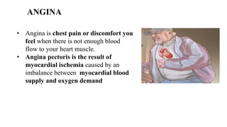
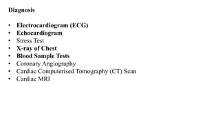
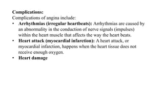
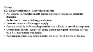
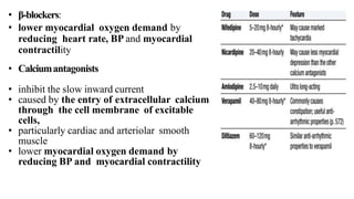
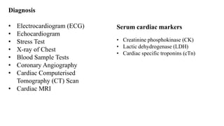
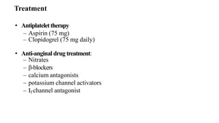
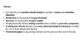
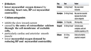
Ischaemic heart disease
➢Ischaemic heart disease (IHD) is defined as an
acute or chronic form of cardiac disability
arising from an imbalance between the
myocardial supply and demand for oxygenated
blood.
• Etiology
• IHD is invariably caused by disease affecting the coronary arteries, the
most prevalent being atherosclerosis accounting for more than 90% of
cases, while other causes are responsible for less than 10% of cases of
IHD. Therefore, it is convenient to consider the etiology of IHD under
three broad headings:
• i) coronary atherosclerosis;
• ii) superadded changes in coronary atherosclerosis; and
• iii) non-atherosclerotic causes.
ANGINA
• Angina is chest pain or discomfort you
feel when there is not enough blood
flow to your heart muscle.
• Angina pectoris is the result of
myocardial ischemia caused by an
imbalance between myocardial blood
supply and oxygen demand
Types:
There are three types of angina:
• Stable angina is the most common type.
• It happens when the heart is working harder than usual — for instance,
during exercise.
• Stable angina has a regular pattern. Rest or medicines relieve the symptoms.
• Unstable angina is the most dangerous.
• It does not follow a regular pattern and can happen without physical
exertion.
• It does not go away with rest or medicine alone.
• It mostly results from atherosclerosis, which involves a blockage preventing
blood from reaching the heart.
• Unstable angina can indicate the risk of a heart attack.
• Variant or Prinzmetal angina is rare.
• It can develop when the body is at rest, often around midnight or the
early morning.
• It happens when a spasm occurs in the coronary arteries. It is a chronic
condition, but medication can help manage it.
• Microvascular angina: Microvascular angina can occur with coronary
microvascular disease.
• This affects the smallest coronary arteries.
• Microvascular angina tends to be more persistent than stable angina.
• It often lasts longer than 10 minutes and sometimes longer than 30
minutes.
CLASS Characteristic
ClassI No angina with ordinary activity.Angina with
strenuousactivity
ClassII Angina during ordinary activity, e.g. walking up hills, walking rapidly
upstairs, with mild limitationof activities
ClassIII Angina with low levels of activity, e.g. walking 50– 100 yards on the
flat, walking up one flight of stairs, with markedrestrictionof activities
ClassIV Angina at restor with any level of exercise
Classification
Causes
• Angina usually results from underlying coronary artery disease.
• The coronary arteries supply the heart with oxygen-rich blood. When
cholesterol collects on the wall of an artery and forms hard plaques,
this effectively narrows the arteries.
• Other factors, including damage to the arteries and smoking, increase
the risk of plaque buildup.
• When the arteries narrow, it becomes harder for oxygen-rich blood to
reach the heart. Also, plaques may break off and form clots that
block the arteries.
• If blood cannot carry oxygen to the heart, the heart muscle cannot work
properly. This causes angina.
Risk factors
Angina can develop as a result of:
• Stress
• An overuse of alcohol
• Smoking
• Exposure to particle pollution, for example, at work
• Low physical activity
• An unhealthful diet
• High cholesterol levels
• Overweight or obesity
• Genetic factors
• Conditions such as heart disease, diabetes, low blood pressure,
metabolic syndrome, and anaemia
• An age of over 45 for males, or 55 for females
Symptoms
• Chest pain is the primary symptom of angina. Angina-related chest pain
makes you feel tight and heavy in the chest. The pain is felt in the left
portion of the body from the back, neck, and jaw and extends up to
the left arm.
• Feeling of breathlessness
• Indigestion-like discomfort in the lower portion of the chest and belly
• Feeling of nausea
• Experiencing severe tiredness from time to time
Pathophysiology of angina
• Angina pectoris is the most common clinical manifestation of myocardial
ischemia.
• Myocardial ischemia is caused by chemical and mechanical stimulation
of sensory afferent nerve endings in the coronary vessels and
myocardium.
• Increases in the heart rate and myocardial contraction result in
increased myocardial oxygen demand.
• Myocardial ischemia can result from a reduction of coronary blood
flow, abnormal constriction of coronary microcirculation, or reduced
oxygen-carrying capacity of the blood.
• Increases in both afterload and preload increase myocardial wall
tension and, therefore, increase myocardial oxygen demand.
• Oxygen supply to any organ system is determined by blood flow. The
ability of the coronary arteries to increase blood flow in response
to increased cardiac metabolic demand is referred to as coronary
flow reserve (CFR).
• Fixed atherosclerotic lesions of at least 90% almost completely
abolish the flow reserve; patients with these lesions may experience
angina at rest or unstable angina.
• Patients with a fixed coronary atherosclerotic lesion of at least 50%
show myocardial ischemia during increased myocardial metabolic
demand, resulting in stable angina.
• These patients are not able to increase their coronary blood flow
during stress to match the increased myocardial demand, thus they
experience angina.
• Prinzmetal angina is defined as resting angina caused by coronary artery
spasm.
• Prinzmetal angina may be due to a deficiency of nitric oxide
production, hyperinsulinemia, low intracellular magnesium levels,
smoking cigarettes, and using cocaine.
Diagnosis
• Electrocardiogram (ECG)
• Echocardiogram
• Stress Test
• X-ray of Chest
• Blood Sample Tests
• Coronary Angiography
• Cardiac Computerised Tomography (CT) Scan
• Cardiac MRI
Complications:
Complications of angina include:
• Arrhythmias (irregular heartbeats): Arrhythmias are caused by
an abnormality in the conduction of nerve signals (impulses)
within the heart muscle that affects the way the heart beats.
• Heart attack (myocardial infarction): A heart attack, or
myocardial infarction, happens when the heart tissue does not
receive enough oxygen.
• Heart damage
• Antiplatelet therapy
– Aspirin (75 mg)
– Clopidogrel (75 mg daily)
• Anti-anginal drug treatment:
– Nitrates
– β-blockers
– calcium antagonists
– potassium channel activators
– If channel antagonist
Treatment
Nitrate
Ex. Glyceryl trinitrate, Isosorbide dinitrate
• Act directly on vascularsmooth muscleto produce venous and arteriolar
dilatation
• Reduction in myocardial oxygen demand
• Increase in myocardial oxygen supply
• Pprophylactically before taking exercise that is liable to provoke symptoms.
• Continuous nitrate therapy can cause pharmacological tolerance avoided
by a 6–8-hour nitrate-free period
• Nocturnalangina:Long-acting nitrates can be given at the end of the day
• β-blockers:
• lower myocardial oxygen demand by
reducing heart rate, BP and myocardial
contractility
• Calciumantagonists
• inhibit the slow inward current
• caused by the entry of extracellular calcium
through the cell membrane of excitable
cells,
• particularly cardiac and arteriolar smooth
muscle
• lower myocardial oxygen demand by
reducing BP and myocardial contractility
Potassiumchannelactivators
• Nicorandil(10–30mg12-hourlyorally)- only drug in this class currently
available for clinical use
• If channel antagonist
• Ivabradineis the first of this class of drug
• Induces bradycardiaby modulating ion channels in the sinusnode
• Comparatively, does not have other cardiovascular effects
• Safe to use in patients with heart failure
• Passing a fine guidewire across a coronarystenosisunder radiographic
control
• Ballonis placed and then inflated to dilate the stenosis
• Then a coronarystentis deployed on a balloon
– maximise and maintain dilatation of a stenosed vessel
– reduces both acute complications and the incidence of
clinically important restenosis
• Mainly used in single or two-vessel disease
Percutaneous Coronary Intervention (PCI)
*
• Stenosed artery is by-passed with
– internal mammaryarteries
– radialarteries
– reversedsegmentsof the patient’s saphenousvein
• Major surgery under cardiopulmonarybypass,
• But in some cases, grafts can be applied to the beating heart: ‘off-pump’
surgery
• Operative mortality is approximately 1.5% but risks are higher
– elderly patients,
– with poor left ventricular function
– those with significant comorbidities, such as renal failure
• 90% of patients are free of angina 1 year after CABG surgery, but
fewer than 60% of patients are asymptomatic after 5 or more years.
Coronary artery bypass grafting
MYOCARDIAL INFARCTION
• MI is defined as a diseased condition that is caused by reduced blood
flow in a coronary artery due to atherosclerosis & occlusion of an
artery by an embolus or thrombus.
• MI or heart attack is the irreversible damage of myocardial tissue
caused by prolonged ischemia and hypoxia.
• Etiology
• Tobacco, smoking
• Hypertension
• Drug abuse
• Obesity
• Stress
• Alcohol
• Age
• Gender
• Diabetes
• Hyperlipoproteinaemia
• Family history of Ischaemic
Heart Disease
• Hyperhomocysteinemia
• Chronic kidney disease
Pathophysiology
• Symptoms of MI include
• Chest pain, which travels from left arm to neck,
• Shortness of breath,
• Sweating,
• Nausea,
• Vomiting,
• Abnormal heart beating,
• Anxiety,
• Fatigue,
• Weakness,
• Stress, depression, and other factors.
Diagnosis
• Electrocardiogram (ECG)
• Echocardiogram
• Stress Test
• X-ray of Chest
• Blood Sample Tests
• Coronary Angiography
• Cardiac Computerised
Tomography (CT) Scan
• Cardiac MRI
Serum cardiac markers
• Creatinine phosphokinase (CK)
• Lactic dehydrogenase (LDH)
• Cardiac specific troponins (cTn)
Complications:
Complications of angina include:
• Arrhythmias (irregular heartbeats): Arrhythmias are caused by
an abnormality in the conduction of nerve signals (impulses)
within the heart muscle that affects the way the heart beats.
• Heart attack (myocardial infarction): A heart attack, or
myocardial infarction, happens when the heart tissue does not
receive enough oxygen.
• Heart damage
• Antiplatelet therapy
– Aspirin (75 mg)
– Clopidogrel (75 mg daily)
• Anti-anginal drug treatment:
– Nitrates
– β-blockers
– calcium antagonists
– potassium channel activators
– If channel antagonist
Treatment
Nitrate
• Act directly on vascularsmooth muscleto produce venous and arteriolar
dilatation
• Reduction in myocardial oxygen demand
• Increase in myocardial oxygen supply
• Prophylactically before taking exercise that is liable to provoke symptoms.
• Continuous nitrate therapy can cause pharmacological tolerance avoided
by a 6–8-hour nitrate-free period
• Nocturnalangina:Long-acting nitrates can be given at the end of the day
• β-blockers:
• lower myocardial oxygen demand by
reducing heart rate, BP and myocardial
contractility
• Calciumantagonists
• inhibit the slow inward current
• caused by the entry of extracellular calcium
through the cell membrane of excitable
cells,
• particularly cardiac and arteriolar smooth
muscle
• lower myocardial oxygen demand by
reducing BP and myocardial contractility
Potassiumchannelactivators
• Nicorandil(10–30mg12-hourlyorally)- only drug in this class currently
available for clinical use
• If channel antagonist
• Ivabradineis the first of this class of drug
• Induces bradycardiaby modulating ion channels in the sinusnode
• Comparatively, does not have other cardiovascular effects
• Safe to use in patients with heart failure
• Passing a fine guidewire across a coronarystenosisunder radiographic
control
• Ballonis placed and then inflated to dilate the stenosis
• Then a coronarystentis deployed on a balloon
– maximise and maintain dilatation of a stenosed vessel
– reduces both acute complications and the incidence of
clinically important restenosis
• Mainly used in single or two-vessel disease
Percutaneous Coronary Intervention (PCI)
*
• Stenosed artery is by-passed with
– internal mammaryarteries
– radialarteries
– reversedsegmentsof the patient’s saphenousvein
• Operative mortality is approximately 1.5% but risks are higher
– elderly patients,
– with poor left ventricular function
– those with significant comorbidities, such as renal failure
• 90% of patients are free of angina 1 year after CABG surgery, but fewer
than 60% of patients are asymptomatic after 5 or more years.
Coronary artery bypass grafting
• Essential not only to relieve distress but also to lower adrenergic
drive
• IV opiates:initially morphine sulphate 5– 10mg or
diamorphine 2.5–5 mg)
• IVAntiemetics:initially metoclopramide 10mg
• Intramuscular injections should be avoided
Analgesics
Antiplatelet therapy
• Aspirin: oral dose of 300mg first tablet within the first 12 hours, followed
by 75 mg.
• Aspirin + clopidogrel 600 mg: Early (within 12 hours), followed by 150
mg daily for 1 week and 75mg daily thereafter
• Ticagrelor (180 mg followed by 90 mg 12-hourly): more effective than
clopidogrel
• Antiplatelet treatment with i.v. glycoprotein IIb/IIIa inhibitors (Tirofiban
and eptifibatide) reduce the combined endpoint of death or MI and are used
in the context of PCI
Antithrombotic Therapy
Anticoagulants
• Reduces the risk of thromboembolic complication
• Prevents reinfarction in the absence of reperfusion therapy or after
successful thrombolysis
• Unfractionated heparin, fractioned (low molecular weight) heparin
or a pentasaccharide.
• Continued for 8 days or until discharge from hospital or coronary
revascularization
Antithrombotic Therapy
Reperfusion Therapy
Thrombolysis
• Reduce hospital mortality by 25–50%
• This survival advantage is maintained for at least 10 years
Alteplase (human tissue plasminogen activator)
• Over 90 minutes (bolus dose of 15 mg)
• Followed by 0.75 mg/kg body weight, but not exceeding 50mg, over 30
mins then 0.5mg/kg body weight but not exceeding 35mg over 60mins better
survival rates than other thrombolytic agents such as
Streptokinase
• Analogues of tPA (Tenecteplase and reteplase): longer plasma half-life than
alteplase and can be given as an intravenous bolus
• Riskstratificationandfurtherinvestigationlifestylemodification
– Cessation of smoking
– Regular exercise
– Diet (weight control, lipid-lowering)
• Secondary preventiondrugtherapy
– Antiplatelet therapy (aspirin and/or clopidogrel)
– β-blocker
– ACE inhibitor
– Statin
– Additional therapy for control of diabetes and hypertension
– Aldosterone receptor antagonis
• Rehabilitation devices:Implantable cardiac defibrillator (high-risk
patients)
Thank
you

Angina and MI PATHOPHYSIOLOGY .pdf

  • 1.
  • 2.
    ➢Ischaemic heart disease(IHD) is defined as an acute or chronic form of cardiac disability arising from an imbalance between the myocardial supply and demand for oxygenated blood.
  • 3.
    • Etiology • IHDis invariably caused by disease affecting the coronary arteries, the most prevalent being atherosclerosis accounting for more than 90% of cases, while other causes are responsible for less than 10% of cases of IHD. Therefore, it is convenient to consider the etiology of IHD under three broad headings: • i) coronary atherosclerosis; • ii) superadded changes in coronary atherosclerosis; and • iii) non-atherosclerotic causes.
  • 4.
    ANGINA • Angina ischest pain or discomfort you feel when there is not enough blood flow to your heart muscle. • Angina pectoris is the result of myocardial ischemia caused by an imbalance between myocardial blood supply and oxygen demand
  • 5.
    Types: There are threetypes of angina: • Stable angina is the most common type. • It happens when the heart is working harder than usual — for instance, during exercise. • Stable angina has a regular pattern. Rest or medicines relieve the symptoms. • Unstable angina is the most dangerous. • It does not follow a regular pattern and can happen without physical exertion. • It does not go away with rest or medicine alone. • It mostly results from atherosclerosis, which involves a blockage preventing blood from reaching the heart. • Unstable angina can indicate the risk of a heart attack.
  • 6.
    • Variant orPrinzmetal angina is rare. • It can develop when the body is at rest, often around midnight or the early morning. • It happens when a spasm occurs in the coronary arteries. It is a chronic condition, but medication can help manage it. • Microvascular angina: Microvascular angina can occur with coronary microvascular disease. • This affects the smallest coronary arteries. • Microvascular angina tends to be more persistent than stable angina. • It often lasts longer than 10 minutes and sometimes longer than 30 minutes.
  • 7.
    CLASS Characteristic ClassI Noangina with ordinary activity.Angina with strenuousactivity ClassII Angina during ordinary activity, e.g. walking up hills, walking rapidly upstairs, with mild limitationof activities ClassIII Angina with low levels of activity, e.g. walking 50– 100 yards on the flat, walking up one flight of stairs, with markedrestrictionof activities ClassIV Angina at restor with any level of exercise Classification
  • 9.
    Causes • Angina usuallyresults from underlying coronary artery disease. • The coronary arteries supply the heart with oxygen-rich blood. When cholesterol collects on the wall of an artery and forms hard plaques, this effectively narrows the arteries. • Other factors, including damage to the arteries and smoking, increase the risk of plaque buildup. • When the arteries narrow, it becomes harder for oxygen-rich blood to reach the heart. Also, plaques may break off and form clots that block the arteries. • If blood cannot carry oxygen to the heart, the heart muscle cannot work properly. This causes angina.
  • 10.
    Risk factors Angina candevelop as a result of: • Stress • An overuse of alcohol • Smoking • Exposure to particle pollution, for example, at work • Low physical activity • An unhealthful diet • High cholesterol levels • Overweight or obesity • Genetic factors • Conditions such as heart disease, diabetes, low blood pressure, metabolic syndrome, and anaemia • An age of over 45 for males, or 55 for females
  • 11.
    Symptoms • Chest painis the primary symptom of angina. Angina-related chest pain makes you feel tight and heavy in the chest. The pain is felt in the left portion of the body from the back, neck, and jaw and extends up to the left arm. • Feeling of breathlessness • Indigestion-like discomfort in the lower portion of the chest and belly • Feeling of nausea • Experiencing severe tiredness from time to time
  • 12.
    Pathophysiology of angina •Angina pectoris is the most common clinical manifestation of myocardial ischemia. • Myocardial ischemia is caused by chemical and mechanical stimulation of sensory afferent nerve endings in the coronary vessels and myocardium. • Increases in the heart rate and myocardial contraction result in increased myocardial oxygen demand. • Myocardial ischemia can result from a reduction of coronary blood flow, abnormal constriction of coronary microcirculation, or reduced oxygen-carrying capacity of the blood.
  • 13.
    • Increases inboth afterload and preload increase myocardial wall tension and, therefore, increase myocardial oxygen demand. • Oxygen supply to any organ system is determined by blood flow. The ability of the coronary arteries to increase blood flow in response to increased cardiac metabolic demand is referred to as coronary flow reserve (CFR). • Fixed atherosclerotic lesions of at least 90% almost completely abolish the flow reserve; patients with these lesions may experience angina at rest or unstable angina.
  • 14.
    • Patients witha fixed coronary atherosclerotic lesion of at least 50% show myocardial ischemia during increased myocardial metabolic demand, resulting in stable angina. • These patients are not able to increase their coronary blood flow during stress to match the increased myocardial demand, thus they experience angina. • Prinzmetal angina is defined as resting angina caused by coronary artery spasm. • Prinzmetal angina may be due to a deficiency of nitric oxide production, hyperinsulinemia, low intracellular magnesium levels, smoking cigarettes, and using cocaine.
  • 15.
    Diagnosis • Electrocardiogram (ECG) •Echocardiogram • Stress Test • X-ray of Chest • Blood Sample Tests • Coronary Angiography • Cardiac Computerised Tomography (CT) Scan • Cardiac MRI
  • 16.
    Complications: Complications of anginainclude: • Arrhythmias (irregular heartbeats): Arrhythmias are caused by an abnormality in the conduction of nerve signals (impulses) within the heart muscle that affects the way the heart beats. • Heart attack (myocardial infarction): A heart attack, or myocardial infarction, happens when the heart tissue does not receive enough oxygen. • Heart damage
  • 17.
    • Antiplatelet therapy –Aspirin (75 mg) – Clopidogrel (75 mg daily) • Anti-anginal drug treatment: – Nitrates – β-blockers – calcium antagonists – potassium channel activators – If channel antagonist Treatment
  • 18.
    Nitrate Ex. Glyceryl trinitrate,Isosorbide dinitrate • Act directly on vascularsmooth muscleto produce venous and arteriolar dilatation • Reduction in myocardial oxygen demand • Increase in myocardial oxygen supply • Pprophylactically before taking exercise that is liable to provoke symptoms. • Continuous nitrate therapy can cause pharmacological tolerance avoided by a 6–8-hour nitrate-free period • Nocturnalangina:Long-acting nitrates can be given at the end of the day
  • 19.
    • β-blockers: • lowermyocardial oxygen demand by reducing heart rate, BP and myocardial contractility • Calciumantagonists • inhibit the slow inward current • caused by the entry of extracellular calcium through the cell membrane of excitable cells, • particularly cardiac and arteriolar smooth muscle • lower myocardial oxygen demand by reducing BP and myocardial contractility
  • 20.
    Potassiumchannelactivators • Nicorandil(10–30mg12-hourlyorally)- onlydrug in this class currently available for clinical use • If channel antagonist • Ivabradineis the first of this class of drug • Induces bradycardiaby modulating ion channels in the sinusnode • Comparatively, does not have other cardiovascular effects • Safe to use in patients with heart failure
  • 21.
    • Passing afine guidewire across a coronarystenosisunder radiographic control • Ballonis placed and then inflated to dilate the stenosis • Then a coronarystentis deployed on a balloon – maximise and maintain dilatation of a stenosed vessel – reduces both acute complications and the incidence of clinically important restenosis • Mainly used in single or two-vessel disease Percutaneous Coronary Intervention (PCI)
  • 22.
  • 23.
    • Stenosed arteryis by-passed with – internal mammaryarteries – radialarteries – reversedsegmentsof the patient’s saphenousvein • Major surgery under cardiopulmonarybypass, • But in some cases, grafts can be applied to the beating heart: ‘off-pump’ surgery • Operative mortality is approximately 1.5% but risks are higher – elderly patients, – with poor left ventricular function – those with significant comorbidities, such as renal failure • 90% of patients are free of angina 1 year after CABG surgery, but fewer than 60% of patients are asymptomatic after 5 or more years. Coronary artery bypass grafting
  • 25.
    MYOCARDIAL INFARCTION • MIis defined as a diseased condition that is caused by reduced blood flow in a coronary artery due to atherosclerosis & occlusion of an artery by an embolus or thrombus. • MI or heart attack is the irreversible damage of myocardial tissue caused by prolonged ischemia and hypoxia.
  • 26.
    • Etiology • Tobacco,smoking • Hypertension • Drug abuse • Obesity • Stress • Alcohol • Age • Gender • Diabetes • Hyperlipoproteinaemia • Family history of Ischaemic Heart Disease • Hyperhomocysteinemia • Chronic kidney disease
  • 27.
  • 28.
    • Symptoms ofMI include • Chest pain, which travels from left arm to neck, • Shortness of breath, • Sweating, • Nausea, • Vomiting, • Abnormal heart beating, • Anxiety, • Fatigue, • Weakness, • Stress, depression, and other factors.
  • 29.
    Diagnosis • Electrocardiogram (ECG) •Echocardiogram • Stress Test • X-ray of Chest • Blood Sample Tests • Coronary Angiography • Cardiac Computerised Tomography (CT) Scan • Cardiac MRI Serum cardiac markers • Creatinine phosphokinase (CK) • Lactic dehydrogenase (LDH) • Cardiac specific troponins (cTn)
  • 30.
    Complications: Complications of anginainclude: • Arrhythmias (irregular heartbeats): Arrhythmias are caused by an abnormality in the conduction of nerve signals (impulses) within the heart muscle that affects the way the heart beats. • Heart attack (myocardial infarction): A heart attack, or myocardial infarction, happens when the heart tissue does not receive enough oxygen. • Heart damage
  • 31.
    • Antiplatelet therapy –Aspirin (75 mg) – Clopidogrel (75 mg daily) • Anti-anginal drug treatment: – Nitrates – β-blockers – calcium antagonists – potassium channel activators – If channel antagonist Treatment
  • 32.
    Nitrate • Act directlyon vascularsmooth muscleto produce venous and arteriolar dilatation • Reduction in myocardial oxygen demand • Increase in myocardial oxygen supply • Prophylactically before taking exercise that is liable to provoke symptoms. • Continuous nitrate therapy can cause pharmacological tolerance avoided by a 6–8-hour nitrate-free period • Nocturnalangina:Long-acting nitrates can be given at the end of the day
  • 33.
    • β-blockers: • lowermyocardial oxygen demand by reducing heart rate, BP and myocardial contractility • Calciumantagonists • inhibit the slow inward current • caused by the entry of extracellular calcium through the cell membrane of excitable cells, • particularly cardiac and arteriolar smooth muscle • lower myocardial oxygen demand by reducing BP and myocardial contractility
  • 34.
    Potassiumchannelactivators • Nicorandil(10–30mg12-hourlyorally)- onlydrug in this class currently available for clinical use • If channel antagonist • Ivabradineis the first of this class of drug • Induces bradycardiaby modulating ion channels in the sinusnode • Comparatively, does not have other cardiovascular effects • Safe to use in patients with heart failure
  • 35.
    • Passing afine guidewire across a coronarystenosisunder radiographic control • Ballonis placed and then inflated to dilate the stenosis • Then a coronarystentis deployed on a balloon – maximise and maintain dilatation of a stenosed vessel – reduces both acute complications and the incidence of clinically important restenosis • Mainly used in single or two-vessel disease Percutaneous Coronary Intervention (PCI)
  • 36.
  • 37.
    • Stenosed arteryis by-passed with – internal mammaryarteries – radialarteries – reversedsegmentsof the patient’s saphenousvein • Operative mortality is approximately 1.5% but risks are higher – elderly patients, – with poor left ventricular function – those with significant comorbidities, such as renal failure • 90% of patients are free of angina 1 year after CABG surgery, but fewer than 60% of patients are asymptomatic after 5 or more years. Coronary artery bypass grafting
  • 39.
    • Essential notonly to relieve distress but also to lower adrenergic drive • IV opiates:initially morphine sulphate 5– 10mg or diamorphine 2.5–5 mg) • IVAntiemetics:initially metoclopramide 10mg • Intramuscular injections should be avoided Analgesics
  • 40.
    Antiplatelet therapy • Aspirin:oral dose of 300mg first tablet within the first 12 hours, followed by 75 mg. • Aspirin + clopidogrel 600 mg: Early (within 12 hours), followed by 150 mg daily for 1 week and 75mg daily thereafter • Ticagrelor (180 mg followed by 90 mg 12-hourly): more effective than clopidogrel • Antiplatelet treatment with i.v. glycoprotein IIb/IIIa inhibitors (Tirofiban and eptifibatide) reduce the combined endpoint of death or MI and are used in the context of PCI Antithrombotic Therapy
  • 41.
    Anticoagulants • Reduces therisk of thromboembolic complication • Prevents reinfarction in the absence of reperfusion therapy or after successful thrombolysis • Unfractionated heparin, fractioned (low molecular weight) heparin or a pentasaccharide. • Continued for 8 days or until discharge from hospital or coronary revascularization Antithrombotic Therapy
  • 42.
    Reperfusion Therapy Thrombolysis • Reducehospital mortality by 25–50% • This survival advantage is maintained for at least 10 years Alteplase (human tissue plasminogen activator) • Over 90 minutes (bolus dose of 15 mg) • Followed by 0.75 mg/kg body weight, but not exceeding 50mg, over 30 mins then 0.5mg/kg body weight but not exceeding 35mg over 60mins better survival rates than other thrombolytic agents such as Streptokinase • Analogues of tPA (Tenecteplase and reteplase): longer plasma half-life than alteplase and can be given as an intravenous bolus
  • 43.
    • Riskstratificationandfurtherinvestigationlifestylemodification – Cessationof smoking – Regular exercise – Diet (weight control, lipid-lowering) • Secondary preventiondrugtherapy – Antiplatelet therapy (aspirin and/or clopidogrel) – β-blocker – ACE inhibitor – Statin – Additional therapy for control of diabetes and hypertension – Aldosterone receptor antagonis • Rehabilitation devices:Implantable cardiac defibrillator (high-risk patients)
  • 44.