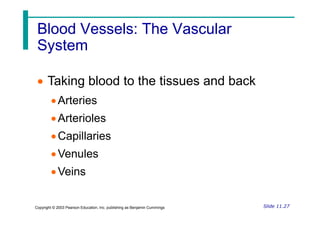
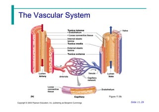
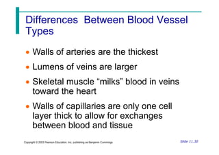
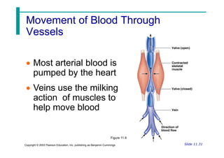
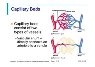
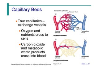
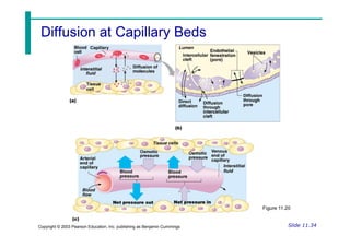
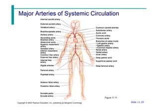
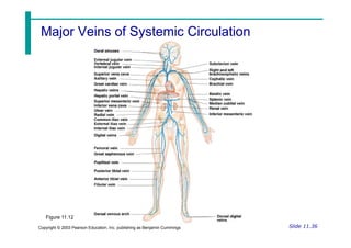
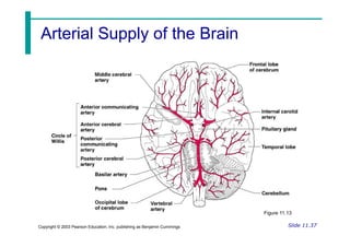
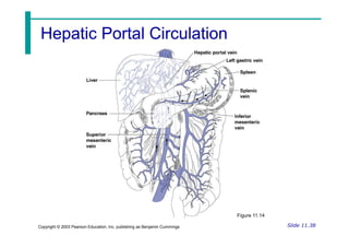
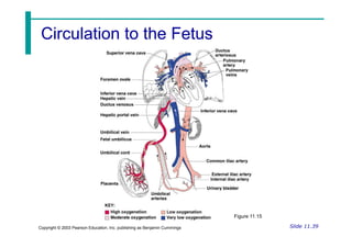
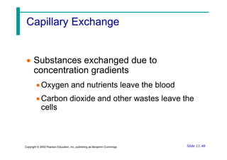
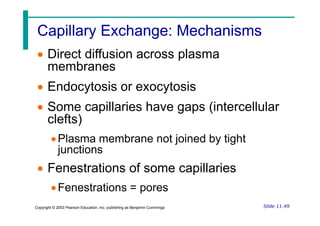
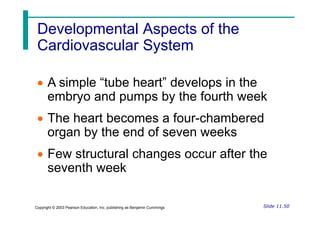
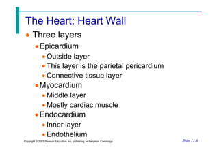
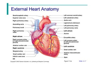
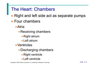
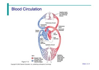
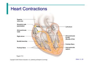
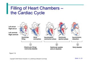
This document summarizes key aspects of the cardiovascular system presented in lecture slides. It describes the closed system of the heart and blood vessels, and the heart's role in pumping blood to circulate oxygen and nutrients to all parts of the body. Key details are provided on the anatomy of the heart, including its chambers, valves, conduction system, and associated blood vessels. The cardiac cycle and regulation of cardiac output through heart rate and stroke volume are also summarized. Finally, the document outlines the different types of blood vessels and their roles in circulation, as well as some major arteries and veins.


![The Heart: Cardiac Output
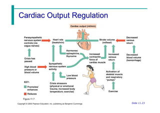
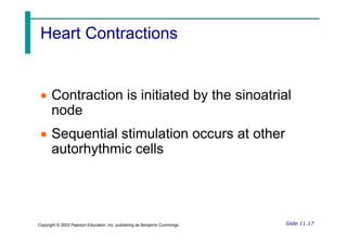
The Heart: Cardiac Output
Cardiac output (CO)
Amount of blood pumped by each side of
the heart in one minute
CO = (heart rate [HR]) x (stroke volume
Slide 11.22
Copyright © 2003 Pearson Education, Inc. publishing as Benjamin Cummings
CO = (heart rate [HR]) x (stroke volume
[SV])
Stroke volume
Volume of blood pumped by each ventricle
in one contraction](https://image.slidesharecdn.com/chapter11cardioppt-231016213804-6e90ec25/85/Chapter-11-cardio-ppt-pdf-22-320.jpg)