Downloaded 429 times


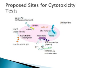


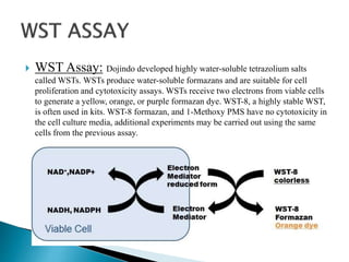


This document discusses methods for evaluating the cytotoxicity of nanoparticles. It describes several common cytotoxicity assays including MTT, WST, trypan blue exclusion, and assays using dehydrogenases. The MTT assay measures mitochondrial activity and is widely used. WST assays use water-soluble reagents and do not require crystal solubilization. Dehydrogenase assays offer high sensitivity by measuring multiple cell elements. The document also provides examples of studies that used these assays to evaluate the cytotoxicity of silver nanoparticles, magnetic nanoparticles, and other nanomaterials.


