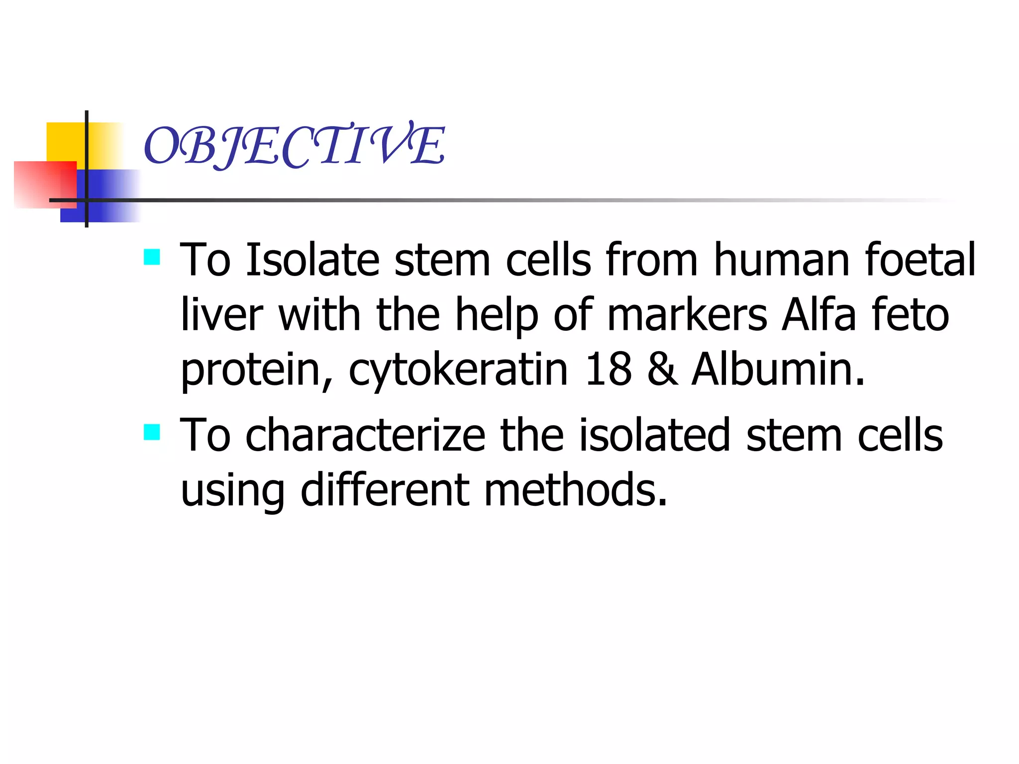
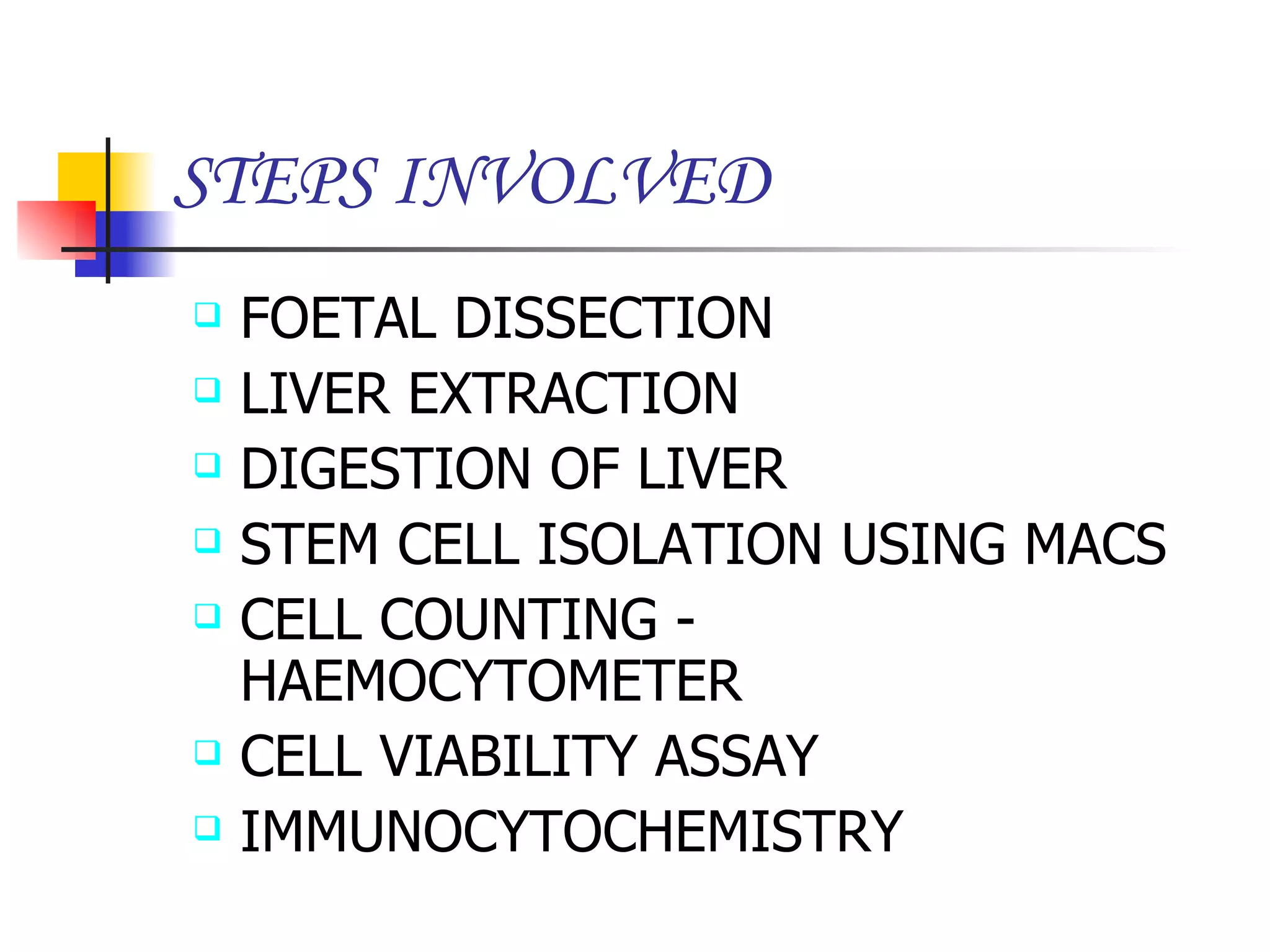
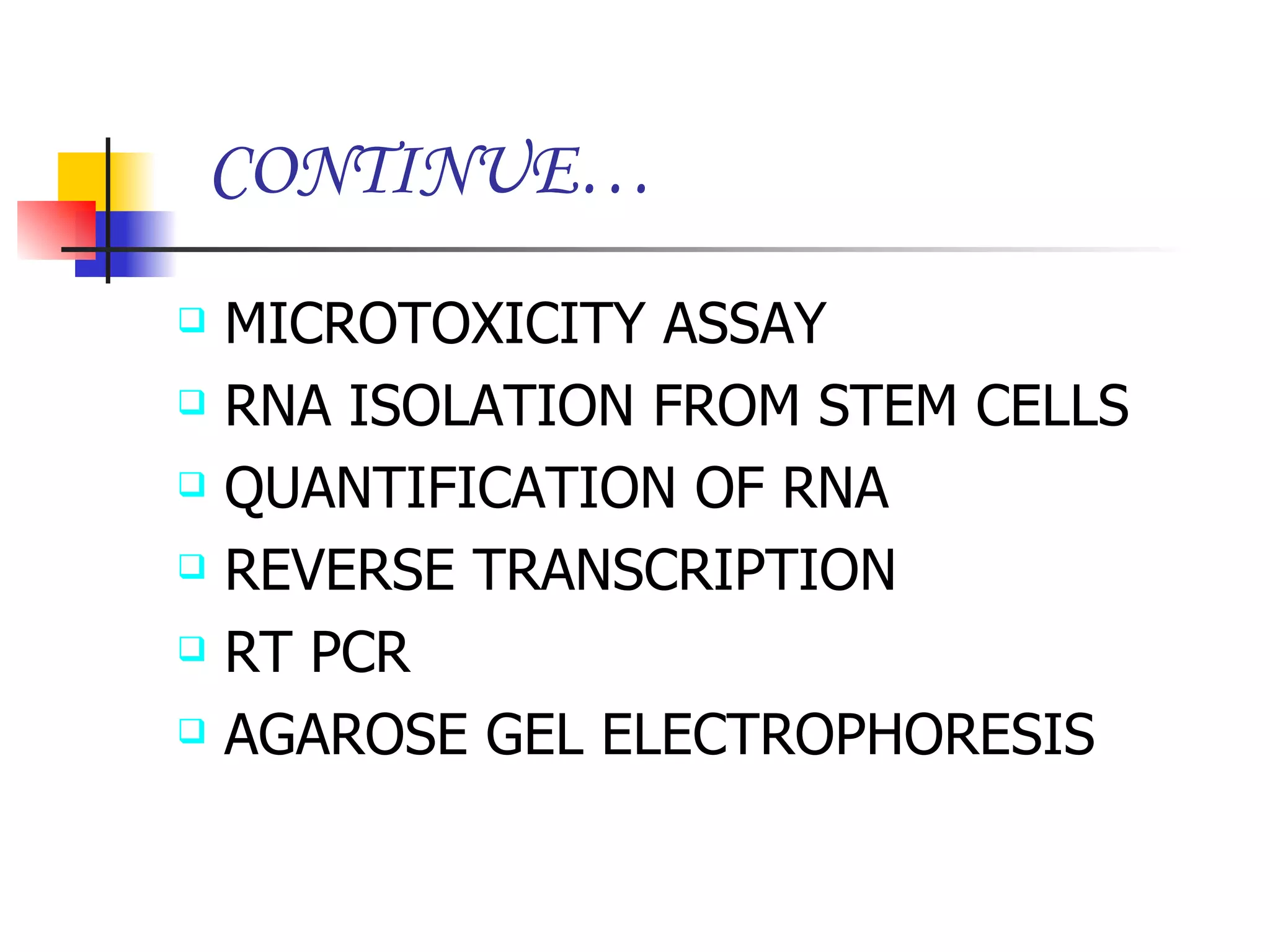
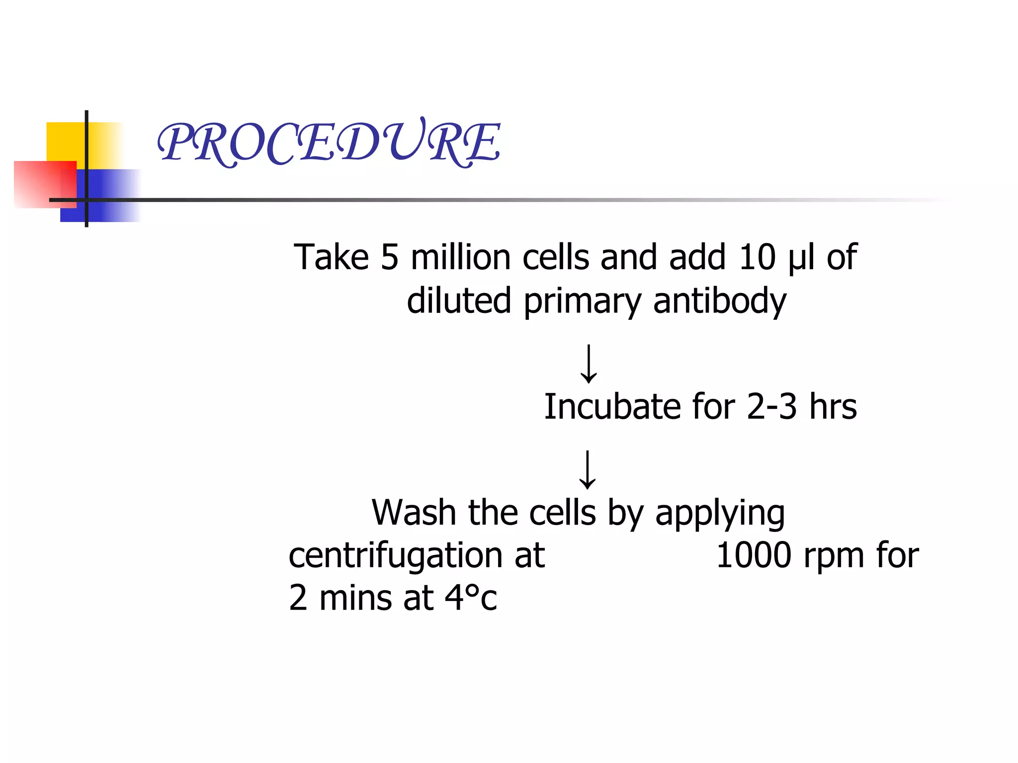
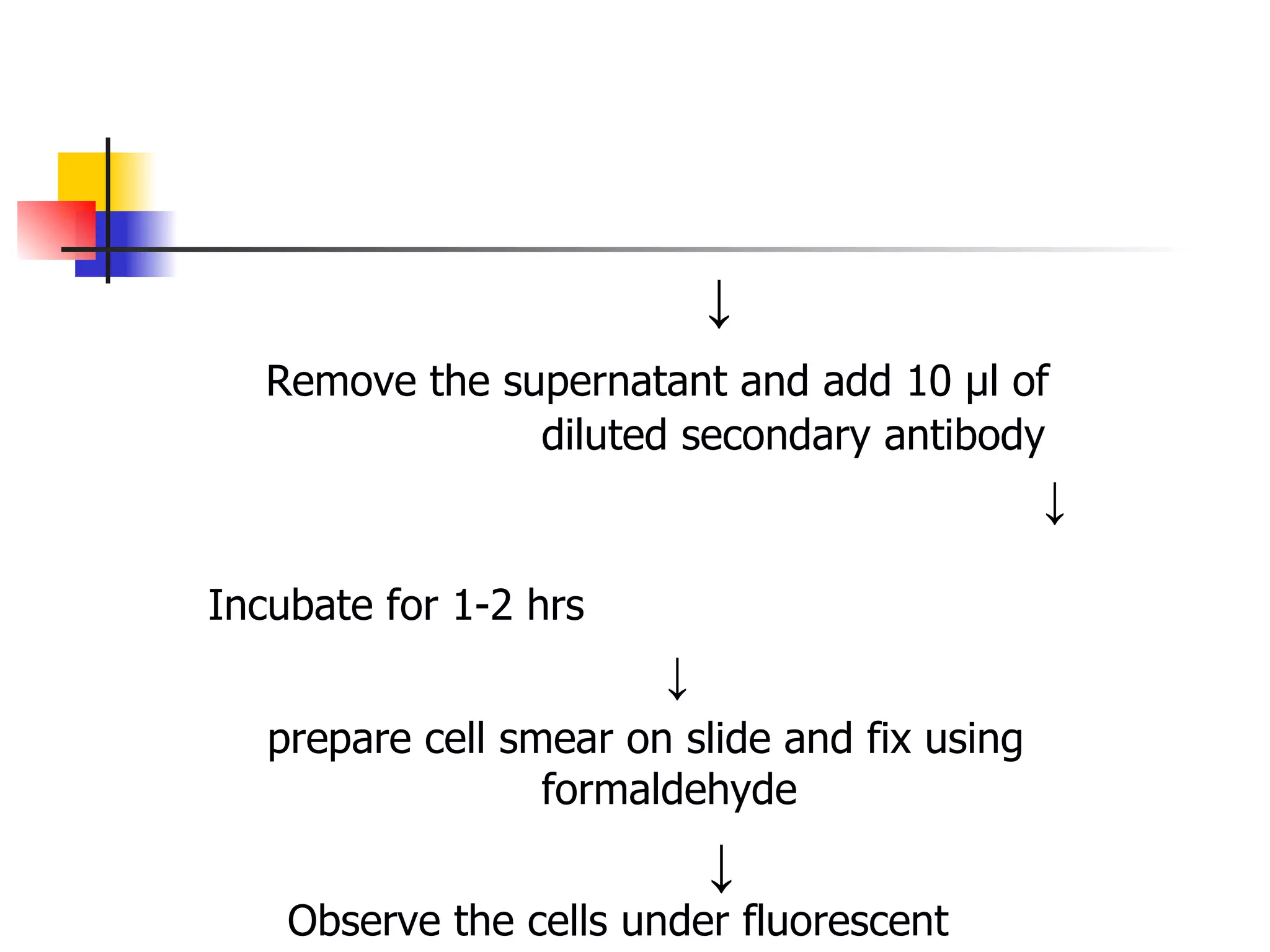
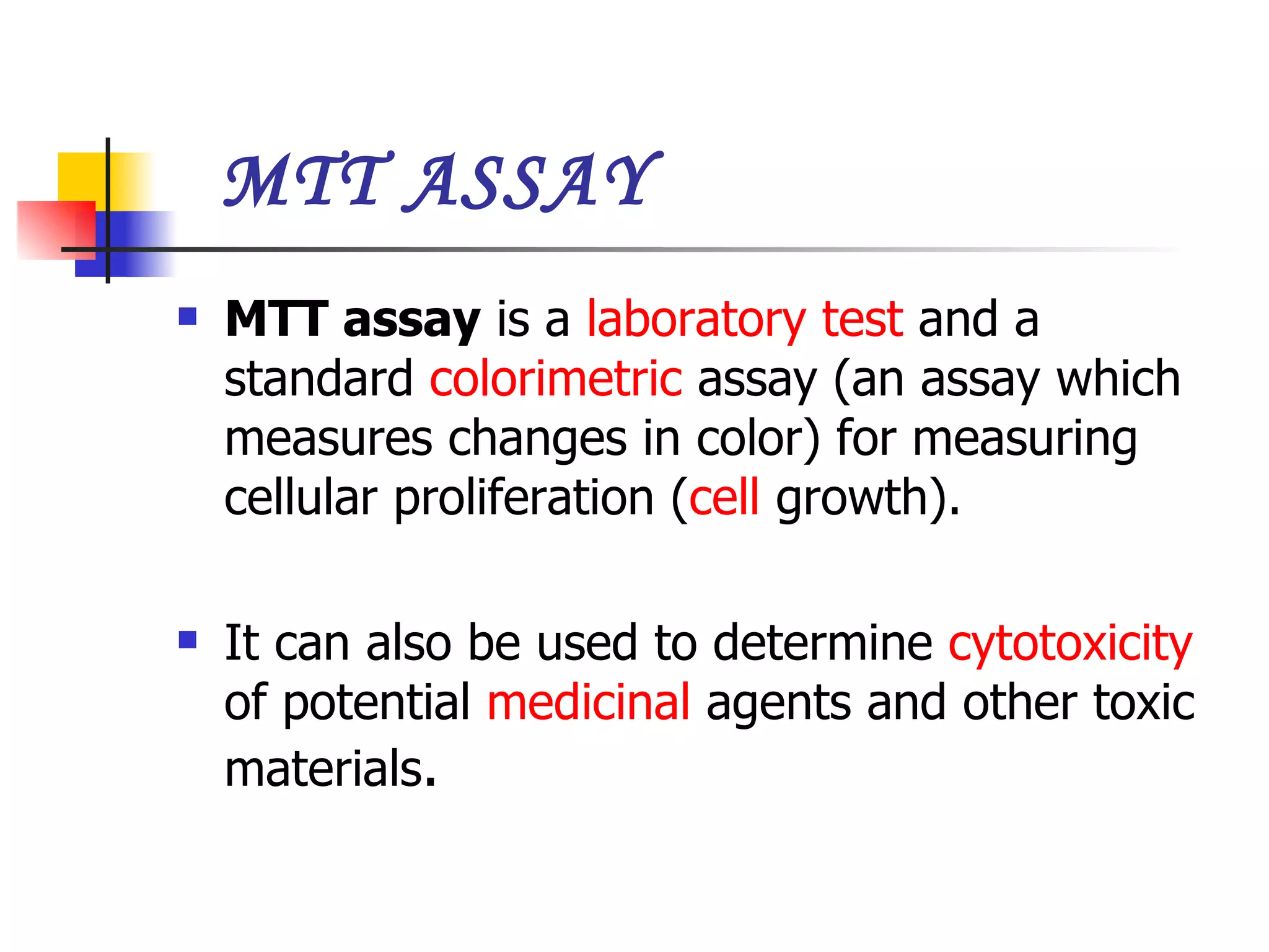
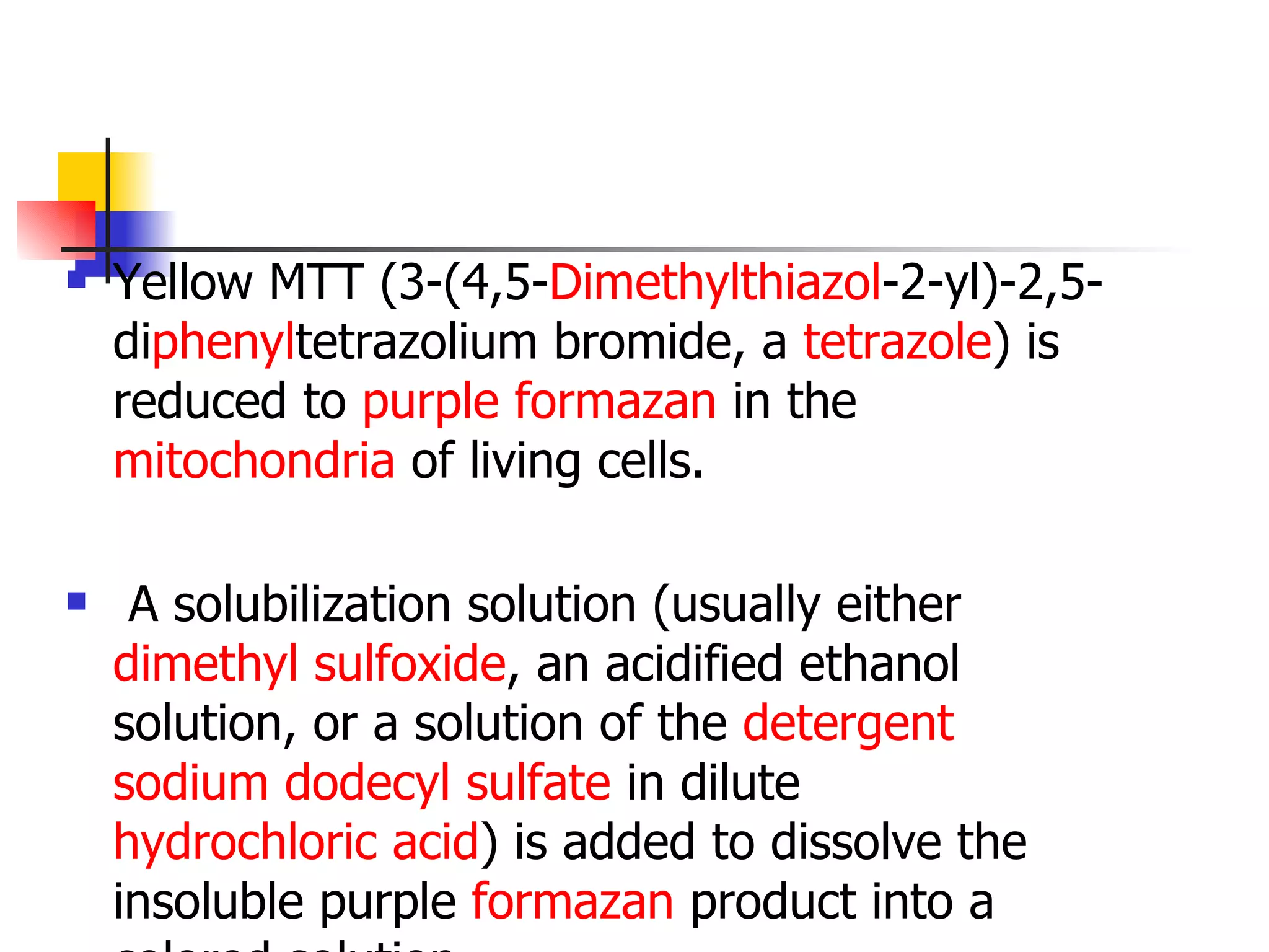
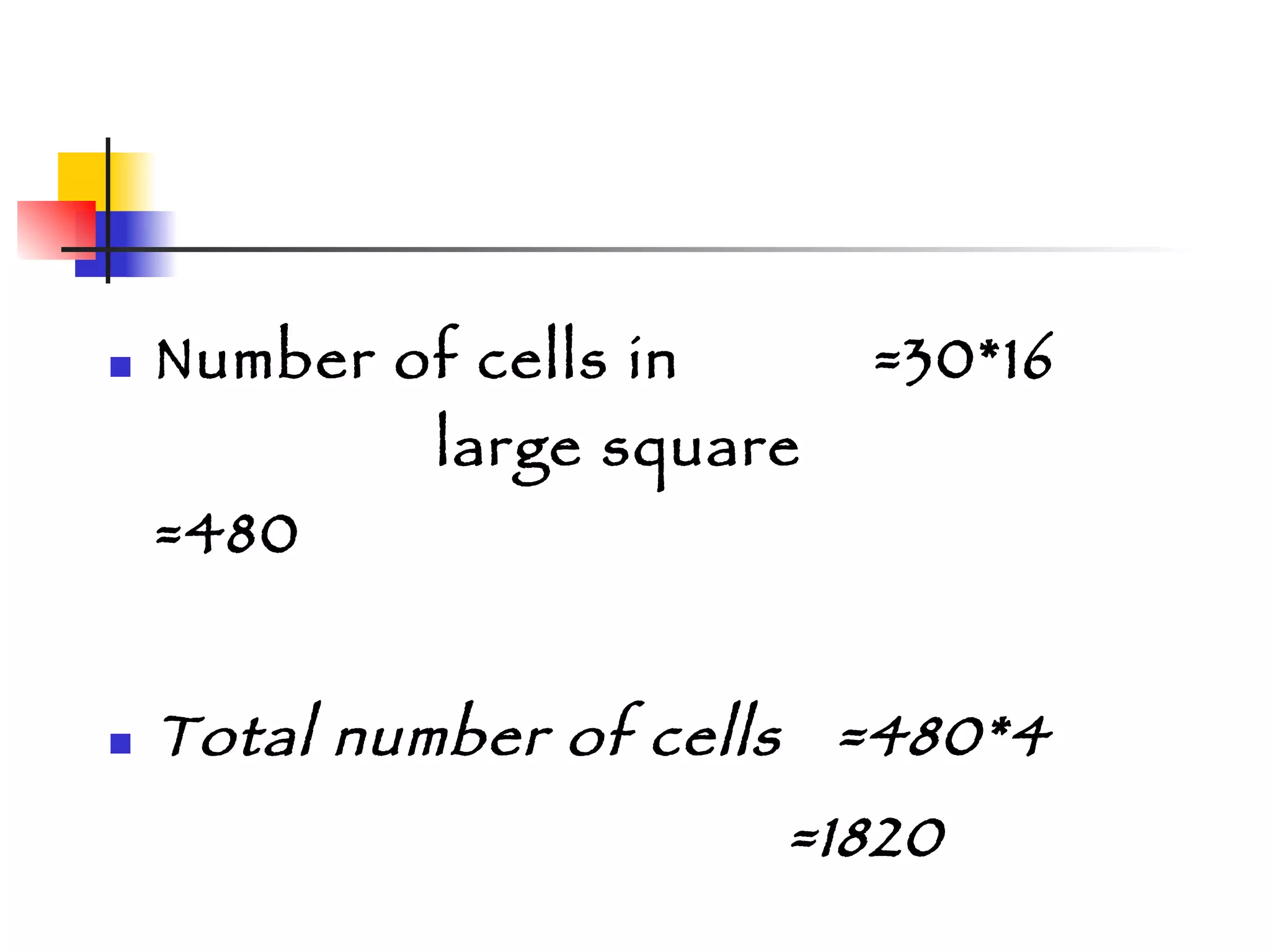
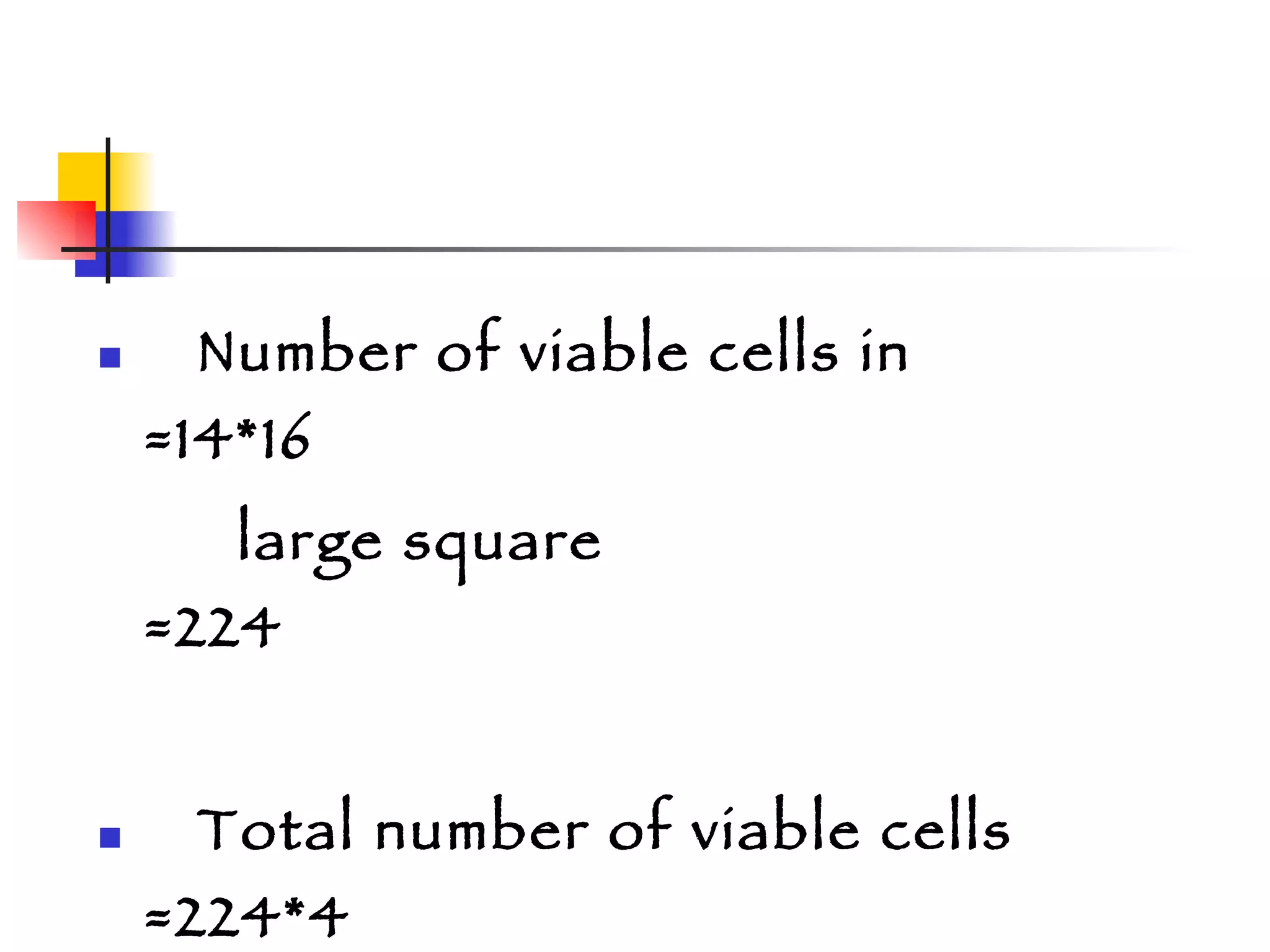
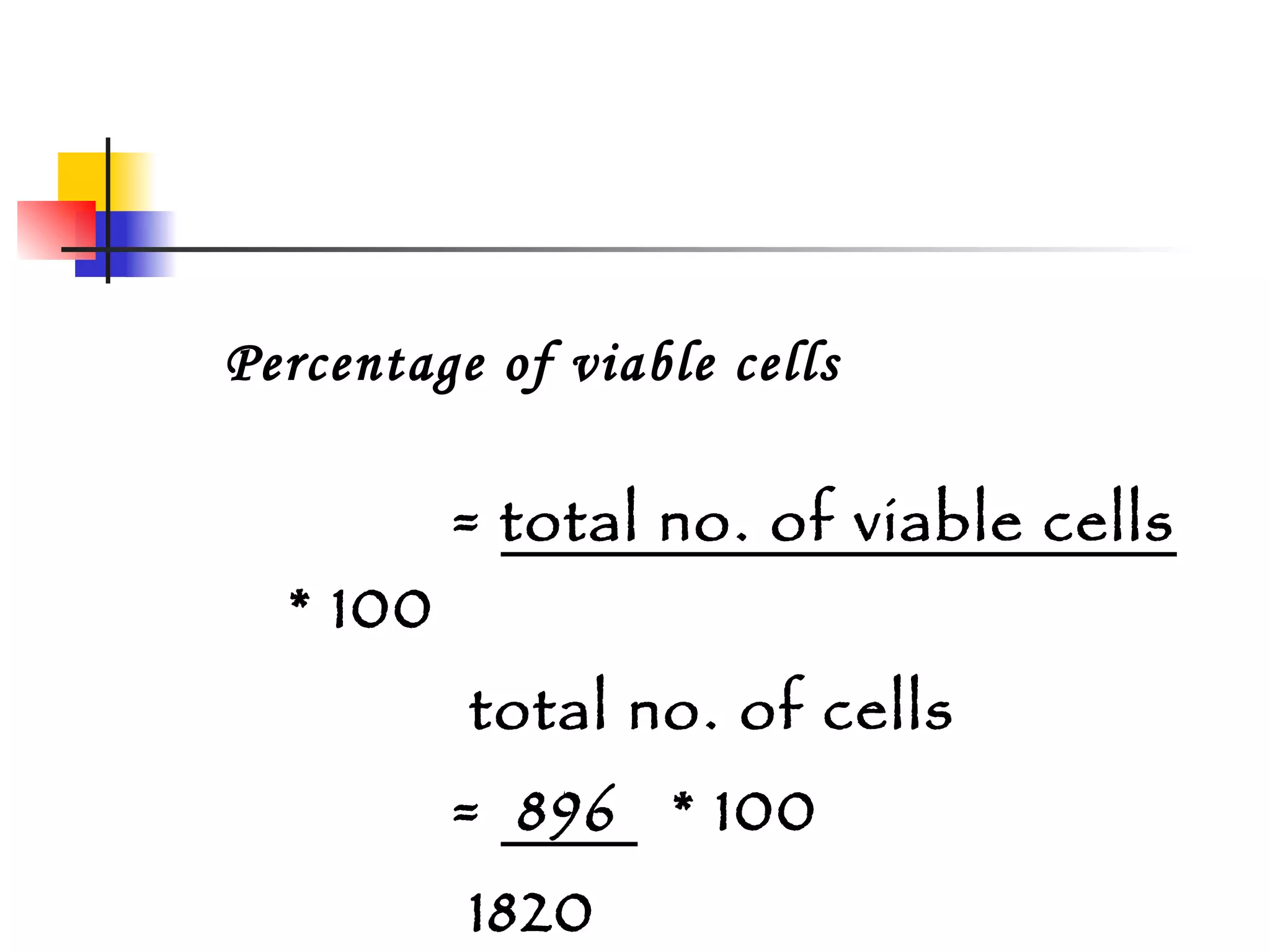
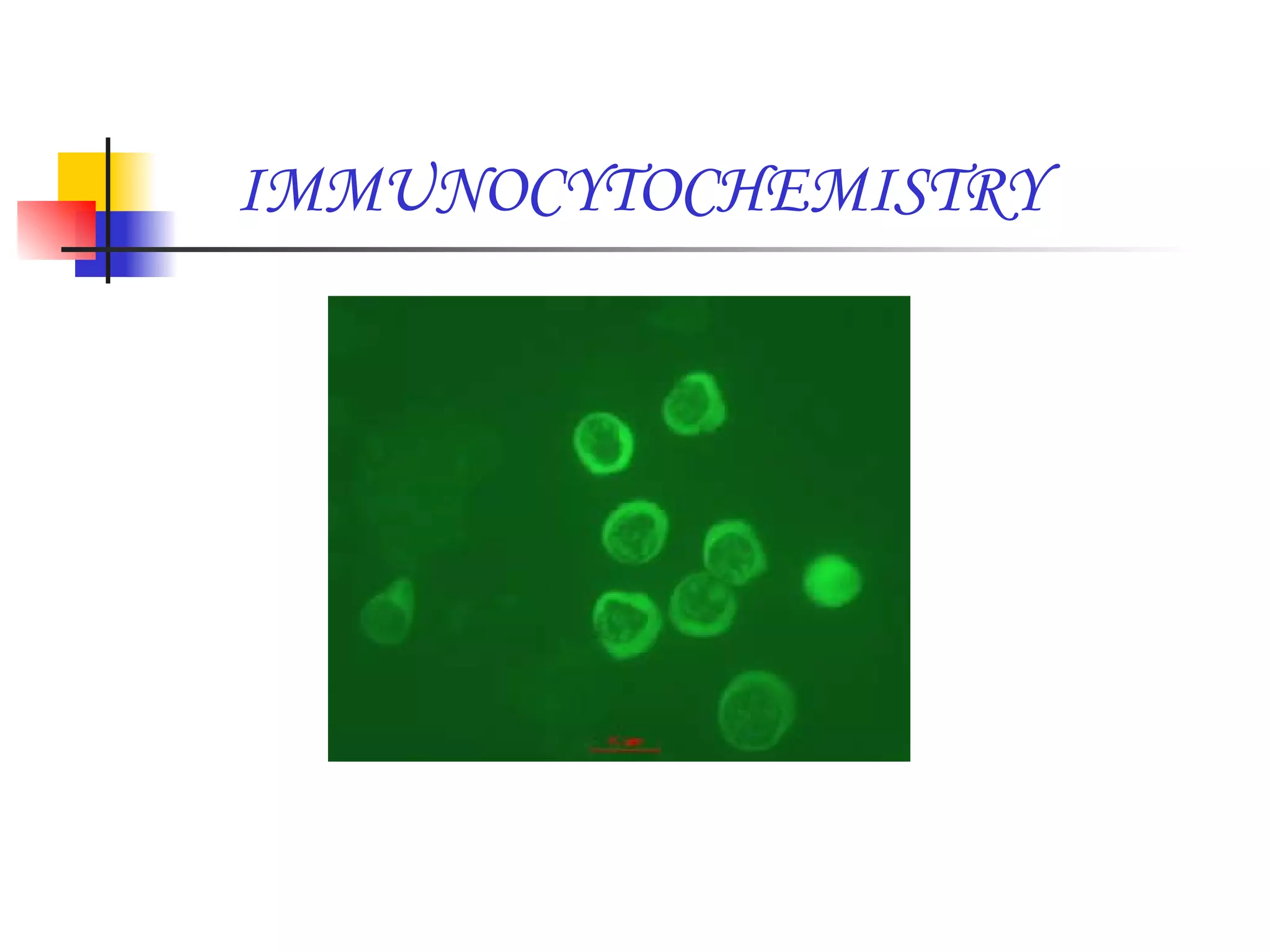
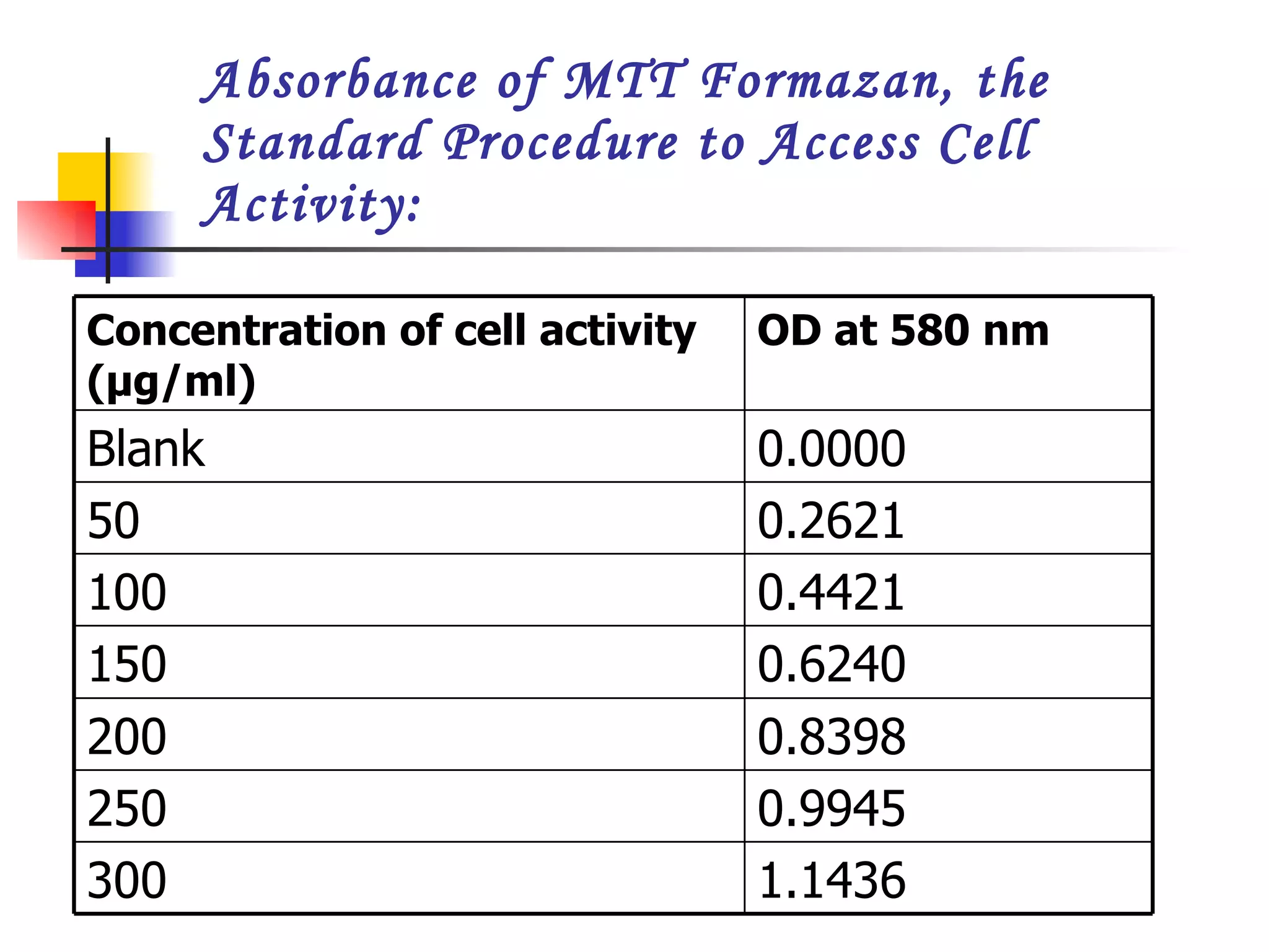
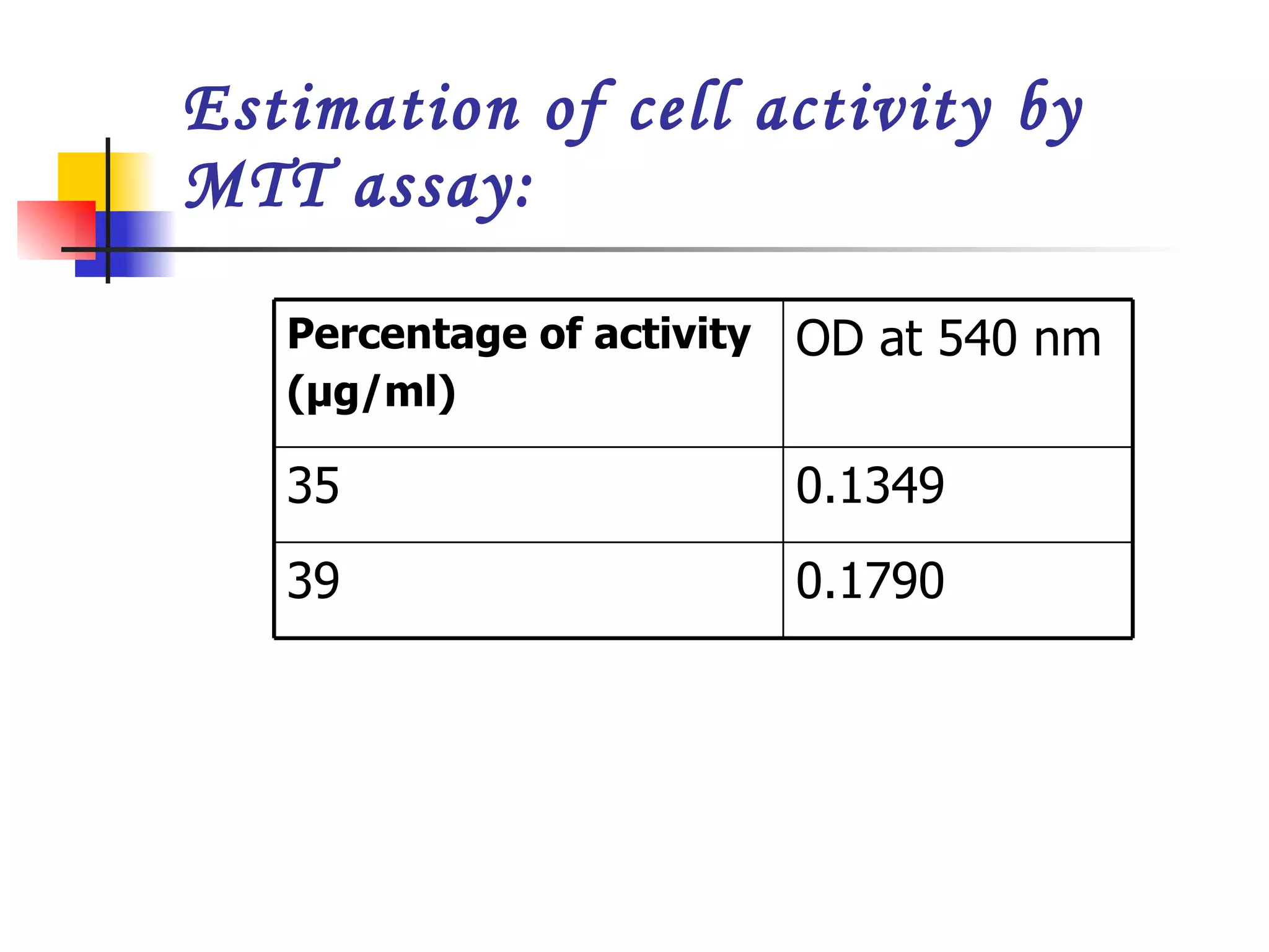
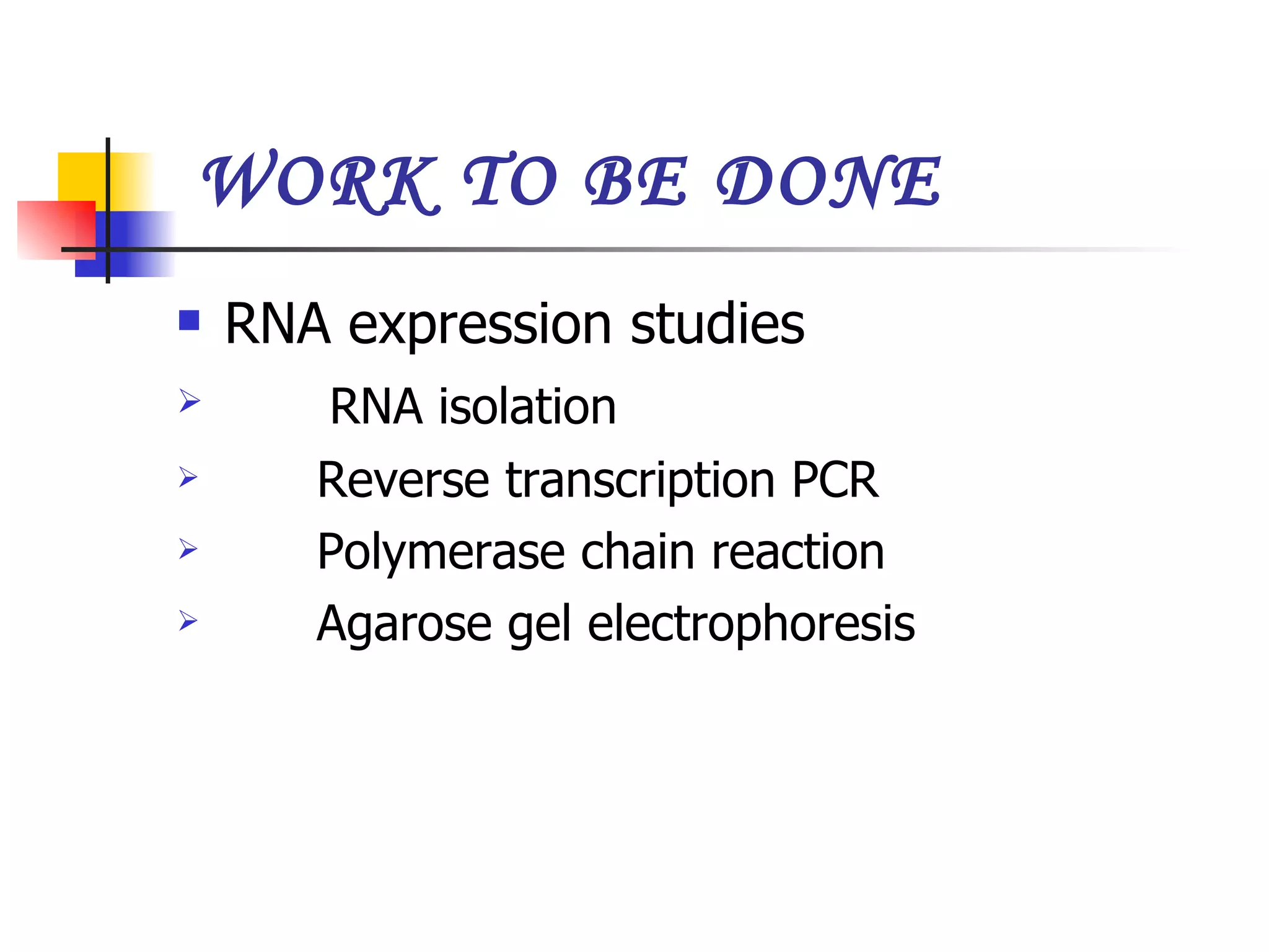
ISOLATION AND CHARACTERISATION OF HUMAN FETAL LIVER STEM CELLS WITH AFP, CK 18 & ALBUMIN    PRESENTED BY   S.EZHIL MANGAI
UNDER THE GUIDANCE OF INTERNAL GUIDE   :  L.G. MOHANA DIVYA   Lecturer   Department Of Biotechnology   KSR College Of Technology  EXTERNAL GUIDE  :  DR. ALEEM KHAN Centre For Liver    Research And Diagnostics    Hyderabad.
INTRODUCTION Stem cells are primal cells common to all multicellular organisms that retain the ability to renew themselves through cell division and can be differentiated into wide range of specialized cell types. There can be two sources of stem cells- Autologous and Allogenic. Autologous embryonic stem cells generated through therapeutic cloning and highly plastic stem cells from the umbilical cord blood or bone marrow from candidates. Allogenic stem cells can be derived from marrow, peripheral blood, cord blood, family donors or HLA typed donors .
PROPERTIES OF STEM CELLS 1.determining precisely how stem cells remain unspecialized and self renewing for many years; and  2.identifying the  signals  that cause stem cells to become specialized cells.
TYPES OF STEM CELLS EMBRYONIC STEM CELL ADULT STEM CELL FETAL LIVER STEM CELL UMBILICAL CORD STEM CELL PANCREATIC STEM CELL
OBJECTIVE To Isolate stem cells from human foetal liver with the help of markers Alfa feto protein, cytokeratin 18 & Albumin. To characterize the isolated stem cells using different methods.
STEPS INVOLVED FOETAL DISSECTION LIVER EXTRACTION DIGESTION OF LIVER STEM CELL ISOLATION USING MACS CELL COUNTING - HAEMOCYTOMETER CELL VIABILITY ASSAY IMMUNOCYTOCHEMISTRY
CONTINUE… MICROTOXICITY ASSAY RNA ISOLATION FROM STEM CELLS QUANTIFICATION OF RNA REVERSE TRANSCRIPTION RT PCR AGAROSE GEL ELECTROPHORESIS
LIVER CELL EXTRACTION Homogenization is done with collagenase 4 for complete extraction of liver cells. Cell washing is done with Hanks buffer 3-4 times.
ISOLATION OF STEM CELLS Isolation of stem cells can be done with MACS and FACS. MACS - Magnetic associated cell sorter and FACS- Fluorescent activated cell sorter MACS is commonly used as fluorescent can cause damage to the cells during isolation.
MAGNETIC ASSOCIATED CELL SORTER 1.  Take the standardized cell sample containing 5 million cells in sterile eppendroff. 2. Add 2 µl of  FCR blocking reagent to the sample and same volume of AFP antibody into the same eppendroff. 3. Incubate for 30-45 mins at temperature of 4 °c.
CONTINUE… 4.  MACS with LS max column is used for separation of stem cells containing AFP+ and AFP- cells . 5. The separation of +ve and –ve cells is due to the magnetic field applied and the binding of specific antibodies with the cells. 6. The cells which do not bind with the specific antibody elute first and are referred as AFP- cells.
CONTINUE… 7 . The remaining cells in the column are extracted by adding Hanks buffer and with the help of pressure syringe. 8. The extracted cells are referred as AFP+ cells. 9. The cells are washed with Hanks Buffer 2-3 times. 10. The cells are preserved in 4 °c with the addition of Hanks buffer .
CELL COUNTING Cell counting is the process of measuring total number of cells per ml. It is done with the help of Haemocytometer The cells are mixed with trypan blue dye in the ratio 1:10 such as dye and cells respectively. The cells are made into exact concentration and placed on the square slide covered with cover slip.
CONTINUE… The slide is placed on the microscope and observed for total number of cells. The calculation is done with the formula number of cells  =  number of  *  dilution    per cubic mm  cells counted  factor
RESULTS CELL COUNTING : Total number of cells  in small square  = 43 Total number of cells  in large square  = 43*16= 688 Total number of cells counted  = 688*4= 2752
APPLICATIONS Stem cells are used in blood transfusions. Used in organ transplantation. Used in cancer studies. Used in diagnostic of diseases such as Parkinson's, diabetes etc.
OBJECTIVE To isolate the fetal liver Cells from the fetus by two step collagenase method. Separation of CD34+ cells  by magnetic activated cell sorting method. Characterization of isolated cells by polymerase chain reaction by using cell markers namely  ALB, AFP, CK18.
VIABILITY ASSAY The cell viability test was performed in total cells and in MACS sorted cells by dye exclusion method.  Trypan blue  is a  vital stain  used to selectively colour dead  tissues  or  cells  blue. It is a  diazo dye   The dead cells intake dye which results in blue colour. Whereas the viable cells remain colorless.
IMMUNOCYTOCHEMISTRY To characterize the isolated stem cells. It visualizes the sub cellular localization of antigen in isolated stem cell populations . Immunocytochemistry refers to a process of localizing proteins in the cells exploiting the binding of antibodies to antigen.  The visual marker may be a fluorophore (like FITC, rhodamine), colloidal metal, hapten, radioactive marker or more commonly for light microscopy an enzyme. Ideally, maximal signal strength along with minimal background or non-specific staining is required to give optimal antigen demonstration.
  PROCEDURE Take 5 million cells and add 10  µl of    diluted primary antibody   ↓       Incubate for 2-3 hrs     ↓  Wash the cells by applying centrifugation at  1000 rpm for 2 mins at 4°c
↓ Remove the supernatant and add 10 µl of    diluted secondary antibody    ↓   Incubate for 1-2 hrs    ↓ prepare cell smear on slide and fix using  formaldehyde ↓    Observe the cells under fluorescent  microscopy
MTT ASSAY MTT assay  is a  laboratory test  and a standard  colorimetric  assay (an assay which measures changes in color) for measuring cellular proliferation ( cell  growth). It can also be used to determine  cytotoxicity  of potential  medicinal  agents and other toxic materials .
Yellow MTT (3-(4,5- Di methyl thiazol -2-yl)-2,5-di phenyl tetrazolium bromide, a  tetrazole ) is reduced to  purple   formazan  in the  mitochondria  of living cells.  A solubilization solution (usually either  dimethyl sulfoxide , an acidified ethanol solution, or a solution of the  detergent   sodium dodecyl sulfate  in dilute  hydrochloric acid ) is added to dissolve the insoluble purple  formazan  product into a colored solution.
The  absorbance  of this colored solution can be quantified by measuring at a certain  wavelength  (usually between 500 and 600 nm) by a  spectrophotometer . The absorption maximum is dependent on the solvent employed.
PROCEDURE   1.5 ml sample is added to 1 ml of MTT      reagent   ↓   Incubation for 2-3 hrs at room temperature   ↓   Add 1 ml of isopropanol   ↓     Vortex the sample
↓ centrifuge at 3000 rpm for 4 mins   ↓ collect the supernatant and calculate OD at  570 nm   ↓     With the readings plot a graph for  absorbance vs. activity of cells.
RESULTS VIABILITY ASSAY Number of cells in small square =30 Number of viable cells  =14 Number of dead cells  =16
Number of cells in  =30*16  large square      =480   Total number of cells  =480*4   =1820
Number of viable cells in  =14*16 large square  =224 Total number of viable cells  =224*4 =896
Percentage of viable cells =  total no. of viable cells   * 100 total no. of cells =  896  * 100 1820 = 49.23%
IMMUNOCYTOCHEMISTRY
MTT ASSAY Absorbance of MTT Formazan, the Standard Procedure to Access Cell Activity: 0.9945 250 1.1436 300 0.8398 200 0.6240 150 0.4421 100 0.2621 50 0.0000 Blank OD at 580 nm   Concentration of cell activity (µg/ml)
  n  Estimation of cell activity by MTT assay:   0.1790 39 0.1349 35 OD at 540 nm Percentage of activity (µg/ml)
 
WORK TO BE DONE RNA expression studies RNA isolation Reverse transcription PCR Polymerase chain reaction Agarose gel electrophoresis
THANK YOU

Ezhil1

  • 1.
    ISOLATION AND CHARACTERISATIONOF HUMAN FETAL LIVER STEM CELLS WITH AFP, CK 18 & ALBUMIN PRESENTED BY S.EZHIL MANGAI
  • 2.
    UNDER THE GUIDANCEOF INTERNAL GUIDE : L.G. MOHANA DIVYA Lecturer Department Of Biotechnology KSR College Of Technology EXTERNAL GUIDE : DR. ALEEM KHAN Centre For Liver Research And Diagnostics Hyderabad.
  • 3.
    INTRODUCTION Stem cellsare primal cells common to all multicellular organisms that retain the ability to renew themselves through cell division and can be differentiated into wide range of specialized cell types. There can be two sources of stem cells- Autologous and Allogenic. Autologous embryonic stem cells generated through therapeutic cloning and highly plastic stem cells from the umbilical cord blood or bone marrow from candidates. Allogenic stem cells can be derived from marrow, peripheral blood, cord blood, family donors or HLA typed donors .
  • 4.
    PROPERTIES OF STEMCELLS 1.determining precisely how stem cells remain unspecialized and self renewing for many years; and 2.identifying the signals that cause stem cells to become specialized cells.
  • 5.
    TYPES OF STEMCELLS EMBRYONIC STEM CELL ADULT STEM CELL FETAL LIVER STEM CELL UMBILICAL CORD STEM CELL PANCREATIC STEM CELL
  • 6.
    OBJECTIVE To Isolatestem cells from human foetal liver with the help of markers Alfa feto protein, cytokeratin 18 & Albumin. To characterize the isolated stem cells using different methods.
  • 7.
    STEPS INVOLVED FOETALDISSECTION LIVER EXTRACTION DIGESTION OF LIVER STEM CELL ISOLATION USING MACS CELL COUNTING - HAEMOCYTOMETER CELL VIABILITY ASSAY IMMUNOCYTOCHEMISTRY
  • 8.
    CONTINUE… MICROTOXICITY ASSAYRNA ISOLATION FROM STEM CELLS QUANTIFICATION OF RNA REVERSE TRANSCRIPTION RT PCR AGAROSE GEL ELECTROPHORESIS
  • 9.
    LIVER CELL EXTRACTIONHomogenization is done with collagenase 4 for complete extraction of liver cells. Cell washing is done with Hanks buffer 3-4 times.
  • 10.
    ISOLATION OF STEMCELLS Isolation of stem cells can be done with MACS and FACS. MACS - Magnetic associated cell sorter and FACS- Fluorescent activated cell sorter MACS is commonly used as fluorescent can cause damage to the cells during isolation.
  • 11.
    MAGNETIC ASSOCIATED CELLSORTER 1. Take the standardized cell sample containing 5 million cells in sterile eppendroff. 2. Add 2 µl of FCR blocking reagent to the sample and same volume of AFP antibody into the same eppendroff. 3. Incubate for 30-45 mins at temperature of 4 °c.
  • 12.
    CONTINUE… 4. MACS with LS max column is used for separation of stem cells containing AFP+ and AFP- cells . 5. The separation of +ve and –ve cells is due to the magnetic field applied and the binding of specific antibodies with the cells. 6. The cells which do not bind with the specific antibody elute first and are referred as AFP- cells.
  • 13.
    CONTINUE… 7 .The remaining cells in the column are extracted by adding Hanks buffer and with the help of pressure syringe. 8. The extracted cells are referred as AFP+ cells. 9. The cells are washed with Hanks Buffer 2-3 times. 10. The cells are preserved in 4 °c with the addition of Hanks buffer .
  • 14.
    CELL COUNTING Cellcounting is the process of measuring total number of cells per ml. It is done with the help of Haemocytometer The cells are mixed with trypan blue dye in the ratio 1:10 such as dye and cells respectively. The cells are made into exact concentration and placed on the square slide covered with cover slip.
  • 15.
    CONTINUE… The slideis placed on the microscope and observed for total number of cells. The calculation is done with the formula number of cells = number of * dilution per cubic mm cells counted factor
  • 16.
    RESULTS CELL COUNTING: Total number of cells in small square = 43 Total number of cells in large square = 43*16= 688 Total number of cells counted = 688*4= 2752
  • 17.
    APPLICATIONS Stem cellsare used in blood transfusions. Used in organ transplantation. Used in cancer studies. Used in diagnostic of diseases such as Parkinson's, diabetes etc.
  • 18.
    OBJECTIVE To isolatethe fetal liver Cells from the fetus by two step collagenase method. Separation of CD34+ cells by magnetic activated cell sorting method. Characterization of isolated cells by polymerase chain reaction by using cell markers namely ALB, AFP, CK18.
  • 19.
    VIABILITY ASSAY Thecell viability test was performed in total cells and in MACS sorted cells by dye exclusion method. Trypan blue is a vital stain used to selectively colour dead tissues or cells blue. It is a diazo dye The dead cells intake dye which results in blue colour. Whereas the viable cells remain colorless.
  • 20.
    IMMUNOCYTOCHEMISTRY To characterizethe isolated stem cells. It visualizes the sub cellular localization of antigen in isolated stem cell populations . Immunocytochemistry refers to a process of localizing proteins in the cells exploiting the binding of antibodies to antigen. The visual marker may be a fluorophore (like FITC, rhodamine), colloidal metal, hapten, radioactive marker or more commonly for light microscopy an enzyme. Ideally, maximal signal strength along with minimal background or non-specific staining is required to give optimal antigen demonstration.
  • 21.
    PROCEDURETake 5 million cells and add 10 µl of diluted primary antibody ↓ Incubate for 2-3 hrs ↓ Wash the cells by applying centrifugation at 1000 rpm for 2 mins at 4°c
  • 22.
    ↓ Remove thesupernatant and add 10 µl of diluted secondary antibody ↓ Incubate for 1-2 hrs ↓ prepare cell smear on slide and fix using formaldehyde ↓ Observe the cells under fluorescent microscopy
  • 23.
    MTT ASSAY MTTassay is a laboratory test and a standard colorimetric assay (an assay which measures changes in color) for measuring cellular proliferation ( cell growth). It can also be used to determine cytotoxicity of potential medicinal agents and other toxic materials .
  • 24.
    Yellow MTT (3-(4,5-Di methyl thiazol -2-yl)-2,5-di phenyl tetrazolium bromide, a tetrazole ) is reduced to purple formazan in the mitochondria of living cells. A solubilization solution (usually either dimethyl sulfoxide , an acidified ethanol solution, or a solution of the detergent sodium dodecyl sulfate in dilute hydrochloric acid ) is added to dissolve the insoluble purple formazan product into a colored solution.
  • 25.
    The absorbance of this colored solution can be quantified by measuring at a certain wavelength (usually between 500 and 600 nm) by a spectrophotometer . The absorption maximum is dependent on the solvent employed.
  • 26.
    PROCEDURE 1.5 ml sample is added to 1 ml of MTT reagent ↓ Incubation for 2-3 hrs at room temperature ↓ Add 1 ml of isopropanol ↓ Vortex the sample
  • 27.
    ↓ centrifuge at3000 rpm for 4 mins ↓ collect the supernatant and calculate OD at 570 nm ↓ With the readings plot a graph for absorbance vs. activity of cells.
  • 28.
    RESULTS VIABILITY ASSAYNumber of cells in small square =30 Number of viable cells =14 Number of dead cells =16
  • 29.
    Number of cellsin =30*16 large square =480 Total number of cells =480*4 =1820
  • 30.
    Number of viablecells in =14*16 large square =224 Total number of viable cells =224*4 =896
  • 31.
    Percentage of viablecells = total no. of viable cells * 100 total no. of cells = 896 * 100 1820 = 49.23%
  • 32.
  • 33.
    MTT ASSAY Absorbanceof MTT Formazan, the Standard Procedure to Access Cell Activity: 0.9945 250 1.1436 300 0.8398 200 0.6240 150 0.4421 100 0.2621 50 0.0000 Blank OD at 580 nm Concentration of cell activity (µg/ml)
  • 34.
    n Estimation of cell activity by MTT assay:   0.1790 39 0.1349 35 OD at 540 nm Percentage of activity (µg/ml)
  • 35.
  • 36.
    WORK TO BEDONE RNA expression studies RNA isolation Reverse transcription PCR Polymerase chain reaction Agarose gel electrophoresis
  • 37.