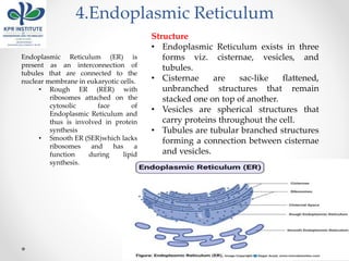
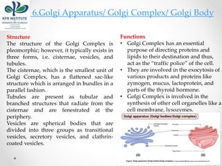
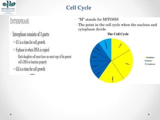
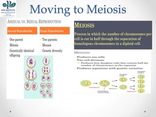
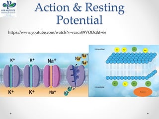
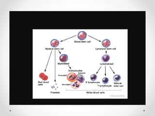
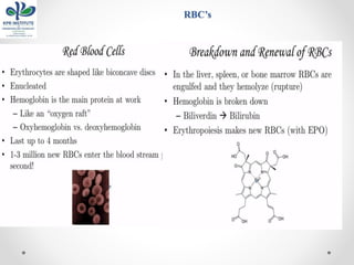
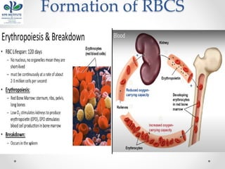
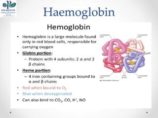
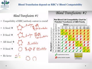
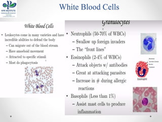
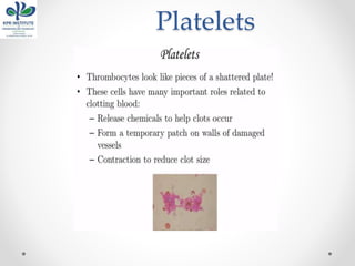
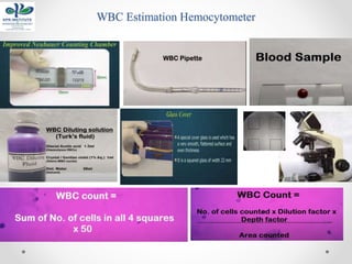
DEPARTMENT OF BIOMEDICAL ENGINEERING
U19BM302 HUMAN ANATOMY AND
PHYSIOLOGY
L T P C
3 0 0 3
Category: PC
Course Content Prepared by: Rajeshwari R AP/BME
Syllabus
Reference & Text Books
Course Outcomes & Mapping
UNIT I
THE CELL & GENERAL PHYSIOLOGY
Introduction to Human Cell
• The cell (from latin cella, meaning "small room") is the basic structural,
functional, and biological unit of all known organisms.
• A cell is the smallest unit of life.
• Cells are often called the "building blocks of life".
• The study of cells is called cell biology, cellular biology, or cytology.
• Two Types of cell: Prokaryotic cell & Eukaryotic Cell
It has been estimated that humans contain
somewhere around 40 trillion (4×10¹³) cells. The
human brain accounts for around 80 billion of
these cells.
Robert Hooke in 1665
Introduction to Human Cell
Structure of Cell
Component & Its Function
1.Plasma Membrane /Cell Membrane
Functions
• The cell membrane provides
mechanical support that facilities the
shape of the cell while enclosing the
cell and its components from the
external environment.
• It regulates what can be allowed to
enter and exit the cell through
channels, acting as a semi-permeable
membrane, which facilities the
exchange of essential compounds
required for the survival of the cell.
• It generates and distributes signals in
and outside of the cell for the proper
functioning of the cell and all the
organelles.
• It allows the interaction between cells
required during tissue formation and
cell fusion.
Structure
• It consists of a phospholipid bilayer
along with two types of proteins viz.
embedded proteins and peripheral
proteins that function in providing shape
and allowing the movement of particles
in and out of the cell.
• The most abundant lipid which is
present in the cell membrane is a
phospholipid which contains a polar
head group attached to two hydrophobic
fatty acid tails.
• The embedded proteins act as channels
for the transfer of particles across the cell
with some proteins acting as receptors
for the binding of various components.
• The peripheral proteins function as to
provide fluidity as well as mechanical
support to the structure of the cell.
A few molecules move freely
Water, Carbon dioxide,
Ammonia, Oxygen
Carrier proteins transport
some molecules
Proteins embedded in
lipid bilayer
Fluid mosaic model –
describes fluid nature of
a lipid bilayer with
proteins
3.Cytoplasm
Cytoplasm refers to everything present inside the cell except the
nucleus.
Structure
• The cytoplasm consists of a cytosol; a
gel-like substance that contains other
matter; cell organelles; smaller cell-like
bodies bound by separate membranes;
and cytoplasmic inclusions; insoluble
molecules that store energy and are not
surrounded by any layer.
• The cytoplasm is colorless and has
about 80% water along with various
nutrients required for the cell.
“It is known to have the properties of both
viscous matters as well as elastic matter.
Under its elasticity, cytoplasm helps in the
movement of materials inside the cell by a
process termed cytoplasmic streaming.”
Functions
• Vital cellular and enzymatic
reactions like cellular respiration and
translation of mRNA into proteins
occur in the cytoplasm.
• It acts as a buffer and protects
genetic materials as well as other
organelles from damage due to
collision or change in the pH of the
cytosol.
• The process called cytoplasmic
streaming helps in the distribution of
various nutrients and facilitates the
movement of cell organelles within
the cell.
3.Cytoskeleton
• Filaments & fibers
• Made of 3 fiber types
o Microfilaments
o Microtubules
o Intermediate filaments
• 3 functions:
o mechanical support
o anchor organelles
o help move substances
4.Centrioles
Structure
• A centriole consists of a cylindrical
structure made with nine triplets
microtubules that surround the
periphery of the centriole while the
center has a Y-shaped linker and a
barrel-like structure that stabilizes the
centriole.
• Another structure called cartwheel is
present in a centriole which is made up
of a central hub with nine
spokes/filaments radiating from it. Each
of these filaments/spokes is connected
to the microtubules through a pinhead.
Functions
• During cell division, centrioles have a
crucial role in forming spindle fibers
which assist the movement of
chromatids towards their respective
sides.
4.Endoplasmic Reticulum
Endoplasmic Reticulum (ER) is
present as an interconnection of
tubules that are connected to the
nuclear membrane in eukaryotic cells.
• Rough ER (RER) with
ribosomes attached on the
cytosolic face of
Endoplasmic Reticulum and
thus is involved in protein
synthesis
• Smooth ER (SER)which lacks
ribosomes and has a
function during lipid
synthesis.
Structure
• Endoplasmic Reticulum exists in three
forms viz. cisternae, vesicles, and
tubules.
• Cisternae are sac-like flattened,
unbranched structures that remain
stacked one on top of another.
• Vesicles are spherical structures that
carry proteins throughout the cell.
• Tubules are tubular branched structures
forming a connection between cisternae
and vesicles.
Functions
• ER contains many of the enzymes required for several metabolic
processes, and the surface of the ER is essential for other operations
like diffusion, osmosis, and active transport.
• One of the crucial functions of ER is the synthesis of lipids like
cholesterol and steroids.
• Rough ER allows for the modification of polypeptides emerging out
of the ribosomes to prepare secondary and tertiary structures of the
protein.
• ER also synthesizes various membrane proteins and has a crucial
role in preparing the nuclear envelope after cell division
5.Lysosomes
• Contain digestive enzymes
• Functions
o Aid in cell renewal
o Break down old cell parts
o Digests invaders
Structure
• The shape of lysozymes is irregular or
pleomorphic; however, mostly, they are
found in the spherical or granular
structure.
• Lysozymes are surrounded by a
lysosomal membrane that contains the
enzymes within the lysosome and
protects the cytosol with the rest of the
cell from the harmful action of the
enzymes.
6.Golgi Apparatus/ Golgi Complex/ Golgi Body
Structure
The structure of the Golgi Complex is
pleomorphic; however, it typically exists in
three forms, i.e. cisternae, vesicles, and
tubules.
The cisternae, which is the smallest unit of
Golgi Complex, has a flattened sac-like
structure which is arranged in bundles in a
parallel fashion.
Tubules are present as tubular and
branched structures that radiate from the
cisternae and are fenestrated at the
periphery.
Vesicles are spherical bodies that are
divided into three groups as transitional
vesicles, secretory vesicles, and clathrin-
coated vesicles.
Functions
• Golgi Complex has an essential
purpose of directing proteins and
lipids to their destination and thus,
act as the “traffic police” of the cell.
• They are involved in the exocytosis of
various products and proteins like
zymogen, mucus, lactoprotein, and
parts of the thyroid hormone.
• Golgi Complex is involved in the
synthesis of other cell organelles like a
cell membrane, lysozymes.
7.Mitochondria
Functions
• The primary function of mitochondria is
the synthesis of energy in the form of
ATP required for the proper functioning
of all the cell organelles.
• Mitochondria also help in balancing the
amount of Ca+ ions within the cell and
assists the process of apoptosis.
• Different segments of hormones and
components of blood are built within
mitochondria.
• Mitochondria in the liver have the
ability to detoxify ammonia.
Structure
• A mitochondrion contains two
membranes with the outer layer being
smooth while the inner layer is marked
with folding and finger-like structures
called cristae.
• The inner mitochondrial membrane
contains various enzymes, coenzymes,
and components of multiple cycles
along with pores for the transport of
substrates, ATP, and phosphate
molecules.
• In addition to these enzymes,
mitochondria are also home to single or
double-stranded DNA called mtDNA
that is capable of producing 10% of the
proteins present in the mitochondria
8.Ribosomes
Structure
• The ribonucleoprotein
consists of two subunits.
• Eukaryotic cells have 80S
ribosomes with 60S larger
subunit and 40S smaller
subunit.
Ribosomes are short-lived as
after the protein synthesis, the
subunits split up and can be
either reused or remain broken
up.
Functions
• Ribosomes are the site of biological
protein synthesis in all living
organisms.
9.Vesicles
There are different types of vesicles like vacuoles, secretory and transport vesicles
based on their function.
Structure
• A vesicle is a structure containing liquid
or cytosol which is enclosed by a lipid
bilayer.
• The outer layer enclosing the liquid is
called a lamellar phase which is similar
to the plasma membrane. One end of
the lipid bilayer it hydrophobic whereas
the other end is hydrophilic
Functions
• Vesicles facilitate the storage and
transport of materials in and outside the
cell. It even allows the exchange of
molecules between two cells.
• Because vesicles are enclosed inside a
lipid bilayer, vesicles also function in
metabolism and enzyme storage.
• They allow temporary storage of food
and also control the buoyancy of the cell
10.Vacoule
Structure
• The vacuole is surrounded by a
membrane called tonoplast, which
encloses fluid containing inorganic
materials like water and organic
materials like nutrients and even
enzymes.
• These are formed by the fusion of
various vesicles, so vacuoles are very
similar to vesicles in structure.
Functions
• Vacuoles act as a storage for nutrients as
well as waste materials to protect the
cell for toxicity.
• They have an essential function of
homeostasis as it allows the balance of
pH of the cell by influx and outflow of
H+ ions to the cytoplasm.
• Vacuoles contain enzymes that play an
important role in different metabolic
processes.
11.Nucleus
Structure
• Structurally, the nucleus consists of a nuclear envelope,
chromatin, and nucleolus.
• The nuclear envelope is similar to the cell membrane in
structure and composition. It has pores that allow the
movement of proteins and RNA in and outside the
nucleus. It enables the interaction with other cell organelles
while keeping nucleoplasm and chromatin within the
envelope.
• The chromatin in the nucleus contains RNA or DNA along
with nuclear proteins, as genetic material that is
responsible for carrying the genetic information from one
generation to another. It is present in a sense and compact
structure which might be visible as chromosome under
powerful magnification.
• The nucleolus is like a nucleus within the nucleus. It is a
membrane-less organelle that is responsible for the
synthesis of rRNA and assembly of ribosomes required for
protein synthesis
Functions
• The nucleus is responsible for storage as well as the transfer of genetic
materials in the form of DNA or RNA.
• It aids in the process of transcription by the synthesis of mRNA molecules.
• The nucleus controls the activity of all other organelles while facilitating
processes like cell growth, cell division and synthesis of proteins.
12.Peroxisomes
Structure
• Peroxisome consists of a single
membrane and granular matrix
scattered in the cytoplasm.
• They exist either in the form of
interconnected tubules or as individual
peroxisomes.
• The compartments within every
peroxisome allow the creation of
optimized conditions for different
metabolic activities.
• They consist of several types of enzymes
with major groups being urate oxidase,
D-amino acid oxidase, and catalase
Functions
• Peroxisomes are involved in the
production and elimination of hydrogen
peroxide during biochemical processes.
• Oxidation of fatty acids takes place
within peroxisomes.
• Additionally, peroxisomes are also
involved in the synthesis of lipid-like
cholesterol and plasmalogens
Protein Synthesis
• https://www.youtube.com/watch?v=gG7uCskUOrA
• Cells has Nucleus
• Nucleus has genome formed by 23 pairs of chromosome
• These 23 pairs of chromosomes have long strands of
DNA .These DNA are tightly packed to protein called
Histine
• DNA contains genes. Genes contains instruction to
produce proteins.
• When particular gene is activated ,RNA polymerase
attach themselves to start of the gene.
Protein Synthesis
• RNA polymerase move along the gene sequence
producing Messenger RNA by adding particular bases
floating in the nucleolus.
• The base sequence to be added is determined by DNA
molecule.
• Before moving out of nucleus ,messenger RNA is
processed.(addition and deletion of
sequence).(transcription)
• On moving out the ribosome's bound them to mRNA.
• Ribosome's reads the code and produces AA.
• Transfer RNA of AA attaches to m-RNA releasing end
protein.
Cellular Reproduction
• The process by which cell divides into 2 new daughter
cells
Cell Cycle
Mitosis
Cytokinesis
Moving to Meiosis
Chromosomes
Mitosis & Meiosis
Meiosis Stages
Action & Resting
Potential
https://www.youtube.com/watch?v=rcacx09VODc&t=6s
Action & Resting Potential
Blood cells
https://www.youtube.com/watch?v=9PFO3ZiPi5A
RBC’s
Formation of RBCS
Haemoglobin
Blood Transfusion depend on RBC’s/ Blood Compatability
White Blood Cells
Types WBC’s
Platelets
Blood Plasma
ESTIMATION OF RBC’S
• The purpose of performing Total Red Blood cell count is to know whether or not you
are suffering from Erythrocytosis or Polycythemia (i.e. the increase in the no. of Red
Blood Cells to more than 6.5 million/mm3) or
• Erythrocytopenia or Erythropenia (i.e. the Decrease in the no. of Red Blood Cells to
less than 3.5 million/mm3).
Normal RBCs have a diameter of 6 - 8 μm.
In Males – 4.8-5.5 million/mm3
In Females – 4.5-5 million/mm3
MICRODILUTION METHOD FOR THE ESTIMATION OF TOTAL
RBCs USING HEMOCYTOMETER
• Blood sample (Capillary blood or EDTA anticoagulated specimen)
• RBC diluting fluid (preferably Hayem’s fluid)
• Gauze piece or Cotton
• RBC pipette
• Hemocytometer Neubauer’s Chamber
• Coverslip
• Microscope
COMPONENTS QUANTITY
Mercuric Chloride 0.25 grams
Sodium sulfate 2.5 grams
Sodium chloride 0.5 grams
Distilled water 100 ml
RBC Estimation Hemocytometer
• The purpose of performing Total Red Blood cell count is to know
whether or not you are suffering from Erythrocytosis or Polycythemia
(i.e. the increase in the no. of Red Blood Cells to more than 6.5
million/mm3) or
• Erythrocytopenia or Erythropenia (i.e. the Decrease in the no. of Red
Blood Cells to less than 3.5 million/mm3).
WBC Estimation Hemocytometer
Blood Platelet Estimation
Immunity
Immunity is the capability of multicellular organisms to resist harmful agents.
•Defense against microbes
•Defense against the growth of tumor cells
•kills the growth of tumor cells
•Homeostasis
•destruction of abnormal or dead cells
(e.g. dead red or white blood cells, antigen-antibody complex)
•Immune system = cells, tissues, and molecules that mediate resistance to
infections
•Immunology = study of structure and function of the immune system
•Immunity = resistance of a host to pathogens and their toxic effects
•Immune response = collective and coordinated response to the introduction
of foreign substances in an individual mediated by the cells and molecules of
the immune system
Organs/Cells/Molecules involved in Immunity
Tonsils and adenoids
Thymus
Lymph nodes
Spleen
Payer’s patches
Appendix
Lymphatic vessels
Bone marrow
Lymphocytes
T-lymphocytes
B-Lymphocytes, plasma cells
natural killer lymphocytes
Monocytes, Macrophage
Granulocytes
neutrophils
eosinophils
basophils
Antibodies
Complement
Cytokines
Interleukines
Interferons
Types of Immunity
Types of Immunity
1. Innate (non-adaptive)
• first line of immune response
• relies on mechanisms that exist before infection
• Based on genetic make-up
• Relies on already formed components
• Rapid response: within minutes of infection
• Not specific
same molecules / cells respond to a range of pathogens
• Has no memory
same response after repeated exposure
• Does not lead to clonal expansion
2.Adaptive Immunity
Second line of response
• Based upon resistance acquired during life
• Relies on genetic events and cellular growth
• Responds more slowly, over few days
• Is specific,
each cell responds to a single epitope on an antigen
• Has past memory
repeated exposure leads to faster, stronger response
• Leads to clonal expansion
• Comes in to effect when natural immunity fails
Mechanism of Adaptive Immunity
•Cell-mediated immune response (CMIR)
•T-lymphocytes
•eliminate intracellular microbes that survive within phagocytes or other
infected cells
•Humoral immune response (HIR)
•B-lymphocytes mediated
•eliminate extra-cellular microbes and their toxins
Cell mediated immune response
Primary response
o production of specific clones of effector T cells and memory clones
o develops in several days
o does not limit the infection
Secondary response
o more pronounced, faster
o more effective at limiting the infection
Example - cytotoxic reactions against intracellular parasites, delayed
hypersensitivity (e.g., Tuberculin test) and allograft rejection
2 types T- lymphocytes
• helper T- lymphocytes (CD4+)
o CD4+ T cells activate phagocytes to kill microbes
• cytolytic T-lymphocyte (CD8+)
o CD8+ T cells destroy infected cells containing microbes or microbial proteins
Antibody Mediated Immune Response
•Humoral immunity is also called antibody-
mediated immunity.
•With assistance from helper T cells, B cells will
differentiate into plasma B cells that can
produce antibodies against a specific antigen.
•The humoral immune system deals with
antigens from pathogens that are freely
circulating, or outside the infected cells.
• Antibodies produced by the B cells will bind to
antigens, neutralizing them, or causing lysis
(dissolution or destruction of cells by a lysin) or
phagocytosis.
Process of Cell & Antibody Mediated Immunity
Summary of Cell & Antibody Mediated Immunity

Human Anatomy & Physiology _ Lecture Notes _1

  • 1.
    DEPARTMENT OF BIOMEDICALENGINEERING U19BM302 HUMAN ANATOMY AND PHYSIOLOGY L T P C 3 0 0 3 Category: PC Course Content Prepared by: Rajeshwari R AP/BME
  • 2.
  • 3.
  • 4.
  • 5.
    UNIT I THE CELL& GENERAL PHYSIOLOGY
  • 6.
    Introduction to HumanCell • The cell (from latin cella, meaning "small room") is the basic structural, functional, and biological unit of all known organisms. • A cell is the smallest unit of life. • Cells are often called the "building blocks of life". • The study of cells is called cell biology, cellular biology, or cytology. • Two Types of cell: Prokaryotic cell & Eukaryotic Cell It has been estimated that humans contain somewhere around 40 trillion (4×10¹³) cells. The human brain accounts for around 80 billion of these cells. Robert Hooke in 1665
  • 9.
  • 10.
  • 11.
    Component & ItsFunction 1.Plasma Membrane /Cell Membrane Functions • The cell membrane provides mechanical support that facilities the shape of the cell while enclosing the cell and its components from the external environment. • It regulates what can be allowed to enter and exit the cell through channels, acting as a semi-permeable membrane, which facilities the exchange of essential compounds required for the survival of the cell. • It generates and distributes signals in and outside of the cell for the proper functioning of the cell and all the organelles. • It allows the interaction between cells required during tissue formation and cell fusion. Structure • It consists of a phospholipid bilayer along with two types of proteins viz. embedded proteins and peripheral proteins that function in providing shape and allowing the movement of particles in and out of the cell. • The most abundant lipid which is present in the cell membrane is a phospholipid which contains a polar head group attached to two hydrophobic fatty acid tails. • The embedded proteins act as channels for the transfer of particles across the cell with some proteins acting as receptors for the binding of various components. • The peripheral proteins function as to provide fluidity as well as mechanical support to the structure of the cell.
  • 12.
    A few moleculesmove freely Water, Carbon dioxide, Ammonia, Oxygen Carrier proteins transport some molecules Proteins embedded in lipid bilayer Fluid mosaic model – describes fluid nature of a lipid bilayer with proteins
  • 13.
    3.Cytoplasm Cytoplasm refers toeverything present inside the cell except the nucleus. Structure • The cytoplasm consists of a cytosol; a gel-like substance that contains other matter; cell organelles; smaller cell-like bodies bound by separate membranes; and cytoplasmic inclusions; insoluble molecules that store energy and are not surrounded by any layer. • The cytoplasm is colorless and has about 80% water along with various nutrients required for the cell. “It is known to have the properties of both viscous matters as well as elastic matter. Under its elasticity, cytoplasm helps in the movement of materials inside the cell by a process termed cytoplasmic streaming.” Functions • Vital cellular and enzymatic reactions like cellular respiration and translation of mRNA into proteins occur in the cytoplasm. • It acts as a buffer and protects genetic materials as well as other organelles from damage due to collision or change in the pH of the cytosol. • The process called cytoplasmic streaming helps in the distribution of various nutrients and facilitates the movement of cell organelles within the cell.
  • 14.
    3.Cytoskeleton • Filaments &fibers • Made of 3 fiber types o Microfilaments o Microtubules o Intermediate filaments • 3 functions: o mechanical support o anchor organelles o help move substances
  • 15.
    4.Centrioles Structure • A centrioleconsists of a cylindrical structure made with nine triplets microtubules that surround the periphery of the centriole while the center has a Y-shaped linker and a barrel-like structure that stabilizes the centriole. • Another structure called cartwheel is present in a centriole which is made up of a central hub with nine spokes/filaments radiating from it. Each of these filaments/spokes is connected to the microtubules through a pinhead. Functions • During cell division, centrioles have a crucial role in forming spindle fibers which assist the movement of chromatids towards their respective sides.
  • 16.
    4.Endoplasmic Reticulum Endoplasmic Reticulum(ER) is present as an interconnection of tubules that are connected to the nuclear membrane in eukaryotic cells. • Rough ER (RER) with ribosomes attached on the cytosolic face of Endoplasmic Reticulum and thus is involved in protein synthesis • Smooth ER (SER)which lacks ribosomes and has a function during lipid synthesis. Structure • Endoplasmic Reticulum exists in three forms viz. cisternae, vesicles, and tubules. • Cisternae are sac-like flattened, unbranched structures that remain stacked one on top of another. • Vesicles are spherical structures that carry proteins throughout the cell. • Tubules are tubular branched structures forming a connection between cisternae and vesicles.
  • 17.
    Functions • ER containsmany of the enzymes required for several metabolic processes, and the surface of the ER is essential for other operations like diffusion, osmosis, and active transport. • One of the crucial functions of ER is the synthesis of lipids like cholesterol and steroids. • Rough ER allows for the modification of polypeptides emerging out of the ribosomes to prepare secondary and tertiary structures of the protein. • ER also synthesizes various membrane proteins and has a crucial role in preparing the nuclear envelope after cell division
  • 18.
    5.Lysosomes • Contain digestiveenzymes • Functions o Aid in cell renewal o Break down old cell parts o Digests invaders Structure • The shape of lysozymes is irregular or pleomorphic; however, mostly, they are found in the spherical or granular structure. • Lysozymes are surrounded by a lysosomal membrane that contains the enzymes within the lysosome and protects the cytosol with the rest of the cell from the harmful action of the enzymes.
  • 19.
    6.Golgi Apparatus/ GolgiComplex/ Golgi Body Structure The structure of the Golgi Complex is pleomorphic; however, it typically exists in three forms, i.e. cisternae, vesicles, and tubules. The cisternae, which is the smallest unit of Golgi Complex, has a flattened sac-like structure which is arranged in bundles in a parallel fashion. Tubules are present as tubular and branched structures that radiate from the cisternae and are fenestrated at the periphery. Vesicles are spherical bodies that are divided into three groups as transitional vesicles, secretory vesicles, and clathrin- coated vesicles. Functions • Golgi Complex has an essential purpose of directing proteins and lipids to their destination and thus, act as the “traffic police” of the cell. • They are involved in the exocytosis of various products and proteins like zymogen, mucus, lactoprotein, and parts of the thyroid hormone. • Golgi Complex is involved in the synthesis of other cell organelles like a cell membrane, lysozymes.
  • 20.
    7.Mitochondria Functions • The primaryfunction of mitochondria is the synthesis of energy in the form of ATP required for the proper functioning of all the cell organelles. • Mitochondria also help in balancing the amount of Ca+ ions within the cell and assists the process of apoptosis. • Different segments of hormones and components of blood are built within mitochondria. • Mitochondria in the liver have the ability to detoxify ammonia. Structure • A mitochondrion contains two membranes with the outer layer being smooth while the inner layer is marked with folding and finger-like structures called cristae. • The inner mitochondrial membrane contains various enzymes, coenzymes, and components of multiple cycles along with pores for the transport of substrates, ATP, and phosphate molecules. • In addition to these enzymes, mitochondria are also home to single or double-stranded DNA called mtDNA that is capable of producing 10% of the proteins present in the mitochondria
  • 21.
    8.Ribosomes Structure • The ribonucleoprotein consistsof two subunits. • Eukaryotic cells have 80S ribosomes with 60S larger subunit and 40S smaller subunit. Ribosomes are short-lived as after the protein synthesis, the subunits split up and can be either reused or remain broken up. Functions • Ribosomes are the site of biological protein synthesis in all living organisms.
  • 22.
    9.Vesicles There are differenttypes of vesicles like vacuoles, secretory and transport vesicles based on their function. Structure • A vesicle is a structure containing liquid or cytosol which is enclosed by a lipid bilayer. • The outer layer enclosing the liquid is called a lamellar phase which is similar to the plasma membrane. One end of the lipid bilayer it hydrophobic whereas the other end is hydrophilic Functions • Vesicles facilitate the storage and transport of materials in and outside the cell. It even allows the exchange of molecules between two cells. • Because vesicles are enclosed inside a lipid bilayer, vesicles also function in metabolism and enzyme storage. • They allow temporary storage of food and also control the buoyancy of the cell
  • 23.
    10.Vacoule Structure • The vacuoleis surrounded by a membrane called tonoplast, which encloses fluid containing inorganic materials like water and organic materials like nutrients and even enzymes. • These are formed by the fusion of various vesicles, so vacuoles are very similar to vesicles in structure. Functions • Vacuoles act as a storage for nutrients as well as waste materials to protect the cell for toxicity. • They have an essential function of homeostasis as it allows the balance of pH of the cell by influx and outflow of H+ ions to the cytoplasm. • Vacuoles contain enzymes that play an important role in different metabolic processes.
  • 24.
    11.Nucleus Structure • Structurally, thenucleus consists of a nuclear envelope, chromatin, and nucleolus. • The nuclear envelope is similar to the cell membrane in structure and composition. It has pores that allow the movement of proteins and RNA in and outside the nucleus. It enables the interaction with other cell organelles while keeping nucleoplasm and chromatin within the envelope. • The chromatin in the nucleus contains RNA or DNA along with nuclear proteins, as genetic material that is responsible for carrying the genetic information from one generation to another. It is present in a sense and compact structure which might be visible as chromosome under powerful magnification. • The nucleolus is like a nucleus within the nucleus. It is a membrane-less organelle that is responsible for the synthesis of rRNA and assembly of ribosomes required for protein synthesis
  • 25.
    Functions • The nucleusis responsible for storage as well as the transfer of genetic materials in the form of DNA or RNA. • It aids in the process of transcription by the synthesis of mRNA molecules. • The nucleus controls the activity of all other organelles while facilitating processes like cell growth, cell division and synthesis of proteins.
  • 26.
    12.Peroxisomes Structure • Peroxisome consistsof a single membrane and granular matrix scattered in the cytoplasm. • They exist either in the form of interconnected tubules or as individual peroxisomes. • The compartments within every peroxisome allow the creation of optimized conditions for different metabolic activities. • They consist of several types of enzymes with major groups being urate oxidase, D-amino acid oxidase, and catalase Functions • Peroxisomes are involved in the production and elimination of hydrogen peroxide during biochemical processes. • Oxidation of fatty acids takes place within peroxisomes. • Additionally, peroxisomes are also involved in the synthesis of lipid-like cholesterol and plasmalogens
  • 27.
    Protein Synthesis • https://www.youtube.com/watch?v=gG7uCskUOrA •Cells has Nucleus • Nucleus has genome formed by 23 pairs of chromosome • These 23 pairs of chromosomes have long strands of DNA .These DNA are tightly packed to protein called Histine • DNA contains genes. Genes contains instruction to produce proteins. • When particular gene is activated ,RNA polymerase attach themselves to start of the gene.
  • 28.
    Protein Synthesis • RNApolymerase move along the gene sequence producing Messenger RNA by adding particular bases floating in the nucleolus. • The base sequence to be added is determined by DNA molecule. • Before moving out of nucleus ,messenger RNA is processed.(addition and deletion of sequence).(transcription) • On moving out the ribosome's bound them to mRNA. • Ribosome's reads the code and produces AA. • Transfer RNA of AA attaches to m-RNA releasing end protein.
  • 29.
    Cellular Reproduction • Theprocess by which cell divides into 2 new daughter cells
  • 30.
  • 31.
  • 32.
  • 33.
  • 34.
  • 35.
  • 36.
  • 37.
  • 38.
  • 39.
  • 41.
  • 42.
  • 43.
  • 44.
    Blood Transfusion dependon RBC’s/ Blood Compatability
  • 45.
  • 46.
  • 47.
  • 48.
  • 49.
    ESTIMATION OF RBC’S •The purpose of performing Total Red Blood cell count is to know whether or not you are suffering from Erythrocytosis or Polycythemia (i.e. the increase in the no. of Red Blood Cells to more than 6.5 million/mm3) or • Erythrocytopenia or Erythropenia (i.e. the Decrease in the no. of Red Blood Cells to less than 3.5 million/mm3). Normal RBCs have a diameter of 6 - 8 μm. In Males – 4.8-5.5 million/mm3 In Females – 4.5-5 million/mm3
  • 50.
    MICRODILUTION METHOD FORTHE ESTIMATION OF TOTAL RBCs USING HEMOCYTOMETER • Blood sample (Capillary blood or EDTA anticoagulated specimen) • RBC diluting fluid (preferably Hayem’s fluid) • Gauze piece or Cotton • RBC pipette • Hemocytometer Neubauer’s Chamber • Coverslip • Microscope COMPONENTS QUANTITY Mercuric Chloride 0.25 grams Sodium sulfate 2.5 grams Sodium chloride 0.5 grams Distilled water 100 ml
  • 51.
  • 52.
    • The purposeof performing Total Red Blood cell count is to know whether or not you are suffering from Erythrocytosis or Polycythemia (i.e. the increase in the no. of Red Blood Cells to more than 6.5 million/mm3) or • Erythrocytopenia or Erythropenia (i.e. the Decrease in the no. of Red Blood Cells to less than 3.5 million/mm3).
  • 53.
  • 55.
  • 57.
    Immunity Immunity is thecapability of multicellular organisms to resist harmful agents. •Defense against microbes •Defense against the growth of tumor cells •kills the growth of tumor cells •Homeostasis •destruction of abnormal or dead cells (e.g. dead red or white blood cells, antigen-antibody complex) •Immune system = cells, tissues, and molecules that mediate resistance to infections •Immunology = study of structure and function of the immune system •Immunity = resistance of a host to pathogens and their toxic effects •Immune response = collective and coordinated response to the introduction of foreign substances in an individual mediated by the cells and molecules of the immune system
  • 58.
    Organs/Cells/Molecules involved inImmunity Tonsils and adenoids Thymus Lymph nodes Spleen Payer’s patches Appendix Lymphatic vessels Bone marrow Lymphocytes T-lymphocytes B-Lymphocytes, plasma cells natural killer lymphocytes Monocytes, Macrophage Granulocytes neutrophils eosinophils basophils Antibodies Complement Cytokines Interleukines Interferons
  • 59.
  • 60.
    Types of Immunity 1.Innate (non-adaptive) • first line of immune response • relies on mechanisms that exist before infection • Based on genetic make-up • Relies on already formed components • Rapid response: within minutes of infection • Not specific same molecules / cells respond to a range of pathogens • Has no memory same response after repeated exposure • Does not lead to clonal expansion
  • 61.
    2.Adaptive Immunity Second lineof response • Based upon resistance acquired during life • Relies on genetic events and cellular growth • Responds more slowly, over few days • Is specific, each cell responds to a single epitope on an antigen • Has past memory repeated exposure leads to faster, stronger response • Leads to clonal expansion • Comes in to effect when natural immunity fails Mechanism of Adaptive Immunity •Cell-mediated immune response (CMIR) •T-lymphocytes •eliminate intracellular microbes that survive within phagocytes or other infected cells •Humoral immune response (HIR) •B-lymphocytes mediated •eliminate extra-cellular microbes and their toxins
  • 62.
    Cell mediated immuneresponse Primary response o production of specific clones of effector T cells and memory clones o develops in several days o does not limit the infection Secondary response o more pronounced, faster o more effective at limiting the infection Example - cytotoxic reactions against intracellular parasites, delayed hypersensitivity (e.g., Tuberculin test) and allograft rejection 2 types T- lymphocytes • helper T- lymphocytes (CD4+) o CD4+ T cells activate phagocytes to kill microbes • cytolytic T-lymphocyte (CD8+) o CD8+ T cells destroy infected cells containing microbes or microbial proteins
  • 63.
    Antibody Mediated ImmuneResponse •Humoral immunity is also called antibody- mediated immunity. •With assistance from helper T cells, B cells will differentiate into plasma B cells that can produce antibodies against a specific antigen. •The humoral immune system deals with antigens from pathogens that are freely circulating, or outside the infected cells. • Antibodies produced by the B cells will bind to antigens, neutralizing them, or causing lysis (dissolution or destruction of cells by a lysin) or phagocytosis.
  • 64.
    Process of Cell& Antibody Mediated Immunity
  • 65.
    Summary of Cell& Antibody Mediated Immunity