SlideShare a Scribd company logo
The Cell
Cell: is the structural and functional unit of life (and of disease processes). Cell also defines
as the smallest unit of protoplasm than can exist independently and reproduce its kind.
Human being possesses billions of cells, arranged in more than 200 types ,but on the
functional similarities, theses cells are classified into 4 basic groups: epithelial, connective,
muscular and nervous tissues.
The cell either:
• Eu-karoytic : contain nuclei.
• Pro-karotic : contain no nuclear envelop, no histones and no membranous
organells.
A- The cytoplasm
1. Cytoplasmic matrix ( cell sap): it is a colloidal gel-like solution of proteins, lipids,
carbohydrates, minerals, enzymes, small molecules, and ions.
2. Cytoplasmic organelles (organoids): these are specialized structures with specific
functions, and they can be enclosed in membranes (membranous organelles) or not (non-
membranes organelles).
3. Cytoplasmic Inclusions: these are
temporary, non-living components
which appear and disappear at
different periods in certain cells,
such as stored food(glycogen, fat),
Pigments(exogenous & endogenous)
and crystals.
Cell Organelles(Organoids)
Membranous organelles: these are permanent components, present in all nucleated
cells, and included:
1. Cell membrane(plasma membrane or plasmalemma).
2. Mitochondria.
3. Golgi Apparatus.
4. Lysosomes.
5. Rough Endoplasmic Reticulum (RER).
6. Smooth Endoplasmic Reticulum (SER).
7. Peroxisomes.
1-Cell membrane(plasma membrane or plasmalemma):
Is the boundary that separates the living cell from it’s non-living surroundings.
Thickness: 7.5-10 nm(75-100 Angstrom) and consequently is visible only in electron
microscope, where it appears as if it’s formed of three layers, therefore it’s called
tri-lamellare membrane. Its outer and inner layers appear as dark lines, while its
middle layer appears as a light area.
Structure of the cell membrane
1-Lipids
a. Phospholipids: It’s composed of:
i. Phosphate head:
• Has affinity for aqueous solution (hydrophilic )
• It's the charged polar head
• Directed outwards
ii. Fatty acid tails:
 Has no affinity for aqueous solution (hydrophobic )
 It's the non- polar (not charged )tail
 Directed inwards
Phosphate head
Fatty acid
Arranged as a bilayer
b. Cholesterol: fit into spaces between phospholipids and prevent water-soluble
molecules from diffusing across the membrane.
2- Protein molecules
a. Extrinsic or Peripheral protein:
 Lie outside lipid bilayer
 forms a non-continuous layer loosely bound to both surface of membrane.
b. Intrinsic or Integral proteins:
-Small intrinsic proteins: small proteins embedded in the lipid bilayer.
-Large intrinsic proteins: called trans membrane proteins, they contain channels
through which ions can pass.
trans membrane proteins
Peripheral protein
Functions of membrane proteins
Outside
Plasma
membrane
Inside
Transporter Cell surface
receptor
Enzyme
activity
Cell surface
identity marker
Attachment to the
cytoskeleton
Cell adhesion
“Antigen”
“Channel”
3- Carbohydrates molecules
Membrane carbohydrates either linked to the protein molecules forming glycoproteins or to
lipid forming glycolipids.
Glycoproteins + Glycolipids = Glycocalyx (Cell Coat), it may be thick or thin according to
function of cell.It helps in adhesion adjacent cells and enables cells to recognize other cells
of their own special kind. ABO antigen. Form part of the basement membrane
Cell adhesion Cell surface identity marker
Dr.
Sabah
A.
AL-Qadasi
The functions of the cell membrane
1. protects the cell by acting as a barrier
2. regulates the transport of substances in and out of the cell(active transport,
selective transport, phagocytosis, pinocytosis and exocytosis)
3. receives chemical messages from other cell
4. acts as receptors
5. cell mobility, secretions, and absorptions of substances
2-Endoplamic Reticulum(ER):
 They are membranous cell organelles formed of communicating wide and narrow
tubules (cisternae).
 They synthesize protein, carbohydrates, lipid and regulate mineral metabolism.
 They are 2 types of ER: Rough and Smooth
i. Rough Endoplasmic Reticulum(RER):
 is studded with ribosomes and is the site of protein synthesis and processing.
 They are basophilic substances (stains well with basic stains)
 Their number increases in protein
secreting cells such as fibroblast, plasma
cell,ameloblast, hepatic and pancreatic cells
 Protects the cytoplasm from the action
Of the hydrolytic enzyme.
ii. Smooth Endoplasmic Reticulum(SER):
 Can’t demonstrated by LM, while by EM
 With EM it appears as anastomosing tubules with no ribosomes.
 It’s the site of phospholipids and carbohydrates synthesis
 Fund in great amount in endocrine cells which synthesize steroid hormones.
 Regulates muscles contraction (calcium ions).
 Play role in platelets formation
 Play role in HCL formation
 Detoxification of excess
drugs or hormones
3- Golgi Apparatus:
 The Golgi structure is a smooth, curvy structure. It is a flattened stack of membranes.
 It has a front end and a back end.
 The front end is called the cis face and the back end is called the trans face.
 Golgi apparatus has cisternae which are the flattened membrane folds and secretory
vesicles which are what the cell discharges.
 Golgi apparatus is responsible for collecting, concentrating, packaging, sorting and
adding specific products to the secretion.
By TEM
• In nerve cells it surrounds the nucleus, while in secretory epithelial cells it’s found
between the nucleus and cells free border.
• With silver stain: it appears as brown
fibrillar or granular network called positive image.
• With H&E: it appears as an unstained area called
negative image in a highly basophilic cells
(e.g. plasma cells).
• With EM: it has 3 forms:
i. Saccules: small sacks arranged one above the other to form stacks.
Each stack has concave mature face and a convex immature face.
ii. Transfer vesicles: small round membranous vesicles containing protein and originate
from RER.
Transfer vesicles fuse with saccules of immature convex face.
iii. Secretor vesicles: when transfer vesicles are concentrated and enveloped by
membrane they known as secretory vesicles.
They arise from the periphery of saccules. Secretory vesicles are discharged outside the
cell by exocytosis, others remain intracellular as lysosomes.
 Functions of Golgi apparatus:
1. Accumulate, concentrate and package secretory products of cells.
2. Rich in sulfotransferase enzyme which adds sulfates to certain secretory products.
3. Rich in sugar transferase enzyme which adds carbohydrates to certain secretory
products.
4. Keep cell membranes and cell coat in good condition.
4- Lysosomes:
• Lysosomes are enzyme-filled sacs, generally spherical, surrounded by a single membrane,
present in all kind of the cells.
• Their main function is an intracellular digestion.
• They are very common in white blood cells, where disease and sickness are fought so a
lot of bacteria needs to be digested.
• Their shape and size vary depending on what material is digested.
• They contain about 40 different enzymes (ex. nucleases, proteases, lipases, and
carbohydrases, phosphatase).
 With EM they appear in 2 forms:
i. Primary lysosomes: homogeneous rounded vesicle.
ii. Secondary lysosomes: heterogeneous rounded
bodies because they contain ingested and digested elements.
 Fate of the primary lysosomes:
These are newly formed lysosomes which have budded off from the Golgi apparatus.
They may circulate in cytoplasm and remain such as or fuse with some foreign particles or
cytoplasmic bodies like old organelles to form secondary lysosomes.
 Secondary lysosomes formed through the following processes:
1. Phagocytosis: (cell eating) the phagocytosed foreign bodies (phagosomes) fused with
primary lysosomes to form digestive vacuole.
2.Pinocytosis: (cell drinking) the pinocytosed foreign bodies (pinocytic vesicles) fused with
primary lysosomes to form multivesicular body.
3. Autophagocytosis: old membrane bound mitochondria fused with primary lysosomes to
form autophagic vacuole. They may be expelled by exocytosis or remain in cytoplasm.
4. Residual bodies: after digestion of the contents of secondary lysosomes diffuse into the
cytoplasm, the remaining vacuole is called
residual body. They may be expelled by
exocytosis or remain in cytoplasm.
Long lifespan cells (nerve& cardiac cells)
contain many residual bodies (lipofuscin) .
They may be expelled by exocytosis or
remain in cytoplasm.
 Functions of the lysosomes:
1. Intracellular digestion
2. Defense the body against invading organisms
3. Digest old mitochondria
4. Facilitate penetration of sperm into the oocyte
5. Concerned with post-mortem changes (suicide bags) such as hypoxia, ischemia and
bacterial infection.
5- Mitochondria(The Powerhouse) :
• Mitos= thread + chondrons = granules, so with LM they appear as rods, granules or
filaments.
• With EM they appear as vesicles surrounded by 2 membranes; an outer smooth, and an
inner rough because it projects into the cavity of the mitochondrion forming shelves called
cristae which increase the surface area.
• Mitochondrial matrix contains respiratory apparatus, DNA &RNA , ribosomes, calcium and
magnesium granules.
• Mitochondrion has its own
strand of DNA, so they can
divide to increase their number.
• They are concerned with
oxidative phosphorylation and ATP
production and also play role in lipid synthesis (except in RBCs)
• Each cell contains a different number of mitochondria. The number present is
dependent upon how much energy the cell requires. The more energy a cell needs the
more mitochondria that will be present. Cells have the ability to produce more
mitochondria as needed.
• Mitochondria are in constant movement and they are able to expand, contract, divide
and fuse to make larger ones (giant mitochondrion) .
• They are sensitive to temperature, PH, osmotic pressure.
• Their life span is about 10 days.
 Functions of the mitochondria:
1. Supply energy (ATP) to all cellular activities.
2. Storage of calcium and magnesium ions as dark granules.
3. Catalyze the enzymatic reactions of mitochondria.
4. Play role in lipid synthesis.
6- Peroxisomes or Microbodies:
• Peroxidase are self-replicating organelles that contain oxidative enzymes.
• They are somewhat larger than primary lysosomes.
• In rats, they are distinguishable from lysosomes by an electron-dense granule nucleoid of
urate oxidase
• They contain more than 40 oxidative enzymes, especially urate oxidase, D-amino acid
oxidase, hydroxyacid oxidase, and catalase.
 Functions of Peroxisomes:
Hydrogen peroxidase (H2O2) degradation: beta oxidation of long chained fatty acids
produces H2O2 a potentially toxic substance to cells that must be eliminated.
Peroxisomes contain catalase enzyme which destroys H2O2. Excess H2O2 that accumulates in
cells from other sources can also be eliminated by peroxisomes.

More Related Content

Similar to Cellular Structure.pdf

Cell - Animal/plant
Cell - Animal/plantCell - Animal/plant
Cell - Animal/plant
ankush9927
 
CELL STRUCTURE AND TRANSPORT.pptx
CELL STRUCTURE AND TRANSPORT.pptxCELL STRUCTURE AND TRANSPORT.pptx
CELL STRUCTURE AND TRANSPORT.pptx
KavithaAnandhan2
 
ppt_bme
ppt_bme ppt_bme
ppt_bme
JemiJavia
 
ppt of cell.pdf
ppt of cell.pdfppt of cell.pdf
ppt of cell.pdf
JemiJavia
 
The Cell........................................................................
The Cell........................................................................The Cell........................................................................
The Cell........................................................................
mariafermani1
 
Cell
CellCell
2 Plant Cell physiology and their role tt
2 Plant Cell physiology and their role tt2 Plant Cell physiology and their role tt
2 Plant Cell physiology and their role tt
Bhim Joshi
 
Chapter 4th cell and tissues
Chapter 4th cell and tissues Chapter 4th cell and tissues
Chapter 4th cell and tissues
TehminaNaz8
 
Functions of cell organells nursery
Functions of cell organells nurseryFunctions of cell organells nursery
Functions of cell organells nursery
dina merzeban
 
Cellular level of organization.pptx
Cellular level of organization.pptxCellular level of organization.pptx
Cellular level of organization.pptx
AbhiDabra
 
cell organelles 4.pdf
cell organelles 4.pdfcell organelles 4.pdf
cell organelles 4.pdf
AsadMoh1
 
CELL-the-unit-of-life-8.pptx
CELL-the-unit-of-life-8.pptxCELL-the-unit-of-life-8.pptx
CELL-the-unit-of-life-8.pptx
MUSharavanaPriyan
 
Cell organelles
Cell organellesCell organelles
Cell organelles
muhammad aleem ijaz
 
Cell
CellCell
cell organisation .pptx
cell organisation .pptxcell organisation .pptx
cell organisation .pptx
chetanadakhare
 
Anatomy presentation (Introduction to Cell Structure) 2 1.pptx
Anatomy presentation (Introduction to Cell Structure) 2 1.pptxAnatomy presentation (Introduction to Cell Structure) 2 1.pptx
Anatomy presentation (Introduction to Cell Structure) 2 1.pptx
MuhammadIbrahim362085
 
Cell hap
Cell hapCell hap
Cell hap
Nisha Mhaske
 
Cell Theory.pdf
Cell Theory.pdfCell Theory.pdf
Cell Theory.pdf
alizain9604
 
cellstructurefunction-190628175900.pdf
cellstructurefunction-190628175900.pdfcellstructurefunction-190628175900.pdf
cellstructurefunction-190628175900.pdf
RCGaur1
 
Cell: Structure & Function
Cell: Structure & FunctionCell: Structure & Function
Cell: Structure & Function
Pradeep Singh Narwat
 

Similar to Cellular Structure.pdf (20)

Cell - Animal/plant
Cell - Animal/plantCell - Animal/plant
Cell - Animal/plant
 
CELL STRUCTURE AND TRANSPORT.pptx
CELL STRUCTURE AND TRANSPORT.pptxCELL STRUCTURE AND TRANSPORT.pptx
CELL STRUCTURE AND TRANSPORT.pptx
 
ppt_bme
ppt_bme ppt_bme
ppt_bme
 
ppt of cell.pdf
ppt of cell.pdfppt of cell.pdf
ppt of cell.pdf
 
The Cell........................................................................
The Cell........................................................................The Cell........................................................................
The Cell........................................................................
 
Cell
CellCell
Cell
 
2 Plant Cell physiology and their role tt
2 Plant Cell physiology and their role tt2 Plant Cell physiology and their role tt
2 Plant Cell physiology and their role tt
 
Chapter 4th cell and tissues
Chapter 4th cell and tissues Chapter 4th cell and tissues
Chapter 4th cell and tissues
 
Functions of cell organells nursery
Functions of cell organells nurseryFunctions of cell organells nursery
Functions of cell organells nursery
 
Cellular level of organization.pptx
Cellular level of organization.pptxCellular level of organization.pptx
Cellular level of organization.pptx
 
cell organelles 4.pdf
cell organelles 4.pdfcell organelles 4.pdf
cell organelles 4.pdf
 
CELL-the-unit-of-life-8.pptx
CELL-the-unit-of-life-8.pptxCELL-the-unit-of-life-8.pptx
CELL-the-unit-of-life-8.pptx
 
Cell organelles
Cell organellesCell organelles
Cell organelles
 
Cell
CellCell
Cell
 
cell organisation .pptx
cell organisation .pptxcell organisation .pptx
cell organisation .pptx
 
Anatomy presentation (Introduction to Cell Structure) 2 1.pptx
Anatomy presentation (Introduction to Cell Structure) 2 1.pptxAnatomy presentation (Introduction to Cell Structure) 2 1.pptx
Anatomy presentation (Introduction to Cell Structure) 2 1.pptx
 
Cell hap
Cell hapCell hap
Cell hap
 
Cell Theory.pdf
Cell Theory.pdfCell Theory.pdf
Cell Theory.pdf
 
cellstructurefunction-190628175900.pdf
cellstructurefunction-190628175900.pdfcellstructurefunction-190628175900.pdf
cellstructurefunction-190628175900.pdf
 
Cell: Structure & Function
Cell: Structure & FunctionCell: Structure & Function
Cell: Structure & Function
 

Recently uploaded

Parabolic antenna alignment system with Real-Time Angle Position Feedback
Parabolic antenna alignment system with Real-Time Angle Position FeedbackParabolic antenna alignment system with Real-Time Angle Position Feedback
Parabolic antenna alignment system with Real-Time Angle Position Feedback
StevenPatrick17
 
PAC_Orientation_Package.ppt Post abortion care Post abortion care Post a...
PAC_Orientation_Package.ppt  Post abortion care  Post abortion care    Post a...PAC_Orientation_Package.ppt  Post abortion care  Post abortion care    Post a...
PAC_Orientation_Package.ppt Post abortion care Post abortion care Post a...
Addis53
 
EE1K.pdf/////////////////////////////////////////////
EE1K.pdf/////////////////////////////////////////////EE1K.pdf/////////////////////////////////////////////
EE1K.pdf/////////////////////////////////////////////
khansultana
 
一比一原版坎特伯雷大学毕业证(UC毕业证书)学历如何办理
一比一原版坎特伯雷大学毕业证(UC毕业证书)学历如何办理一比一原版坎特伯雷大学毕业证(UC毕业证书)学历如何办理
一比一原版坎特伯雷大学毕业证(UC毕业证书)学历如何办理
cenaws
 
在线办理(UOIT毕业证书)安大略省理工大学毕业证在读证明一模一样
在线办理(UOIT毕业证书)安大略省理工大学毕业证在读证明一模一样在线办理(UOIT毕业证书)安大略省理工大学毕业证在读证明一模一样
在线办理(UOIT毕业证书)安大略省理工大学毕业证在读证明一模一样
yhkox
 
欧洲杯买球-欧洲杯买球买球推荐-欧洲杯买球买球推荐网站|【​网址​🎉ac10.net🎉​】
欧洲杯买球-欧洲杯买球买球推荐-欧洲杯买球买球推荐网站|【​网址​🎉ac10.net🎉​】欧洲杯买球-欧洲杯买球买球推荐-欧洲杯买球买球推荐网站|【​网址​🎉ac10.net🎉​】
欧洲杯买球-欧洲杯买球买球推荐-欧洲杯买球买球推荐网站|【​网址​🎉ac10.net🎉​】
ahmedendrise81
 
按照学校原版(UofT文凭证书)多伦多大学毕业证快速办理
按照学校原版(UofT文凭证书)多伦多大学毕业证快速办理按照学校原版(UofT文凭证书)多伦多大学毕业证快速办理
按照学校原版(UofT文凭证书)多伦多大学毕业证快速办理
evnum
 
一比一原版(surrey毕业证书)英国萨里大学毕业证成绩单修改如何办理
一比一原版(surrey毕业证书)英国萨里大学毕业证成绩单修改如何办理一比一原版(surrey毕业证书)英国萨里大学毕业证成绩单修改如何办理
一比一原版(surrey毕业证书)英国萨里大学毕业证成绩单修改如何办理
gnokue
 
一比一原版美国罗格斯大学毕业证(rutgers学位证)如何办理
一比一原版美国罗格斯大学毕业证(rutgers学位证)如何办理一比一原版美国罗格斯大学毕业证(rutgers学位证)如何办理
一比一原版美国罗格斯大学毕业证(rutgers学位证)如何办理
lyurzi7r
 
一比一原版(uwm毕业证书)美国威斯康星大学密尔沃基分校毕业证如何办理
一比一原版(uwm毕业证书)美国威斯康星大学密尔沃基分校毕业证如何办理一比一原版(uwm毕业证书)美国威斯康星大学密尔沃基分校毕业证如何办理
一比一原版(uwm毕业证书)美国威斯康星大学密尔沃基分校毕业证如何办理
aweuwyo
 
欧洲杯足彩-欧洲杯足彩买球平台-正规欧洲杯足彩买球平台|【​网址​🎉ac99.net🎉​】
欧洲杯足彩-欧洲杯足彩买球平台-正规欧洲杯足彩买球平台|【​网址​🎉ac99.net🎉​】欧洲杯足彩-欧洲杯足彩买球平台-正规欧洲杯足彩买球平台|【​网址​🎉ac99.net🎉​】
欧洲杯足彩-欧洲杯足彩买球平台-正规欧洲杯足彩买球平台|【​网址​🎉ac99.net🎉​】
brunasordi905
 
Federalism-invert.pdf. S s s. S. S s s s s s s s
Federalism-invert.pdf.             S s s. S. S s s s s s s sFederalism-invert.pdf.             S s s. S. S s s s s s s s
Federalism-invert.pdf. S s s. S. S s s s s s s s
rankawatf
 
欧洲杯足彩-欧洲杯足彩投注网-欧洲杯足彩投注网站|【​网址​🎉ac123.net🎉​】
欧洲杯足彩-欧洲杯足彩投注网-欧洲杯足彩投注网站|【​网址​🎉ac123.net🎉​】欧洲杯足彩-欧洲杯足彩投注网-欧洲杯足彩投注网站|【​网址​🎉ac123.net🎉​】
欧洲杯足彩-欧洲杯足彩投注网-欧洲杯足彩投注网站|【​网址​🎉ac123.net🎉​】
ahmedendrise81
 
GUIA_LEGAL_CHAPTER_12_GOVERNMENT PROCUREMENT.pdf
GUIA_LEGAL_CHAPTER_12_GOVERNMENT PROCUREMENT.pdfGUIA_LEGAL_CHAPTER_12_GOVERNMENT PROCUREMENT.pdf
GUIA_LEGAL_CHAPTER_12_GOVERNMENT PROCUREMENT.pdf
ProexportColombia1
 
欧洲杯足彩-欧洲杯足彩体育投注-欧洲杯足彩投注网站|【​网址​🎉ac99.net🎉​】
欧洲杯足彩-欧洲杯足彩体育投注-欧洲杯足彩投注网站|【​网址​🎉ac99.net🎉​】欧洲杯足彩-欧洲杯足彩体育投注-欧洲杯足彩投注网站|【​网址​🎉ac99.net🎉​】
欧洲杯足彩-欧洲杯足彩体育投注-欧洲杯足彩投注网站|【​网址​🎉ac99.net🎉​】
lemike859
 
按照学校原版(ArtEZ文凭证书)ArtEZ艺术学院毕业证快速办理
按照学校原版(ArtEZ文凭证书)ArtEZ艺术学院毕业证快速办理按照学校原版(ArtEZ文凭证书)ArtEZ艺术学院毕业证快速办理
按照学校原版(ArtEZ文凭证书)ArtEZ艺术学院毕业证快速办理
evnum
 
NOSE CANCER Yashita lesson plan Health & Cancer Related Topics.pdf
NOSE CANCER Yashita lesson plan Health & Cancer Related Topics.pdfNOSE CANCER Yashita lesson plan Health & Cancer Related Topics.pdf
NOSE CANCER Yashita lesson plan Health & Cancer Related Topics.pdf
SanskariYuvi
 
一比一原版(kcl毕业证书)英国金斯顿大学毕业证如何办理
一比一原版(kcl毕业证书)英国金斯顿大学毕业证如何办理一比一原版(kcl毕业证书)英国金斯顿大学毕业证如何办理
一比一原版(kcl毕业证书)英国金斯顿大学毕业证如何办理
zukou
 
Are College Degrees Necessary for everyone.pptx
Are College Degrees Necessary for everyone.pptxAre College Degrees Necessary for everyone.pptx
Are College Degrees Necessary for everyone.pptx
Kunal Kumar Gupta
 
一比一原版(lincoln学位证书)英国林肯大学毕业证如何办理
一比一原版(lincoln学位证书)英国林肯大学毕业证如何办理一比一原版(lincoln学位证书)英国林肯大学毕业证如何办理
一比一原版(lincoln学位证书)英国林肯大学毕业证如何办理
eaoko
 

Recently uploaded (20)

Parabolic antenna alignment system with Real-Time Angle Position Feedback
Parabolic antenna alignment system with Real-Time Angle Position FeedbackParabolic antenna alignment system with Real-Time Angle Position Feedback
Parabolic antenna alignment system with Real-Time Angle Position Feedback
 
PAC_Orientation_Package.ppt Post abortion care Post abortion care Post a...
PAC_Orientation_Package.ppt  Post abortion care  Post abortion care    Post a...PAC_Orientation_Package.ppt  Post abortion care  Post abortion care    Post a...
PAC_Orientation_Package.ppt Post abortion care Post abortion care Post a...
 
EE1K.pdf/////////////////////////////////////////////
EE1K.pdf/////////////////////////////////////////////EE1K.pdf/////////////////////////////////////////////
EE1K.pdf/////////////////////////////////////////////
 
一比一原版坎特伯雷大学毕业证(UC毕业证书)学历如何办理
一比一原版坎特伯雷大学毕业证(UC毕业证书)学历如何办理一比一原版坎特伯雷大学毕业证(UC毕业证书)学历如何办理
一比一原版坎特伯雷大学毕业证(UC毕业证书)学历如何办理
 
在线办理(UOIT毕业证书)安大略省理工大学毕业证在读证明一模一样
在线办理(UOIT毕业证书)安大略省理工大学毕业证在读证明一模一样在线办理(UOIT毕业证书)安大略省理工大学毕业证在读证明一模一样
在线办理(UOIT毕业证书)安大略省理工大学毕业证在读证明一模一样
 
欧洲杯买球-欧洲杯买球买球推荐-欧洲杯买球买球推荐网站|【​网址​🎉ac10.net🎉​】
欧洲杯买球-欧洲杯买球买球推荐-欧洲杯买球买球推荐网站|【​网址​🎉ac10.net🎉​】欧洲杯买球-欧洲杯买球买球推荐-欧洲杯买球买球推荐网站|【​网址​🎉ac10.net🎉​】
欧洲杯买球-欧洲杯买球买球推荐-欧洲杯买球买球推荐网站|【​网址​🎉ac10.net🎉​】
 
按照学校原版(UofT文凭证书)多伦多大学毕业证快速办理
按照学校原版(UofT文凭证书)多伦多大学毕业证快速办理按照学校原版(UofT文凭证书)多伦多大学毕业证快速办理
按照学校原版(UofT文凭证书)多伦多大学毕业证快速办理
 
一比一原版(surrey毕业证书)英国萨里大学毕业证成绩单修改如何办理
一比一原版(surrey毕业证书)英国萨里大学毕业证成绩单修改如何办理一比一原版(surrey毕业证书)英国萨里大学毕业证成绩单修改如何办理
一比一原版(surrey毕业证书)英国萨里大学毕业证成绩单修改如何办理
 
一比一原版美国罗格斯大学毕业证(rutgers学位证)如何办理
一比一原版美国罗格斯大学毕业证(rutgers学位证)如何办理一比一原版美国罗格斯大学毕业证(rutgers学位证)如何办理
一比一原版美国罗格斯大学毕业证(rutgers学位证)如何办理
 
一比一原版(uwm毕业证书)美国威斯康星大学密尔沃基分校毕业证如何办理
一比一原版(uwm毕业证书)美国威斯康星大学密尔沃基分校毕业证如何办理一比一原版(uwm毕业证书)美国威斯康星大学密尔沃基分校毕业证如何办理
一比一原版(uwm毕业证书)美国威斯康星大学密尔沃基分校毕业证如何办理
 
欧洲杯足彩-欧洲杯足彩买球平台-正规欧洲杯足彩买球平台|【​网址​🎉ac99.net🎉​】
欧洲杯足彩-欧洲杯足彩买球平台-正规欧洲杯足彩买球平台|【​网址​🎉ac99.net🎉​】欧洲杯足彩-欧洲杯足彩买球平台-正规欧洲杯足彩买球平台|【​网址​🎉ac99.net🎉​】
欧洲杯足彩-欧洲杯足彩买球平台-正规欧洲杯足彩买球平台|【​网址​🎉ac99.net🎉​】
 
Federalism-invert.pdf. S s s. S. S s s s s s s s
Federalism-invert.pdf.             S s s. S. S s s s s s s sFederalism-invert.pdf.             S s s. S. S s s s s s s s
Federalism-invert.pdf. S s s. S. S s s s s s s s
 
欧洲杯足彩-欧洲杯足彩投注网-欧洲杯足彩投注网站|【​网址​🎉ac123.net🎉​】
欧洲杯足彩-欧洲杯足彩投注网-欧洲杯足彩投注网站|【​网址​🎉ac123.net🎉​】欧洲杯足彩-欧洲杯足彩投注网-欧洲杯足彩投注网站|【​网址​🎉ac123.net🎉​】
欧洲杯足彩-欧洲杯足彩投注网-欧洲杯足彩投注网站|【​网址​🎉ac123.net🎉​】
 
GUIA_LEGAL_CHAPTER_12_GOVERNMENT PROCUREMENT.pdf
GUIA_LEGAL_CHAPTER_12_GOVERNMENT PROCUREMENT.pdfGUIA_LEGAL_CHAPTER_12_GOVERNMENT PROCUREMENT.pdf
GUIA_LEGAL_CHAPTER_12_GOVERNMENT PROCUREMENT.pdf
 
欧洲杯足彩-欧洲杯足彩体育投注-欧洲杯足彩投注网站|【​网址​🎉ac99.net🎉​】
欧洲杯足彩-欧洲杯足彩体育投注-欧洲杯足彩投注网站|【​网址​🎉ac99.net🎉​】欧洲杯足彩-欧洲杯足彩体育投注-欧洲杯足彩投注网站|【​网址​🎉ac99.net🎉​】
欧洲杯足彩-欧洲杯足彩体育投注-欧洲杯足彩投注网站|【​网址​🎉ac99.net🎉​】
 
按照学校原版(ArtEZ文凭证书)ArtEZ艺术学院毕业证快速办理
按照学校原版(ArtEZ文凭证书)ArtEZ艺术学院毕业证快速办理按照学校原版(ArtEZ文凭证书)ArtEZ艺术学院毕业证快速办理
按照学校原版(ArtEZ文凭证书)ArtEZ艺术学院毕业证快速办理
 
NOSE CANCER Yashita lesson plan Health & Cancer Related Topics.pdf
NOSE CANCER Yashita lesson plan Health & Cancer Related Topics.pdfNOSE CANCER Yashita lesson plan Health & Cancer Related Topics.pdf
NOSE CANCER Yashita lesson plan Health & Cancer Related Topics.pdf
 
一比一原版(kcl毕业证书)英国金斯顿大学毕业证如何办理
一比一原版(kcl毕业证书)英国金斯顿大学毕业证如何办理一比一原版(kcl毕业证书)英国金斯顿大学毕业证如何办理
一比一原版(kcl毕业证书)英国金斯顿大学毕业证如何办理
 
Are College Degrees Necessary for everyone.pptx
Are College Degrees Necessary for everyone.pptxAre College Degrees Necessary for everyone.pptx
Are College Degrees Necessary for everyone.pptx
 
一比一原版(lincoln学位证书)英国林肯大学毕业证如何办理
一比一原版(lincoln学位证书)英国林肯大学毕业证如何办理一比一原版(lincoln学位证书)英国林肯大学毕业证如何办理
一比一原版(lincoln学位证书)英国林肯大学毕业证如何办理
 

Cellular Structure.pdf

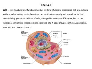
  • 1. The Cell Cell: is the structural and functional unit of life (and of disease processes). Cell also defines as the smallest unit of protoplasm than can exist independently and reproduce its kind. Human being possesses billions of cells, arranged in more than 200 types ,but on the functional similarities, theses cells are classified into 4 basic groups: epithelial, connective, muscular and nervous tissues.
  • 2. The cell either: • Eu-karoytic : contain nuclei. • Pro-karotic : contain no nuclear envelop, no histones and no membranous organells.
  • 3.
  • 4.
  • 5. A- The cytoplasm 1. Cytoplasmic matrix ( cell sap): it is a colloidal gel-like solution of proteins, lipids, carbohydrates, minerals, enzymes, small molecules, and ions. 2. Cytoplasmic organelles (organoids): these are specialized structures with specific functions, and they can be enclosed in membranes (membranous organelles) or not (non- membranes organelles). 3. Cytoplasmic Inclusions: these are temporary, non-living components which appear and disappear at different periods in certain cells, such as stored food(glycogen, fat), Pigments(exogenous & endogenous) and crystals.
  • 6. Cell Organelles(Organoids) Membranous organelles: these are permanent components, present in all nucleated cells, and included: 1. Cell membrane(plasma membrane or plasmalemma). 2. Mitochondria. 3. Golgi Apparatus. 4. Lysosomes. 5. Rough Endoplasmic Reticulum (RER). 6. Smooth Endoplasmic Reticulum (SER). 7. Peroxisomes.
  • 7. 1-Cell membrane(plasma membrane or plasmalemma): Is the boundary that separates the living cell from it’s non-living surroundings. Thickness: 7.5-10 nm(75-100 Angstrom) and consequently is visible only in electron microscope, where it appears as if it’s formed of three layers, therefore it’s called tri-lamellare membrane. Its outer and inner layers appear as dark lines, while its middle layer appears as a light area.
  • 8. Structure of the cell membrane
  • 9. 1-Lipids a. Phospholipids: It’s composed of: i. Phosphate head: • Has affinity for aqueous solution (hydrophilic ) • It's the charged polar head • Directed outwards ii. Fatty acid tails:  Has no affinity for aqueous solution (hydrophobic )  It's the non- polar (not charged )tail  Directed inwards Phosphate head Fatty acid Arranged as a bilayer
  • 10. b. Cholesterol: fit into spaces between phospholipids and prevent water-soluble molecules from diffusing across the membrane.
  • 11. 2- Protein molecules a. Extrinsic or Peripheral protein:  Lie outside lipid bilayer  forms a non-continuous layer loosely bound to both surface of membrane. b. Intrinsic or Integral proteins: -Small intrinsic proteins: small proteins embedded in the lipid bilayer. -Large intrinsic proteins: called trans membrane proteins, they contain channels through which ions can pass. trans membrane proteins Peripheral protein
  • 12. Functions of membrane proteins Outside Plasma membrane Inside Transporter Cell surface receptor Enzyme activity Cell surface identity marker Attachment to the cytoskeleton Cell adhesion “Antigen” “Channel”
  • 13. 3- Carbohydrates molecules Membrane carbohydrates either linked to the protein molecules forming glycoproteins or to lipid forming glycolipids. Glycoproteins + Glycolipids = Glycocalyx (Cell Coat), it may be thick or thin according to function of cell.It helps in adhesion adjacent cells and enables cells to recognize other cells of their own special kind. ABO antigen. Form part of the basement membrane Cell adhesion Cell surface identity marker Dr. Sabah A. AL-Qadasi
  • 14. The functions of the cell membrane 1. protects the cell by acting as a barrier 2. regulates the transport of substances in and out of the cell(active transport, selective transport, phagocytosis, pinocytosis and exocytosis) 3. receives chemical messages from other cell 4. acts as receptors 5. cell mobility, secretions, and absorptions of substances
  • 15.
  • 16. 2-Endoplamic Reticulum(ER):  They are membranous cell organelles formed of communicating wide and narrow tubules (cisternae).  They synthesize protein, carbohydrates, lipid and regulate mineral metabolism.  They are 2 types of ER: Rough and Smooth i. Rough Endoplasmic Reticulum(RER):  is studded with ribosomes and is the site of protein synthesis and processing.  They are basophilic substances (stains well with basic stains)  Their number increases in protein secreting cells such as fibroblast, plasma cell,ameloblast, hepatic and pancreatic cells  Protects the cytoplasm from the action Of the hydrolytic enzyme.
  • 17. ii. Smooth Endoplasmic Reticulum(SER):  Can’t demonstrated by LM, while by EM  With EM it appears as anastomosing tubules with no ribosomes.  It’s the site of phospholipids and carbohydrates synthesis  Fund in great amount in endocrine cells which synthesize steroid hormones.  Regulates muscles contraction (calcium ions).  Play role in platelets formation  Play role in HCL formation  Detoxification of excess drugs or hormones
  • 18. 3- Golgi Apparatus:  The Golgi structure is a smooth, curvy structure. It is a flattened stack of membranes.  It has a front end and a back end.  The front end is called the cis face and the back end is called the trans face.  Golgi apparatus has cisternae which are the flattened membrane folds and secretory vesicles which are what the cell discharges.  Golgi apparatus is responsible for collecting, concentrating, packaging, sorting and adding specific products to the secretion. By TEM
  • 19. • In nerve cells it surrounds the nucleus, while in secretory epithelial cells it’s found between the nucleus and cells free border. • With silver stain: it appears as brown fibrillar or granular network called positive image. • With H&E: it appears as an unstained area called negative image in a highly basophilic cells (e.g. plasma cells). • With EM: it has 3 forms: i. Saccules: small sacks arranged one above the other to form stacks. Each stack has concave mature face and a convex immature face. ii. Transfer vesicles: small round membranous vesicles containing protein and originate from RER. Transfer vesicles fuse with saccules of immature convex face.
  • 20. iii. Secretor vesicles: when transfer vesicles are concentrated and enveloped by membrane they known as secretory vesicles. They arise from the periphery of saccules. Secretory vesicles are discharged outside the cell by exocytosis, others remain intracellular as lysosomes.  Functions of Golgi apparatus: 1. Accumulate, concentrate and package secretory products of cells. 2. Rich in sulfotransferase enzyme which adds sulfates to certain secretory products. 3. Rich in sugar transferase enzyme which adds carbohydrates to certain secretory products. 4. Keep cell membranes and cell coat in good condition.
  • 21. 4- Lysosomes: • Lysosomes are enzyme-filled sacs, generally spherical, surrounded by a single membrane, present in all kind of the cells. • Their main function is an intracellular digestion. • They are very common in white blood cells, where disease and sickness are fought so a lot of bacteria needs to be digested. • Their shape and size vary depending on what material is digested. • They contain about 40 different enzymes (ex. nucleases, proteases, lipases, and carbohydrases, phosphatase).
  • 22.  With EM they appear in 2 forms: i. Primary lysosomes: homogeneous rounded vesicle. ii. Secondary lysosomes: heterogeneous rounded bodies because they contain ingested and digested elements.  Fate of the primary lysosomes: These are newly formed lysosomes which have budded off from the Golgi apparatus. They may circulate in cytoplasm and remain such as or fuse with some foreign particles or cytoplasmic bodies like old organelles to form secondary lysosomes.
  • 23.  Secondary lysosomes formed through the following processes: 1. Phagocytosis: (cell eating) the phagocytosed foreign bodies (phagosomes) fused with primary lysosomes to form digestive vacuole. 2.Pinocytosis: (cell drinking) the pinocytosed foreign bodies (pinocytic vesicles) fused with primary lysosomes to form multivesicular body. 3. Autophagocytosis: old membrane bound mitochondria fused with primary lysosomes to form autophagic vacuole. They may be expelled by exocytosis or remain in cytoplasm. 4. Residual bodies: after digestion of the contents of secondary lysosomes diffuse into the cytoplasm, the remaining vacuole is called residual body. They may be expelled by exocytosis or remain in cytoplasm. Long lifespan cells (nerve& cardiac cells) contain many residual bodies (lipofuscin) . They may be expelled by exocytosis or remain in cytoplasm.
  • 24.  Functions of the lysosomes: 1. Intracellular digestion 2. Defense the body against invading organisms 3. Digest old mitochondria 4. Facilitate penetration of sperm into the oocyte 5. Concerned with post-mortem changes (suicide bags) such as hypoxia, ischemia and bacterial infection.
  • 25. 5- Mitochondria(The Powerhouse) : • Mitos= thread + chondrons = granules, so with LM they appear as rods, granules or filaments. • With EM they appear as vesicles surrounded by 2 membranes; an outer smooth, and an inner rough because it projects into the cavity of the mitochondrion forming shelves called cristae which increase the surface area. • Mitochondrial matrix contains respiratory apparatus, DNA &RNA , ribosomes, calcium and magnesium granules. • Mitochondrion has its own strand of DNA, so they can divide to increase their number. • They are concerned with oxidative phosphorylation and ATP production and also play role in lipid synthesis (except in RBCs)
  • 26. • Each cell contains a different number of mitochondria. The number present is dependent upon how much energy the cell requires. The more energy a cell needs the more mitochondria that will be present. Cells have the ability to produce more mitochondria as needed. • Mitochondria are in constant movement and they are able to expand, contract, divide and fuse to make larger ones (giant mitochondrion) . • They are sensitive to temperature, PH, osmotic pressure. • Their life span is about 10 days.  Functions of the mitochondria: 1. Supply energy (ATP) to all cellular activities. 2. Storage of calcium and magnesium ions as dark granules. 3. Catalyze the enzymatic reactions of mitochondria. 4. Play role in lipid synthesis.
  • 27. 6- Peroxisomes or Microbodies: • Peroxidase are self-replicating organelles that contain oxidative enzymes. • They are somewhat larger than primary lysosomes. • In rats, they are distinguishable from lysosomes by an electron-dense granule nucleoid of urate oxidase • They contain more than 40 oxidative enzymes, especially urate oxidase, D-amino acid oxidase, hydroxyacid oxidase, and catalase.  Functions of Peroxisomes: Hydrogen peroxidase (H2O2) degradation: beta oxidation of long chained fatty acids produces H2O2 a potentially toxic substance to cells that must be eliminated. Peroxisomes contain catalase enzyme which destroys H2O2. Excess H2O2 that accumulates in cells from other sources can also be eliminated by peroxisomes.