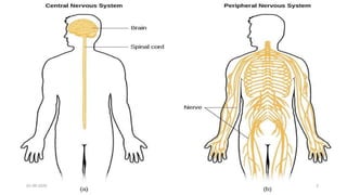
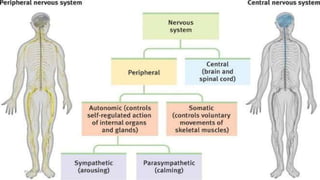
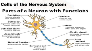
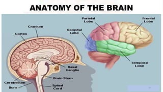
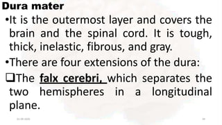
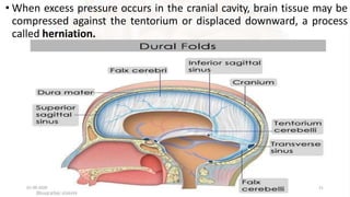
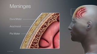
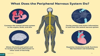
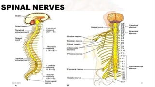
The nervous system is made up of the central nervous system (brain and spinal cord) and the peripheral nervous system. The central nervous system contains approximately 10 million sensory neurons that send information to the brain and 500,000 motor neurons that control muscles and glands. It is protected by three membranes (dura mater, arachnoid, and pia mater) and cerebrospinal fluid. The brain and spinal cord work together to coordinate actions and transmit sensory information through neural signals.


![17 [chapter 17 the special senses]](https://cdn.slidesharecdn.com/ss_thumbnails/17chapter17thespecialsenses-170828041636-thumbnail.jpg?width=640&height=640&fit=bounds)


