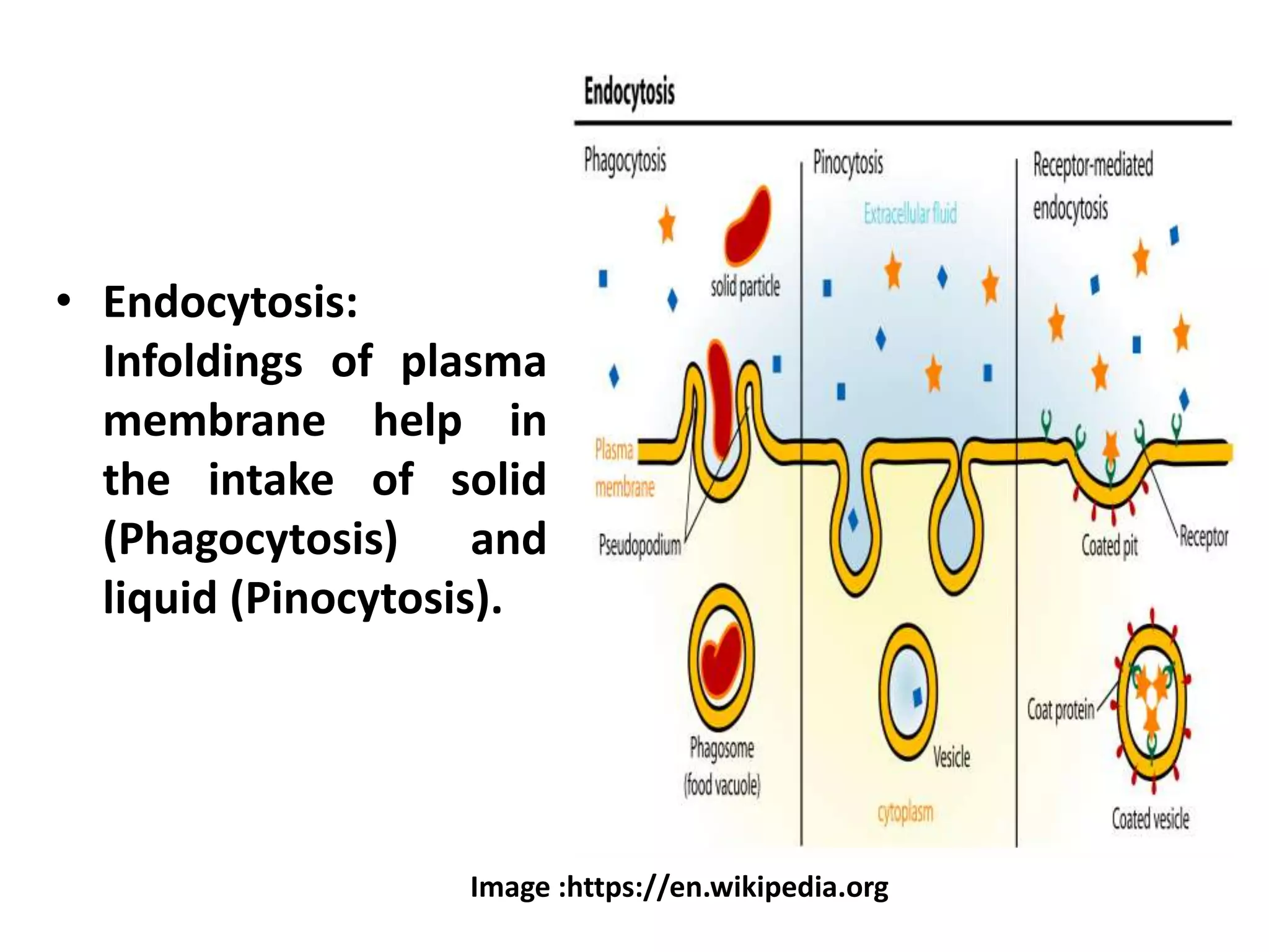
CELL MEMBRANE
Dr. Saji Mariam George
Associate Professor (Retired)
Assumption College Autonomous
Changanacherry
Cell membrane (Plasma membrane ).
• All cells are surrounded by a thin, elastic,
semi- permeable living membrane called Cell
membrane (Plasma membrane or plasma
lemma).
• It serves as the boundary of the cytoplasm.
• Keeps the cell contents in place
• Gives form to the cell
• The thickness varies from 100 – 215 A°.
• Membranes from different sources vary in
their composition.
• Chemically , it is composed of proteins, lipids
and carbohydrates.
• Proteins represent the main component of
most biological membranes.
On the basis of the degree of association with
the membrane , membrane proteins are
classified into
i) Peripheral (Extrinsic ) proteins –
 Have a weaker association with the
membrane
 Soluble in aqueous solutions
 Usually , they are free of lipids .
ii ) Integral (Intrinsic ) proteins
 Represent more than 70% of the membrane
proteins.
 Firmly associated with the membrane
 Insoluble in aqueous solutions
 Associated with lipids – The main lipid
components of plasma membrane -
phospholipids, cholesterol and galactolipids.
 Lipids are arranged in parallel chains in a
double (bimolecular) layer with their
hydrophobic, non polar ends adjoining each
other and their hydrophilic polar ends
associated with proteins.
 More than 30 enzymes have been detected
in isolated plasma membranes like Mg ++
ATPase, Alkaline phosphatase, Adenyl
cyclase, Na+ K+ATPase etc.
MOLECULAR MODELS
OF PLASMA MEMBRANE
1. UNIT MEMBRANE MODEL
 Proposed by Robertson (1960)
 The unit membrane is a trilaminar
membrane with protein – lipid – protein
arrangement.
 The outer and inner dense layers represent
the proteins , each about 20 A° thick.
 The interior less dense region of about 35 A°
thickness represents the bimolecular layer
of phospholipid.
 The total thickness of the plasma membrane
is 75 A° - thickness may vary from
membrane to membrane.
Unit membrane model of Plasma
membrane
Image:http://www.expertsmind.com
Image:https://www.mrgscience.com
Transmission electron micrograph of membrane showing
trilaminar appearance
• The lipid and protein molecules are held
together electrostatically.
• At some places in the membrane , there are
10 A° wide pores.
• Trilaminar composition is found in most of
the membranes of cellular organelles like
Endoplasmic reticulum, Golgi complex,
Lysosomes, Plastids etc.
Limitations of Unit membrane model
Unit membrane model is not sufficient to
explain the structure and function of all
biological membranes which are much more
complex .
2. FLUID MOSAIC MODEL
• Proposed by Singer and Nicholson (1972)
• Generally accepted model
• Described as ‘protein icebergs’ in a sea of
lipids.
Important Postulates :
1. The lipid and integral proteins are disposed
in a kind of mosaic arrangement.
2. The biological membranes are quasi- fluid
structures in which both the lipids and the
integral proteins are able to perform
translational movements within the bilayer.
Fluid mosaic model of Plasma
membrane
• The main components of the membrane viz.
lipids and proteins are held together by
relatively weak interactions – ionic and H –
bonds .
• Fluidity (both lipids and proteins have
freedom of lateral movement within the
bilayer ) is achieved with slightest disturbance.
• Intrinsic proteins and lipids are amphipathic
molecules , having both hydrophilic and
hydrophobic groups within the same
molecule .
• This explains that transmembrane protein
traversing through the membrane will have
hydrophilic groups on either side of the
membrane and hydrophobic group in the
interior of the membrane which is in contact
with the lipids.
• Such traversing proteins may be in contact
with the aqueous solvent on both sides of
the membrane.
• The membranes are dynamic not static.
• Small sized pores , with 0.7 nm diameter
are present.
Functions of Plasma membrane
• Controls the entry and exit of molecules and ions
into and out of the cell - helps to maintain a
balance between the osmotic pressure of the
intracellular fluid and that of the interstitial fluid.
• Osmosis - Plasma membrane is semi- permeable.
The to and fro movement of water molecules
through the plasma membrane occurs due to the
differences in the concentration of the solutes on
either sides.
• Endocytosis:
Infoldings of plasma
membrane help in
the intake of solid
(Phagocytosis) and
liquid (Pinocytosis).
Image :https://en.wikipedia.org
• Exocytosis :
It permits exit of
secretions and wastes.
THANK YOU

CELL MEMBRANE SMG

  • 1.
    CELL MEMBRANE Dr. SajiMariam George Associate Professor (Retired) Assumption College Autonomous Changanacherry
  • 2.
    Cell membrane (Plasmamembrane ). • All cells are surrounded by a thin, elastic, semi- permeable living membrane called Cell membrane (Plasma membrane or plasma lemma). • It serves as the boundary of the cytoplasm. • Keeps the cell contents in place • Gives form to the cell • The thickness varies from 100 – 215 A°.
  • 3.
    • Membranes fromdifferent sources vary in their composition. • Chemically , it is composed of proteins, lipids and carbohydrates. • Proteins represent the main component of most biological membranes.
  • 4.
    On the basisof the degree of association with the membrane , membrane proteins are classified into i) Peripheral (Extrinsic ) proteins –  Have a weaker association with the membrane  Soluble in aqueous solutions  Usually , they are free of lipids .
  • 5.
    ii ) Integral(Intrinsic ) proteins  Represent more than 70% of the membrane proteins.  Firmly associated with the membrane  Insoluble in aqueous solutions  Associated with lipids – The main lipid components of plasma membrane - phospholipids, cholesterol and galactolipids.
  • 6.
     Lipids arearranged in parallel chains in a double (bimolecular) layer with their hydrophobic, non polar ends adjoining each other and their hydrophilic polar ends associated with proteins.  More than 30 enzymes have been detected in isolated plasma membranes like Mg ++ ATPase, Alkaline phosphatase, Adenyl cyclase, Na+ K+ATPase etc.
  • 7.
    MOLECULAR MODELS OF PLASMAMEMBRANE 1. UNIT MEMBRANE MODEL  Proposed by Robertson (1960)  The unit membrane is a trilaminar membrane with protein – lipid – protein arrangement.  The outer and inner dense layers represent the proteins , each about 20 A° thick.
  • 8.
     The interiorless dense region of about 35 A° thickness represents the bimolecular layer of phospholipid.  The total thickness of the plasma membrane is 75 A° - thickness may vary from membrane to membrane.
  • 9.
    Unit membrane modelof Plasma membrane Image:http://www.expertsmind.com
  • 10.
  • 11.
    • The lipidand protein molecules are held together electrostatically. • At some places in the membrane , there are 10 A° wide pores. • Trilaminar composition is found in most of the membranes of cellular organelles like Endoplasmic reticulum, Golgi complex, Lysosomes, Plastids etc.
  • 12.
    Limitations of Unitmembrane model Unit membrane model is not sufficient to explain the structure and function of all biological membranes which are much more complex .
  • 13.
    2. FLUID MOSAICMODEL • Proposed by Singer and Nicholson (1972) • Generally accepted model • Described as ‘protein icebergs’ in a sea of lipids. Important Postulates : 1. The lipid and integral proteins are disposed in a kind of mosaic arrangement. 2. The biological membranes are quasi- fluid structures in which both the lipids and the integral proteins are able to perform translational movements within the bilayer.
  • 14.
    Fluid mosaic modelof Plasma membrane
  • 15.
    • The maincomponents of the membrane viz. lipids and proteins are held together by relatively weak interactions – ionic and H – bonds . • Fluidity (both lipids and proteins have freedom of lateral movement within the bilayer ) is achieved with slightest disturbance.
  • 16.
    • Intrinsic proteinsand lipids are amphipathic molecules , having both hydrophilic and hydrophobic groups within the same molecule . • This explains that transmembrane protein traversing through the membrane will have hydrophilic groups on either side of the membrane and hydrophobic group in the interior of the membrane which is in contact with the lipids.
  • 17.
    • Such traversingproteins may be in contact with the aqueous solvent on both sides of the membrane. • The membranes are dynamic not static. • Small sized pores , with 0.7 nm diameter are present.
  • 18.
    Functions of Plasmamembrane • Controls the entry and exit of molecules and ions into and out of the cell - helps to maintain a balance between the osmotic pressure of the intracellular fluid and that of the interstitial fluid. • Osmosis - Plasma membrane is semi- permeable. The to and fro movement of water molecules through the plasma membrane occurs due to the differences in the concentration of the solutes on either sides.
  • 19.
    • Endocytosis: Infoldings ofplasma membrane help in the intake of solid (Phagocytosis) and liquid (Pinocytosis). Image :https://en.wikipedia.org
  • 20.
    • Exocytosis : Itpermits exit of secretions and wastes.
  • 21.