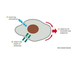
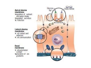
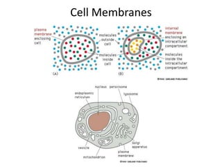
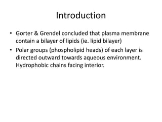
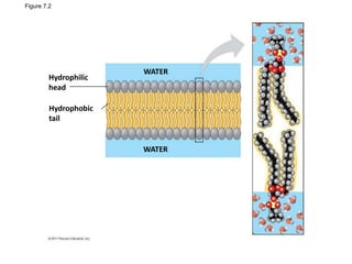
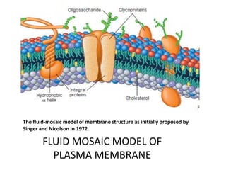
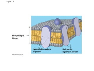
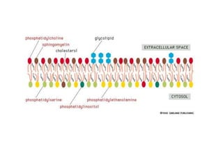
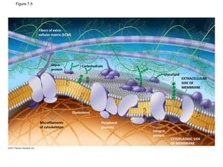
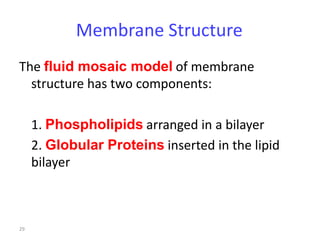
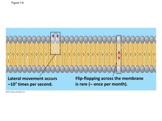
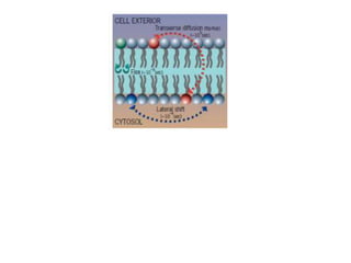
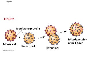
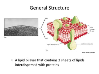
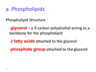
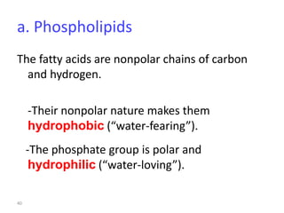
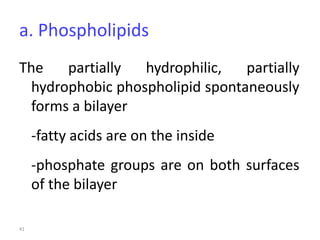
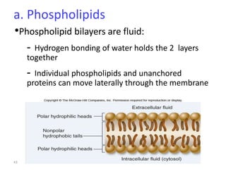
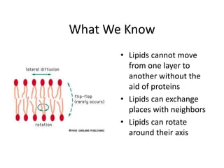
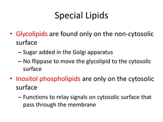
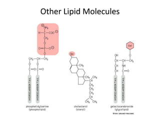
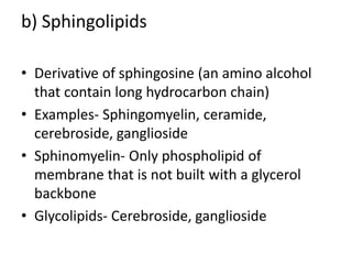
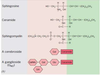
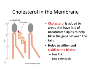
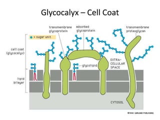
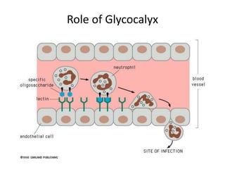
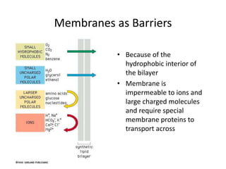
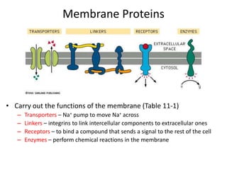
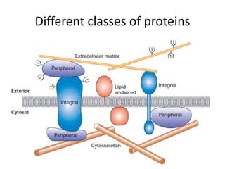
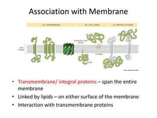
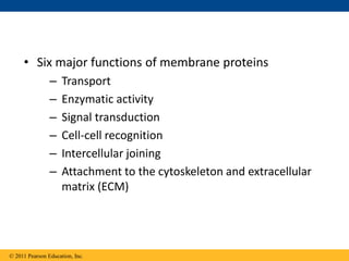
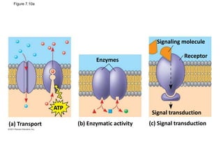
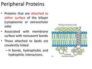
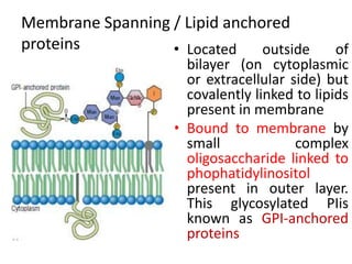
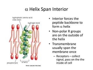
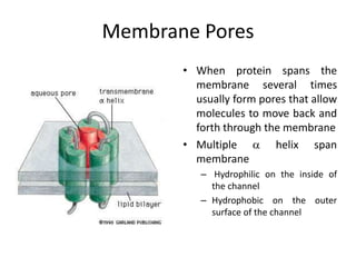
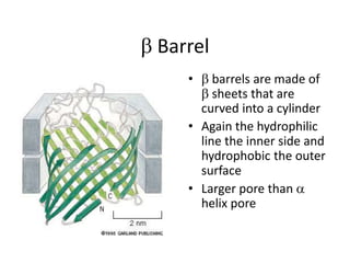
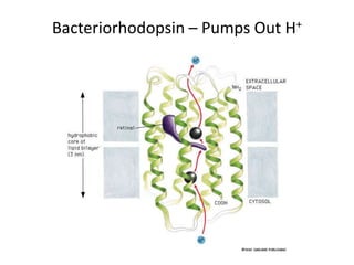
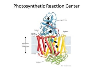
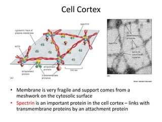
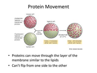
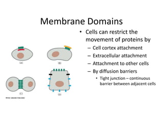
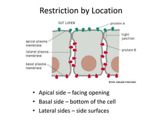
Membrane Structures
Plasma Membrane
• The ‘container’ for the cell
– Holds the cytoplasm and organelles together
• Barrier for the cell
– Bacteria have a single membrane
– Eukaryotes have outer plasma membrane and
internal membranes
• Endoplasmic reticulum
• Nuclear membrane
• Membrane-bound organelles
• Cells are separated from the external world by
a thin, fragile structure called the plasma
membrane that is only 5 to 10 nm wide
Functions of Plasma membranes
1.Compartmentalization- Membranes are continuous, unbroken
sheets and, enclose compartments. The plasma membrane
encloses the contents of the entire cell, whereas the nuclear
and cytoplasmic membranes enclose diverse intracellular
spaces. The various membrane-bounded compartments of a
cell possess different contents. Membrane
compartmentalization allows specialized activities to proceed
without external interference and enables cellular activities to
be regulated independently of one another.
2. Scaffold for biochemical activities. Membranes not only
enclose compartments but are also a distinct compartment
themselves. As long as reactants are present in solution
their relative positions cannot be stabilized and their
interactions are dependent on random collisions.
Because of their construction, membranes provide
the cell with an extensive framework or scaffolding
within which components can be ordered for
effective interaction
3. Providing a selectively permeable barrier.
• Membranes prevent the unrestricted exchange of
molecules from one side to the other.
• At the same time, membranes provide the means of
communication between the compartments they
separate.
• The plasma membrane, which encircles a cell, can be
compared to a moat around a castle: both serve as a
general barrier, yet both have gated “bridges” that
promote the movement of select elements into and
out of the enclosed living space.
4. Transporting solutes.
• The plasma membrane contains the machinery for
physically transporting substances from one side of the
membrane to another, often from a region where the
solute is present at low concentration into a region where
that solute is present at much higher concentration.
• The membrane’s transport machinery allows a cell to
accumulate substances, such as sugars and amino acids,
that are necessary to fuel its metabolism and build its
macromolecules.
• The plasma membrane is also able to transport specific
ions, thereby establishing ionic gradients across itself. This
capability is especially critical for nerve and muscle cells.
5. Responding to external signals.
• The plasma membrane respond to external stimuli, a
process known as signal transduction.
• Membranes possess receptors that combine with specific
molecules (or ligands) having a complementary structure.
Different types of cells have membranes with different
receptors and are, therefore, capable of recognizing and
responding to different ligands in their environment.
• The interaction of a plasma membrane receptor with an
external ligand may cause the membrane to generate a
signal that stimulates or inhibits internal activities.
• For example, signals generated at the plasma membrane
may tell a cell to manufacture more glycogen, to prepare
for cell division, to move toward a higher concentration of a
particular compound, to release calcium from internal
stores, or possibly to commit suicide
6. Intercellular interaction.
• Situated at the outer edge of every living cell, the plasma
membrane of multicellular organisms mediates the
interactions between a cell and its neighbours.
• The plasma membrane allows cells to recognize and signal
one another, to adhere when appropriate, and to
exchange materials and information.
7. Energy transduction.
• Membranes are intimately involved in the processes by
which one type of energy is converted to another type
(energy transduction).
• The most fundamental energy transduction occurs during
photosynthesis when energy in sunlight is absorbed by
membrane-bound pigments, converted into chemical
energy, and stored in carbohydrates. Membranes are also
involved in the transfer of chemical energy from
carbohydrates and fats to ATP.
• In eukaryotes, the machinery for these energy conversions
is contained within membranes of chloroplasts and
mitochondria
Membrane functions in plant cells
Cell Membranes
Introduction
• Gorter & Grendel concluded that plasma membrane
contain a bilayer of lipids (ie. lipid bilayer)
• Polar groups (phospholipid heads) of each layer is
directed outward towards aqueous environment.
Hydrophobic chains facing interior.
Figure 7.2
Hydrophilic
head
Hydrophobic
tail
WATER
WATER
© 2011 Pearson Education, Inc.
• In 1935, Davson and Danielli proposed a sandwich model in
which the phospholipid bilayer lies between two layers of
globular proteins
• Later studies found problems with this model, particularly
the placement of membrane proteins, which have
hydrophilic and hydrophobic regions
• In 1972, S. J. Singer and G. Nicolson proposed that the
membrane is a mosaic of proteins dispersed within the
bilayer, with only the hydrophilic regions exposed to water
1954 Davson-Danielli model
showing the lipid bilayer, which is lined on both surfaces by a
monomolecular layer of proteins that extends through the
membrane to form protein-lined pores.
The fluid-mosaic model of membrane structure as initially proposed by
Singer and Nicolson in 1972.
FLUID MOSAIC MODEL OF
PLASMA MEMBRANE
Figure 7.3
Phospholipid
bilayer
Hydrophobic regions
of protein
Hydrophilic
regions of protein
• Bilayer is present in a fluid state and individual lipid
molecules can move laterally within plane of
membrane (mosaic movement)
• Bilayer contain different types of lipids.
• External surfaces of membrane proteins &
phospholipids contain short chain of sugars making
them glycoproteins & glycolipids.
• Outer leaflet may have microdomains consisting
clusters of specific lipids:RAFTS
• Patches of cholesterol & sphingolipid assemble
themselves into microdomains. These are more
gelated & highly ordered
Rafts
• Freeze-fracture studies of the plasma membrane
supported the fluid mosaic model
• Freeze-fracture is a specialized preparation
technique that splits a membrane along the middle
of the phospholipid bilayer
© 2011 Pearson Education, Inc.
Figure 7.4
Knife
Plasma membrane Cytoplasmic layer
Proteins
Extracellular
layer
Inside of extracellular layer Inside of cytoplasmic layer
TECHNIQUE
RESULTS
The Fluidity of Membranes
• Phospholipids in the plasma membrane can move
within the bilayer
• Most of the lipids, and some proteins, drift laterally
• Rarely does a molecule flip-flop transversely across
the membrane
© 2011 Pearson Education, Inc.
Figure 7.5
Glyco-
protein
Carbohydrate
Glycolipid
Microfilaments
of cytoskeleton
EXTRACELLULAR
SIDE OF
MEMBRANE
CYTOPLASMIC SIDE
OF MEMBRANE
Integral
protein
Peripheral
proteins
Cholesterol
Fibers of extra-
cellular matrix (ECM)
29
Membrane Structure
The fluid mosaic model of membrane
structure has two components:
1. Phospholipids arranged in a bilayer
2. Globular Proteins inserted in the lipid
bilayer
30
Figure 7.6
Lateral movement occurs
107 times per second.
Flip-flopping across the membrane
is rare ( once per month).
Figure 7.7
Membrane proteins
Mouse cell
Human cell
Hybrid cell
Mixed proteins
after 1 hour
RESULTS
Evolution of Differences in Membrane
Lipid Composition
• Variations in lipid composition of cell membranes of
many species appear to be adaptations to specific
environmental conditions
• Ability to change the lipid compositions in response
to temperature changes has evolved in organisms
that live where temperatures vary
© 2011 Pearson Education, Inc.
General Structure
• A lipid bilayer that contains 2 sheets of lipids
interdispersed with proteins
Membrane chemistry
• Membranes are lipid-protein assemblies in which the
components are held together in thin sheet by non-
covalent bonds.
• Components of Membrane:
1. Lipids
2. Proteins
3. Carbohydrates
• Lipid bilayer serves as barrier for entry of water-soluble
materials in & out of the cell.
• Proteins carry out many functions.
• Lipid to protein ration varies with cell membrane, cell type,
organism.
• This ratio depends on function of each cell type
• E.g.Inner mitochondrial membrane –high
protein/lipid ration compared to RBCs & myelin
sheath around nerve cells.
• E.g.Inner mitochondrial membrane contain
protein carrier of ETC. myelin sheath acts as
electrical insulation for nerve cell it encloses so it
has high lipid content
• Carbohydrates are also present in membrane
which are attached to lipids & proteins
1. Membrane Lipids
• Ampipathic lipids- contain both hydrophilic &
hydrophobic regions.
• Types of lipids present in membrane-
a) Phosphoglycerides
b) Sphingolipids
c) Cholesterol
39
a. Phospholipids
Phospholipid Structure
-glycerol – a 3-carbon polyalcohol acting as a
backbone for the phospholipid
-2 fatty acids attached to the glycerol
-phosphate group attached to the glycerol
40
a. Phospholipids
The fatty acids are nonpolar chains of carbon
and hydrogen.
-Their nonpolar nature makes them
hydrophobic (“water-fearing”).
-The phosphate group is polar and
hydrophilic (“water-loving”).
41
a. Phospholipids
The partially hydrophilic, partially
hydrophobic phospholipid spontaneously
forms a bilayer
-fatty acids are on the inside
-phosphate groups are on both surfaces
of the bilayer
42
43
a. Phospholipids
•Phospholipid bilayers are fluid:
- Hydrogen bonding of water holds the 2 layers
together
- Individual phospholipids and unanchored
proteins can move laterally through the membrane
44
a. Phospholipids
•Phospholipid bilayers are fluid:
- Saturated fatty acids make the membrane less
fluid than unsaturated fatty acids
- Cholesterols make the membrane more rigid
- Warm temperatures make the membrane more
fluid than cold temperatures
Review of Lipids
Lipids
• Most abundant lipid is
the phospholipid
• Phospholipids have a
PO4 group in the 3rd –
OH group of the
glycerol instead of
hydrocarbon
• This can attach a
hydrophilic group
– Choline –
phosphyltidylcholine
– Polar amino acids like
serine -
phosphatidylserine
Importance of Hydrocarbon
• Hydrocarbon tail will determine the fluidity
of the membrane just as it does in fats and
oils
• 2 components are important
– Length of hydrocarbon chain
• 14 to 24 C but usually 18 to 20 C per tail
– Level of unsaturation (# of C=C bonds)
• 1 tail has 1 or more C=C bonds (unsaturated)
• Other tail is saturated (no C=C bonds)
Unsaturated Hydrocarbons
• Each C=C bond causes
a kink or bend in the
tail
– Can’t pack tightly in
the layer
– More lipids that have
unsaturated tails the
more fluid the
membrane
87
Fatty Acids
Lipid Structure
• Hydrophilic head – H2O
loving – due to polar
group in the head
• Hydrophobic tail – H2O
hating – due to the long
hydrocarbon tails
Membrane
• Amphipathic molecules have both components so the
hydrophilic head molecules interact with the aqueous
solution and the hydrophobic tails will interact with
each other
Lipid Bilayer
• Due to amphipathic property the
membrane can reseal after an ‘injury’
• Bilayer is fluid – the orientation of the lipids
and the outer aqueous surroundings keeps
the lipids in the bilayer
– The lipid can move around the layer – like one
person moving in a crowded room
• Not the same as flexible – entire membrane
bending
Liposomes
• Can study membranes by using artificial
membrane structures called liposomes
• Can follow the movement of lipids in each of the
layers
What We Know
• Lipids cannot move
from one layer to
another without the
aid of proteins
• Lipids can exchange
places with neighbors
• Lipids can rotate
around their axis
New Membrane
• New lipids are added on one side of the membrane
• Enzyme called flippase used to put the lipid in the
other half of the bilayer
– Flippase may be selective for the type of lipids that it puts
on either surface
Asymmetry
• New membrane comes from
the SER
• Vesicle buds off the SER and
when fuses with the plasma
membrane, the orientation is
maintained
• Membranes have distinct
inner and outer surface
– Inner – cytosolic face
• Adjacent to the cytosol
– Outer – non-cytosolic face
• Adjacent to the cell exterior or
the interior of an organelle
a) Phosphoglycerides
• Most membrane lipids contain a phosphate group, which
makes them phospholipids.
• Membrane phospholipids are built on a glycerol backbone
they are called as phosphoglycerides.
• Examples-
1. Phosphatidylcoline/lecithin (PC)
2. Phosphatidylethanolamine/cephalin (PE)
3. Phosphotidylserine (PS),
4. Phosphatidylinositol (PI)
• At physiological pH-
PS and PI are negatively charged,
PC and PE are neutral.
Membrane Fluidity
• Enables the membrane proteins to
diffuse rapidly
• Simple means of distributing lipids and
proteins
• Allows membranes to fuse with one
another
• Evenly distributed during daughter cell
formation
Membranes are Asymmetrical
• Inner surface is different from the outer
surface
– Types of lipids in each layer
• Proteins in the bilayer have a specific
orientation due to its function
The asymmetry of membrane lipids
• The lipid bilayer consists of two distinct
leaflets that have a distinctly different lipid
composition.
• The appearance of PS on the outer surface of
aging lymphocytes marks the cells for
destruction by macrophages, whereas its
appearance on the outer surface of platelets
leads to blood coagulation.
• All the glycolipids of the plasma membrane
are in the outer leaflet where they often serve
as receptors for extracellular ligands.
Special Lipids
• Glycolipids are found only on the non-cytosolic
surface
– Sugar added in the Golgi apparatus
– No flippase to move the glycolipid to the cytosolic
surface
• Inositol phospholipids are only on the cytosolic
surface
– Functions to relay signals on cytosolic surface that
pass through the membrane
Other Lipid Molecules
Reminder
• Hydrophilic molecules can dissolve in H2O due to
the polarity of both of these molecules
– H bonds and other non-covalent interactions may aid
in this
Reminder
• Hydrophobic molecules will be “caged” by the polar
molecules – requires energy
• Why when fats or oils are placed in water that they
usually sit as a glob on the surface
b) Sphingolipids
• Derivative of sphingosine (an amino alcohol
that contain long hydrocarbon chain)
• Examples- Sphingomyelin, ceramide,
cerebroside, ganglioside
• Sphinomyelin- Only phospholipid of
membrane that is not built with a glycerol
backbone
• Glycolipids- Cerebroside, ganglioside
c) Cholesterol
• May contribute up to 50% in animal
• Absent in plant and bacterial pm
• Interfere with the movements of the fatty
acids tails of the phospholipids.
• Maintain the fluidity of the membrane
Cholesterol in the Membrane
• Cholesterol is added to
areas that have lots of
unsaturated lipids to help
fill in the gaps between the
tails
• Helps to stiffen and
stabilize the bilayer
– Less fluid
– Less permeable
• As temperatures cool, membranes switch from a
fluid state to a solid state
• The temperature at which a membrane solidifies
depends on the types of lipids
• Membranes rich in unsaturated fatty acids are
more fluid than those rich in saturated fatty
acids
• Membranes must be fluid to work properly; they
are usually about as fluid as salad oil
© 2011 Pearson Education, Inc.
• The steroid cholesterol has different effects on
membrane fluidity at different temperatures
• At warm temperatures (such as 37°C), cholesterol
restrains movement of phospholipids
• At cool temperatures, it maintains fluidity by
preventing tight packing
© 2011 Pearson Education, Inc.
Figure 7.8
Fluid
Unsaturated hydrocarbon
tails
Viscous
Saturated hydrocarbon tails
(a) Unsaturated versus saturated hydrocarbon tails
(b) Cholesterol within the animal
cell membrane
Cholesterol
Membranes are Asymmetrical
• Inner surface is different from the outer
surface
– Types of lipids in each layer
• Proteins in the bilayer have a specific
orientation due to its function
Importance of lipid bilayer
• To maintain the proper internal composition
of cell
• Separating electric charges across the plasma
membrane.
• It facilitate the regulated fusion or budding of
membranes.
• Example- cytoplasmic vesicles fuse to the
plasma membrane, fertilization where two
cells fuse to form single cell.
• Liposome- Phospholipid molecule assemble to form
fluid-filled spherical vesicle
Example – a small amount of phosphatidylcholine is
dispersed in aqueous solution, the phospholipid
molecules assemble spontaneously form the walls of
fluid-filled vesicles, called liposomes.
• It is valuable in membrane resarch- Membrane
proteins can be inserted into liposomes and their
function studied in a much simpler environment than
that of a natural membrane.
• Liposomes have also been developed as vehicles to
deliver drugs or DNA molecules within the body
Liposome
b) Membrane carbohydrates
• 2-10% by weight
• 90% are linked to proteins to form glycoproteins
• Remaining attached to lipids to form glycolipids
• Face to extracellular space
• Play major role in interacting with environment &
protein sorting to different cell compartments
• Glycolipids on RBCs membrane determine blood
group
• Blood gr. A- NAG
• Blood gr. B- Galactose
• Blood gr. O-NO terminal
sugar attached
Carbohydrates on Cell Surface
• Many of the plasma membrane proteins have
sugars attached to them
– Short oligosaccharides – glycoproteins
– Long polysaccharides - proteoglycans
• Sugars on the surface make up the glycocalyx
– Keeps cells moist and slippery
– Used as cell recognition (lectins) and adhesion molecules
Glycocalyx – Cell Coat
Role of Glycocalyx
Membranes as Barriers
• Because of the
hydrophobic interior of
the bilayer
• Membrane is
impermeable to ions and
large charged molecules
and require special
membrane proteins to
transport across
Membrane Proteins
• Carry out the functions of the membrane (Table 11-1)
– Transporters – Na+ pump to move Na+ across
– Linkers – integrins to link intercellular components to extracellular ones
– Receptors – to bind a compound that sends a signal to the rest of the cell
– Enzymes – perform chemical reactions in the membrane
Different classes of proteins
Association with Membrane
• Transmembrane/ integral proteins – span the entire
membrane
• Linked by lipids – on either surface of the membrane
• Interaction with transmembrane proteins
• Six major functions of membrane proteins
– Transport
– Enzymatic activity
– Signal transduction
– Cell-cell recognition
– Intercellular joining
– Attachment to the cytoskeleton and extracellular
matrix (ECM)
© 2011 Pearson Education, Inc.
Figure 7.10
Enzymes
Signaling molecule
Receptor
Signal transduction
Glyco-
protein
ATP
(a) Transport (b) Enzymatic activity (c) Signal transduction
(d) Cell-cell recognition (e) Intercellular joining (f) Attachment to
the cytoskeleton
and extracellular
matrix (ECM)
Figure 7.10a
Enzymes
Signaling molecule
Receptor
Signal transduction
ATP
(a) Transport (b) Enzymatic activity (c) Signal transduction
Figure 7.10b
Glyco-
protein
(d) Cell-cell recognition (e) Intercellular joining (f) Attachment to
the cytoskeleton
and extracellular
matrix (ECM)
Integral Proteins
• Penetrate lipid bilayer
• Also called as
transmembrane proteins i.e.
they pass entirely through
bilayer.
• Domains protrude from both
extracellular & cytoplasmic
sides of membrane.
Functions
• as receptors that bind specific substance at
membrane
• channels or transporter in movement of ions
& solutes across membrane
• Agent that transfer electron during
photosynthesis & respiration
Integral membrane protein in RBC
• Band3 and glycophorin A
• Band3- homodimer, Each subunit spans the
membrane at least a dozen times. It serves as
a channel for passive exchange of anions
across the membrane.
• H2O + CO2 H2CO3 HCO3 + H+
The biocarbonate enter the RBC in exchange for
chloride ions.
Glycophorin A-
 The first membrane protein to have its amino acid
sequence determined.
 Dimer
 Span the membrane once
 Glycophorin acts as receptor for protozoan that
causes malaria, providing a path for entry into the
blood cell. Consequently, individuals whose
erythrocytes lack glycophorin A and B are thought to
be protected from acquiring malaria. Differences in
glycophorin amino acid sequence determine
whether a person has an MM, MN,or NN blood type.
Peripheral Proteins
• Proteins that are attached to
either surface of the bilayer
(cytoplasmic or extracellular
side)
• Associated with membrane
surface with noncavent bonds
• Those attached to lipids are
covalently linked
– H bonds, hydrophobic and
hydrophilic interactions
• Act as membrane skeletal by forming fibrillar
network.
• Provide mechanical support
• Functions as anchor for integral membrane
proteins
• Some functions as enzymes, specialized coats
or factor that transmit transmembrane signals
Membrane Spanning / Lipid anchored
proteins • Located outside of
bilayer (on cytoplasmic
or extracellular side) but
covalently linked to lipids
present in membrane
• Bound to membrane by
small complex
oligosaccharide linked to
phophatidylinositol
present in outer layer.
This glycosylated PIis
known as GPI-anchored
proteins
 Helix Span Interior
• Interior forces the
peptide backbone to
form  helix
• Non-polar R groups
are on the outside of
the helix
• Transmembrane
usually span the
membrane once
– Receptors – collect
signal, pass on the the
inside of cell
Membrane Pores
• When protein spans the
membrane several times
usually form pores that allow
molecules to move back and
forth through the membrane
• Multiple  helix span
membrane
– Hydrophilic on the inside of
the channel
– Hydrophobic on the outer
surface of the channel
 Barrel
•  barrels are made of
 sheets that are
curved into a cylinder
• Again the hydrophilic
line the inner side and
hydrophobic the outer
surface
• Larger pore than 
helix pore
Detergents
• Used to remove the proteins
from the membrane
• Amphipathic molecules
• Have a single hydrocarbon tail
• Form small clusters in
aqueous solutions called
micelles
• SDS and Triton X-100
common in the laboratory
Removal of Proteins
Bacteriorhodopsin – Pumps Out H+
Photosynthetic Reaction Center
Cell Cortex
• Membrane is very fragile and support comes from a
meshwork on the cytosolic surface
• Spectrin is an important protein in the cell cortex – links with
transmembrane proteins by an attachment protein
Protein Movement
• Proteins can move through the layer of the
membrane similar to the lipids
• Can’t flip from one side to the other
Membrane Domains
• Cells can restrict the
movement of proteins by
– Cell cortex attachment
– Extracellular attachment
– Attachment to other cells
– By diffusion barriers
• Tight junction – continuous
barrier between adjacent cells
Restriction by Location
• Apical side – facing opening
• Basal side – bottom of the cell
• Lateral sides – side surfaces
Membrane Structure and
Function
Chapter 7

Membrane structure and membrane chemistry.pptx

  • 1.
  • 2.
    Plasma Membrane • The‘container’ for the cell – Holds the cytoplasm and organelles together • Barrier for the cell – Bacteria have a single membrane – Eukaryotes have outer plasma membrane and internal membranes • Endoplasmic reticulum • Nuclear membrane • Membrane-bound organelles
  • 3.
    • Cells areseparated from the external world by a thin, fragile structure called the plasma membrane that is only 5 to 10 nm wide
  • 4.
    Functions of Plasmamembranes 1.Compartmentalization- Membranes are continuous, unbroken sheets and, enclose compartments. The plasma membrane encloses the contents of the entire cell, whereas the nuclear and cytoplasmic membranes enclose diverse intracellular spaces. The various membrane-bounded compartments of a cell possess different contents. Membrane compartmentalization allows specialized activities to proceed without external interference and enables cellular activities to be regulated independently of one another. 2. Scaffold for biochemical activities. Membranes not only enclose compartments but are also a distinct compartment themselves. As long as reactants are present in solution
  • 5.
    their relative positionscannot be stabilized and their interactions are dependent on random collisions. Because of their construction, membranes provide the cell with an extensive framework or scaffolding within which components can be ordered for effective interaction
  • 6.
    3. Providing aselectively permeable barrier. • Membranes prevent the unrestricted exchange of molecules from one side to the other. • At the same time, membranes provide the means of communication between the compartments they separate. • The plasma membrane, which encircles a cell, can be compared to a moat around a castle: both serve as a general barrier, yet both have gated “bridges” that promote the movement of select elements into and out of the enclosed living space.
  • 7.
    4. Transporting solutes. •The plasma membrane contains the machinery for physically transporting substances from one side of the membrane to another, often from a region where the solute is present at low concentration into a region where that solute is present at much higher concentration. • The membrane’s transport machinery allows a cell to accumulate substances, such as sugars and amino acids, that are necessary to fuel its metabolism and build its macromolecules. • The plasma membrane is also able to transport specific ions, thereby establishing ionic gradients across itself. This capability is especially critical for nerve and muscle cells.
  • 8.
    5. Responding toexternal signals. • The plasma membrane respond to external stimuli, a process known as signal transduction. • Membranes possess receptors that combine with specific molecules (or ligands) having a complementary structure. Different types of cells have membranes with different receptors and are, therefore, capable of recognizing and responding to different ligands in their environment. • The interaction of a plasma membrane receptor with an external ligand may cause the membrane to generate a signal that stimulates or inhibits internal activities.
  • 9.
    • For example,signals generated at the plasma membrane may tell a cell to manufacture more glycogen, to prepare for cell division, to move toward a higher concentration of a particular compound, to release calcium from internal stores, or possibly to commit suicide
  • 10.
    6. Intercellular interaction. •Situated at the outer edge of every living cell, the plasma membrane of multicellular organisms mediates the interactions between a cell and its neighbours. • The plasma membrane allows cells to recognize and signal one another, to adhere when appropriate, and to exchange materials and information.
  • 11.
    7. Energy transduction. •Membranes are intimately involved in the processes by which one type of energy is converted to another type (energy transduction). • The most fundamental energy transduction occurs during photosynthesis when energy in sunlight is absorbed by membrane-bound pigments, converted into chemical energy, and stored in carbohydrates. Membranes are also involved in the transfer of chemical energy from carbohydrates and fats to ATP. • In eukaryotes, the machinery for these energy conversions is contained within membranes of chloroplasts and mitochondria
  • 12.
  • 15.
  • 16.
    Introduction • Gorter &Grendel concluded that plasma membrane contain a bilayer of lipids (ie. lipid bilayer) • Polar groups (phospholipid heads) of each layer is directed outward towards aqueous environment. Hydrophobic chains facing interior.
  • 17.
  • 18.
    © 2011 PearsonEducation, Inc. • In 1935, Davson and Danielli proposed a sandwich model in which the phospholipid bilayer lies between two layers of globular proteins • Later studies found problems with this model, particularly the placement of membrane proteins, which have hydrophilic and hydrophobic regions • In 1972, S. J. Singer and G. Nicolson proposed that the membrane is a mosaic of proteins dispersed within the bilayer, with only the hydrophilic regions exposed to water
  • 19.
    1954 Davson-Danielli model showingthe lipid bilayer, which is lined on both surfaces by a monomolecular layer of proteins that extends through the membrane to form protein-lined pores.
  • 20.
    The fluid-mosaic modelof membrane structure as initially proposed by Singer and Nicolson in 1972. FLUID MOSAIC MODEL OF PLASMA MEMBRANE
  • 21.
    Figure 7.3 Phospholipid bilayer Hydrophobic regions ofprotein Hydrophilic regions of protein
  • 23.
    • Bilayer ispresent in a fluid state and individual lipid molecules can move laterally within plane of membrane (mosaic movement) • Bilayer contain different types of lipids. • External surfaces of membrane proteins & phospholipids contain short chain of sugars making them glycoproteins & glycolipids. • Outer leaflet may have microdomains consisting clusters of specific lipids:RAFTS • Patches of cholesterol & sphingolipid assemble themselves into microdomains. These are more gelated & highly ordered
  • 24.
  • 25.
    • Freeze-fracture studiesof the plasma membrane supported the fluid mosaic model • Freeze-fracture is a specialized preparation technique that splits a membrane along the middle of the phospholipid bilayer © 2011 Pearson Education, Inc.
  • 26.
    Figure 7.4 Knife Plasma membraneCytoplasmic layer Proteins Extracellular layer Inside of extracellular layer Inside of cytoplasmic layer TECHNIQUE RESULTS
  • 27.
    The Fluidity ofMembranes • Phospholipids in the plasma membrane can move within the bilayer • Most of the lipids, and some proteins, drift laterally • Rarely does a molecule flip-flop transversely across the membrane © 2011 Pearson Education, Inc.
  • 28.
    Figure 7.5 Glyco- protein Carbohydrate Glycolipid Microfilaments of cytoskeleton EXTRACELLULAR SIDEOF MEMBRANE CYTOPLASMIC SIDE OF MEMBRANE Integral protein Peripheral proteins Cholesterol Fibers of extra- cellular matrix (ECM)
  • 29.
    29 Membrane Structure The fluidmosaic model of membrane structure has two components: 1. Phospholipids arranged in a bilayer 2. Globular Proteins inserted in the lipid bilayer
  • 30.
  • 31.
    Figure 7.6 Lateral movementoccurs 107 times per second. Flip-flopping across the membrane is rare ( once per month).
  • 33.
    Figure 7.7 Membrane proteins Mousecell Human cell Hybrid cell Mixed proteins after 1 hour RESULTS
  • 34.
    Evolution of Differencesin Membrane Lipid Composition • Variations in lipid composition of cell membranes of many species appear to be adaptations to specific environmental conditions • Ability to change the lipid compositions in response to temperature changes has evolved in organisms that live where temperatures vary © 2011 Pearson Education, Inc.
  • 35.
    General Structure • Alipid bilayer that contains 2 sheets of lipids interdispersed with proteins
  • 36.
    Membrane chemistry • Membranesare lipid-protein assemblies in which the components are held together in thin sheet by non- covalent bonds. • Components of Membrane: 1. Lipids 2. Proteins 3. Carbohydrates • Lipid bilayer serves as barrier for entry of water-soluble materials in & out of the cell. • Proteins carry out many functions. • Lipid to protein ration varies with cell membrane, cell type, organism.
  • 37.
    • This ratiodepends on function of each cell type • E.g.Inner mitochondrial membrane –high protein/lipid ration compared to RBCs & myelin sheath around nerve cells. • E.g.Inner mitochondrial membrane contain protein carrier of ETC. myelin sheath acts as electrical insulation for nerve cell it encloses so it has high lipid content • Carbohydrates are also present in membrane which are attached to lipids & proteins
  • 38.
    1. Membrane Lipids •Ampipathic lipids- contain both hydrophilic & hydrophobic regions. • Types of lipids present in membrane- a) Phosphoglycerides b) Sphingolipids c) Cholesterol
  • 39.
    39 a. Phospholipids Phospholipid Structure -glycerol– a 3-carbon polyalcohol acting as a backbone for the phospholipid -2 fatty acids attached to the glycerol -phosphate group attached to the glycerol
  • 40.
    40 a. Phospholipids The fattyacids are nonpolar chains of carbon and hydrogen. -Their nonpolar nature makes them hydrophobic (“water-fearing”). -The phosphate group is polar and hydrophilic (“water-loving”).
  • 41.
    41 a. Phospholipids The partiallyhydrophilic, partially hydrophobic phospholipid spontaneously forms a bilayer -fatty acids are on the inside -phosphate groups are on both surfaces of the bilayer
  • 42.
  • 43.
    43 a. Phospholipids •Phospholipid bilayersare fluid: - Hydrogen bonding of water holds the 2 layers together - Individual phospholipids and unanchored proteins can move laterally through the membrane
  • 44.
    44 a. Phospholipids •Phospholipid bilayersare fluid: - Saturated fatty acids make the membrane less fluid than unsaturated fatty acids - Cholesterols make the membrane more rigid - Warm temperatures make the membrane more fluid than cold temperatures
  • 45.
  • 46.
    Lipids • Most abundantlipid is the phospholipid • Phospholipids have a PO4 group in the 3rd – OH group of the glycerol instead of hydrocarbon • This can attach a hydrophilic group – Choline – phosphyltidylcholine – Polar amino acids like serine - phosphatidylserine
  • 47.
    Importance of Hydrocarbon •Hydrocarbon tail will determine the fluidity of the membrane just as it does in fats and oils • 2 components are important – Length of hydrocarbon chain • 14 to 24 C but usually 18 to 20 C per tail – Level of unsaturation (# of C=C bonds) • 1 tail has 1 or more C=C bonds (unsaturated) • Other tail is saturated (no C=C bonds)
  • 48.
    Unsaturated Hydrocarbons • EachC=C bond causes a kink or bend in the tail – Can’t pack tightly in the layer – More lipids that have unsaturated tails the more fluid the membrane
  • 49.
  • 50.
    Lipid Structure • Hydrophilichead – H2O loving – due to polar group in the head • Hydrophobic tail – H2O hating – due to the long hydrocarbon tails
  • 51.
    Membrane • Amphipathic moleculeshave both components so the hydrophilic head molecules interact with the aqueous solution and the hydrophobic tails will interact with each other
  • 52.
    Lipid Bilayer • Dueto amphipathic property the membrane can reseal after an ‘injury’ • Bilayer is fluid – the orientation of the lipids and the outer aqueous surroundings keeps the lipids in the bilayer – The lipid can move around the layer – like one person moving in a crowded room • Not the same as flexible – entire membrane bending
  • 53.
    Liposomes • Can studymembranes by using artificial membrane structures called liposomes • Can follow the movement of lipids in each of the layers
  • 54.
    What We Know •Lipids cannot move from one layer to another without the aid of proteins • Lipids can exchange places with neighbors • Lipids can rotate around their axis
  • 55.
    New Membrane • Newlipids are added on one side of the membrane • Enzyme called flippase used to put the lipid in the other half of the bilayer – Flippase may be selective for the type of lipids that it puts on either surface
  • 56.
    Asymmetry • New membranecomes from the SER • Vesicle buds off the SER and when fuses with the plasma membrane, the orientation is maintained • Membranes have distinct inner and outer surface – Inner – cytosolic face • Adjacent to the cytosol – Outer – non-cytosolic face • Adjacent to the cell exterior or the interior of an organelle
  • 57.
    a) Phosphoglycerides • Mostmembrane lipids contain a phosphate group, which makes them phospholipids. • Membrane phospholipids are built on a glycerol backbone they are called as phosphoglycerides. • Examples- 1. Phosphatidylcoline/lecithin (PC) 2. Phosphatidylethanolamine/cephalin (PE) 3. Phosphotidylserine (PS), 4. Phosphatidylinositol (PI) • At physiological pH- PS and PI are negatively charged, PC and PE are neutral.
  • 59.
    Membrane Fluidity • Enablesthe membrane proteins to diffuse rapidly • Simple means of distributing lipids and proteins • Allows membranes to fuse with one another • Evenly distributed during daughter cell formation
  • 60.
    Membranes are Asymmetrical •Inner surface is different from the outer surface – Types of lipids in each layer • Proteins in the bilayer have a specific orientation due to its function
  • 61.
    The asymmetry ofmembrane lipids
  • 62.
    • The lipidbilayer consists of two distinct leaflets that have a distinctly different lipid composition. • The appearance of PS on the outer surface of aging lymphocytes marks the cells for destruction by macrophages, whereas its appearance on the outer surface of platelets leads to blood coagulation. • All the glycolipids of the plasma membrane are in the outer leaflet where they often serve as receptors for extracellular ligands.
  • 63.
    Special Lipids • Glycolipidsare found only on the non-cytosolic surface – Sugar added in the Golgi apparatus – No flippase to move the glycolipid to the cytosolic surface • Inositol phospholipids are only on the cytosolic surface – Functions to relay signals on cytosolic surface that pass through the membrane
  • 64.
  • 65.
    Reminder • Hydrophilic moleculescan dissolve in H2O due to the polarity of both of these molecules – H bonds and other non-covalent interactions may aid in this
  • 66.
    Reminder • Hydrophobic moleculeswill be “caged” by the polar molecules – requires energy • Why when fats or oils are placed in water that they usually sit as a glob on the surface
  • 67.
    b) Sphingolipids • Derivativeof sphingosine (an amino alcohol that contain long hydrocarbon chain) • Examples- Sphingomyelin, ceramide, cerebroside, ganglioside • Sphinomyelin- Only phospholipid of membrane that is not built with a glycerol backbone • Glycolipids- Cerebroside, ganglioside
  • 69.
    c) Cholesterol • Maycontribute up to 50% in animal • Absent in plant and bacterial pm • Interfere with the movements of the fatty acids tails of the phospholipids. • Maintain the fluidity of the membrane
  • 70.
    Cholesterol in theMembrane • Cholesterol is added to areas that have lots of unsaturated lipids to help fill in the gaps between the tails • Helps to stiffen and stabilize the bilayer – Less fluid – Less permeable
  • 71.
    • As temperaturescool, membranes switch from a fluid state to a solid state • The temperature at which a membrane solidifies depends on the types of lipids • Membranes rich in unsaturated fatty acids are more fluid than those rich in saturated fatty acids • Membranes must be fluid to work properly; they are usually about as fluid as salad oil © 2011 Pearson Education, Inc.
  • 72.
    • The steroidcholesterol has different effects on membrane fluidity at different temperatures • At warm temperatures (such as 37°C), cholesterol restrains movement of phospholipids • At cool temperatures, it maintains fluidity by preventing tight packing © 2011 Pearson Education, Inc.
  • 73.
    Figure 7.8 Fluid Unsaturated hydrocarbon tails Viscous Saturatedhydrocarbon tails (a) Unsaturated versus saturated hydrocarbon tails (b) Cholesterol within the animal cell membrane Cholesterol
  • 75.
    Membranes are Asymmetrical •Inner surface is different from the outer surface – Types of lipids in each layer • Proteins in the bilayer have a specific orientation due to its function
  • 76.
    Importance of lipidbilayer • To maintain the proper internal composition of cell • Separating electric charges across the plasma membrane. • It facilitate the regulated fusion or budding of membranes. • Example- cytoplasmic vesicles fuse to the plasma membrane, fertilization where two cells fuse to form single cell.
  • 77.
    • Liposome- Phospholipidmolecule assemble to form fluid-filled spherical vesicle Example – a small amount of phosphatidylcholine is dispersed in aqueous solution, the phospholipid molecules assemble spontaneously form the walls of fluid-filled vesicles, called liposomes. • It is valuable in membrane resarch- Membrane proteins can be inserted into liposomes and their function studied in a much simpler environment than that of a natural membrane. • Liposomes have also been developed as vehicles to deliver drugs or DNA molecules within the body
  • 78.
  • 79.
    b) Membrane carbohydrates •2-10% by weight • 90% are linked to proteins to form glycoproteins • Remaining attached to lipids to form glycolipids • Face to extracellular space • Play major role in interacting with environment & protein sorting to different cell compartments • Glycolipids on RBCs membrane determine blood group
  • 80.
    • Blood gr.A- NAG • Blood gr. B- Galactose • Blood gr. O-NO terminal sugar attached
  • 81.
    Carbohydrates on CellSurface • Many of the plasma membrane proteins have sugars attached to them – Short oligosaccharides – glycoproteins – Long polysaccharides - proteoglycans • Sugars on the surface make up the glycocalyx – Keeps cells moist and slippery – Used as cell recognition (lectins) and adhesion molecules
  • 82.
  • 83.
  • 84.
    Membranes as Barriers •Because of the hydrophobic interior of the bilayer • Membrane is impermeable to ions and large charged molecules and require special membrane proteins to transport across
  • 85.
    Membrane Proteins • Carryout the functions of the membrane (Table 11-1) – Transporters – Na+ pump to move Na+ across – Linkers – integrins to link intercellular components to extracellular ones – Receptors – to bind a compound that sends a signal to the rest of the cell – Enzymes – perform chemical reactions in the membrane
  • 86.
  • 87.
    Association with Membrane •Transmembrane/ integral proteins – span the entire membrane • Linked by lipids – on either surface of the membrane • Interaction with transmembrane proteins
  • 88.
    • Six majorfunctions of membrane proteins – Transport – Enzymatic activity – Signal transduction – Cell-cell recognition – Intercellular joining – Attachment to the cytoskeleton and extracellular matrix (ECM) © 2011 Pearson Education, Inc.
  • 89.
    Figure 7.10 Enzymes Signaling molecule Receptor Signaltransduction Glyco- protein ATP (a) Transport (b) Enzymatic activity (c) Signal transduction (d) Cell-cell recognition (e) Intercellular joining (f) Attachment to the cytoskeleton and extracellular matrix (ECM)
  • 90.
    Figure 7.10a Enzymes Signaling molecule Receptor Signaltransduction ATP (a) Transport (b) Enzymatic activity (c) Signal transduction
  • 91.
    Figure 7.10b Glyco- protein (d) Cell-cellrecognition (e) Intercellular joining (f) Attachment to the cytoskeleton and extracellular matrix (ECM)
  • 92.
    Integral Proteins • Penetratelipid bilayer • Also called as transmembrane proteins i.e. they pass entirely through bilayer. • Domains protrude from both extracellular & cytoplasmic sides of membrane.
  • 93.
    Functions • as receptorsthat bind specific substance at membrane • channels or transporter in movement of ions & solutes across membrane • Agent that transfer electron during photosynthesis & respiration
  • 94.
    Integral membrane proteinin RBC • Band3 and glycophorin A • Band3- homodimer, Each subunit spans the membrane at least a dozen times. It serves as a channel for passive exchange of anions across the membrane. • H2O + CO2 H2CO3 HCO3 + H+ The biocarbonate enter the RBC in exchange for chloride ions.
  • 95.
    Glycophorin A-  Thefirst membrane protein to have its amino acid sequence determined.  Dimer  Span the membrane once  Glycophorin acts as receptor for protozoan that causes malaria, providing a path for entry into the blood cell. Consequently, individuals whose erythrocytes lack glycophorin A and B are thought to be protected from acquiring malaria. Differences in glycophorin amino acid sequence determine whether a person has an MM, MN,or NN blood type.
  • 96.
    Peripheral Proteins • Proteinsthat are attached to either surface of the bilayer (cytoplasmic or extracellular side) • Associated with membrane surface with noncavent bonds • Those attached to lipids are covalently linked – H bonds, hydrophobic and hydrophilic interactions
  • 97.
    • Act asmembrane skeletal by forming fibrillar network. • Provide mechanical support • Functions as anchor for integral membrane proteins • Some functions as enzymes, specialized coats or factor that transmit transmembrane signals
  • 98.
    Membrane Spanning /Lipid anchored proteins • Located outside of bilayer (on cytoplasmic or extracellular side) but covalently linked to lipids present in membrane • Bound to membrane by small complex oligosaccharide linked to phophatidylinositol present in outer layer. This glycosylated PIis known as GPI-anchored proteins
  • 99.
     Helix SpanInterior • Interior forces the peptide backbone to form  helix • Non-polar R groups are on the outside of the helix • Transmembrane usually span the membrane once – Receptors – collect signal, pass on the the inside of cell
  • 100.
    Membrane Pores • Whenprotein spans the membrane several times usually form pores that allow molecules to move back and forth through the membrane • Multiple  helix span membrane – Hydrophilic on the inside of the channel – Hydrophobic on the outer surface of the channel
  • 101.
     Barrel • barrels are made of  sheets that are curved into a cylinder • Again the hydrophilic line the inner side and hydrophobic the outer surface • Larger pore than  helix pore
  • 102.
    Detergents • Used toremove the proteins from the membrane • Amphipathic molecules • Have a single hydrocarbon tail • Form small clusters in aqueous solutions called micelles • SDS and Triton X-100 common in the laboratory
  • 103.
  • 104.
  • 105.
  • 106.
    Cell Cortex • Membraneis very fragile and support comes from a meshwork on the cytosolic surface • Spectrin is an important protein in the cell cortex – links with transmembrane proteins by an attachment protein
  • 107.
    Protein Movement • Proteinscan move through the layer of the membrane similar to the lipids • Can’t flip from one side to the other
  • 108.
    Membrane Domains • Cellscan restrict the movement of proteins by – Cell cortex attachment – Extracellular attachment – Attachment to other cells – By diffusion barriers • Tight junction – continuous barrier between adjacent cells
  • 109.
    Restriction by Location •Apical side – facing opening • Basal side – bottom of the cell • Lateral sides – side surfaces
  • 110.