Plasma Membrane
The plasmamembrane, also known
as the cell membrane, is a thin,
flexible barrier found in all cells.
It separates the interior of the cell
from the external environment and
plays a key role in maintaining the
cell's integrity and function.
In plant and bacterial cells, the cell
wall is located outside the plasma
membrane, providing additional
support and protection.
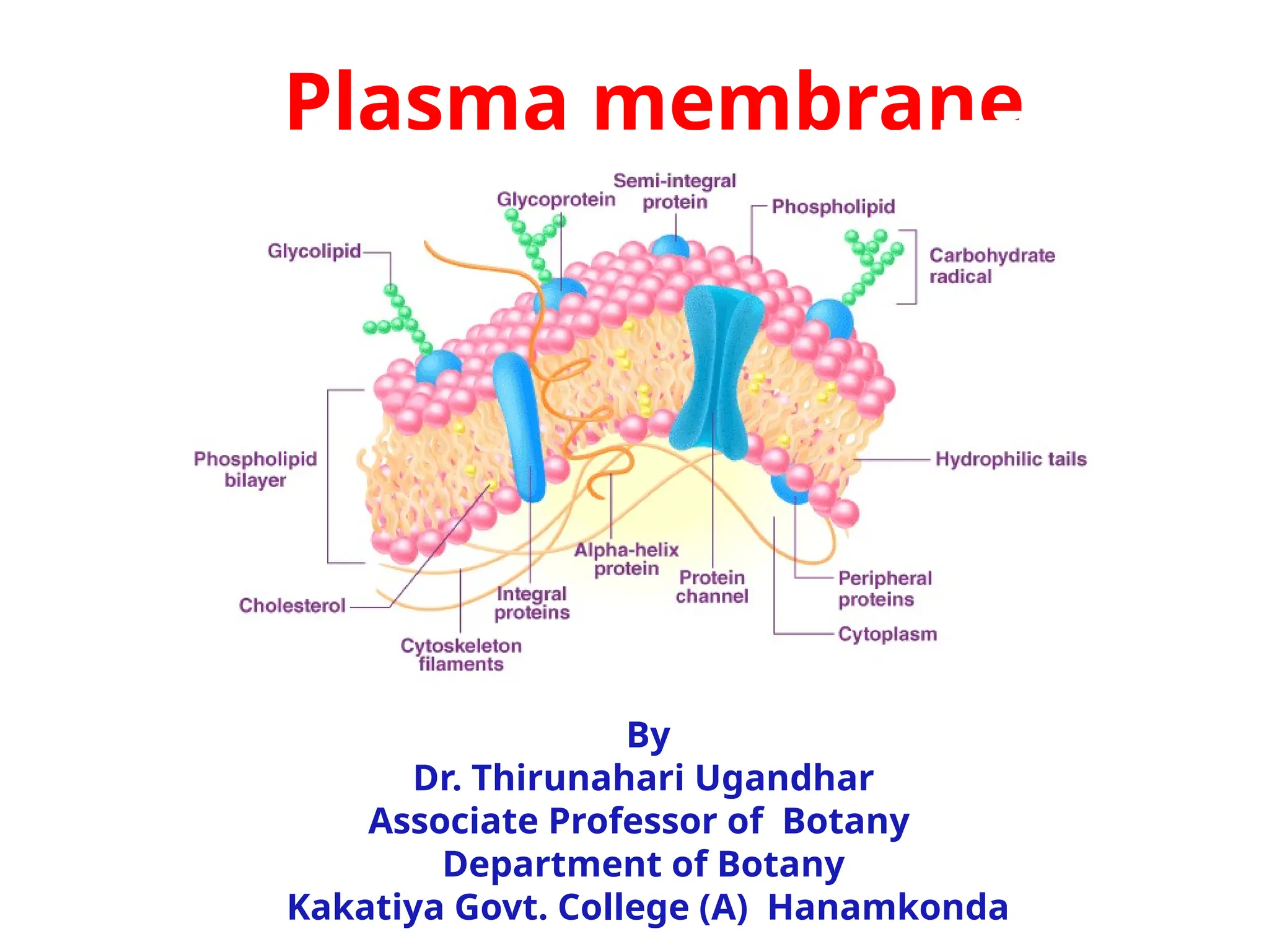
The plasma membrane is primarily
composed of a phospholipid bilayer,
which makes it semipermeable,
allowing selective movement of
substances in and out of the cell.
This helps regulate the transport of
nutrients, ions, and waste products.
5.
It also containsmembrane proteins, which serve
various functions:
• Integral proteins span the membrane and often
function as channels or transporters.
• Peripheral proteins are attached to the surface and
may act as enzymes or provide structural support.
The fluidity of the membrane is maintained by the
arrangement of phospholipids and the presence of
cholesterol (in animal cells), allowing it to function
across different temperatures.
Overall, the plasma membrane plays a critical role in:
• Controlling the movement of substances
• Facilitating communication between cells
• Providing structural support
• Helping maintain homeostasis
6.
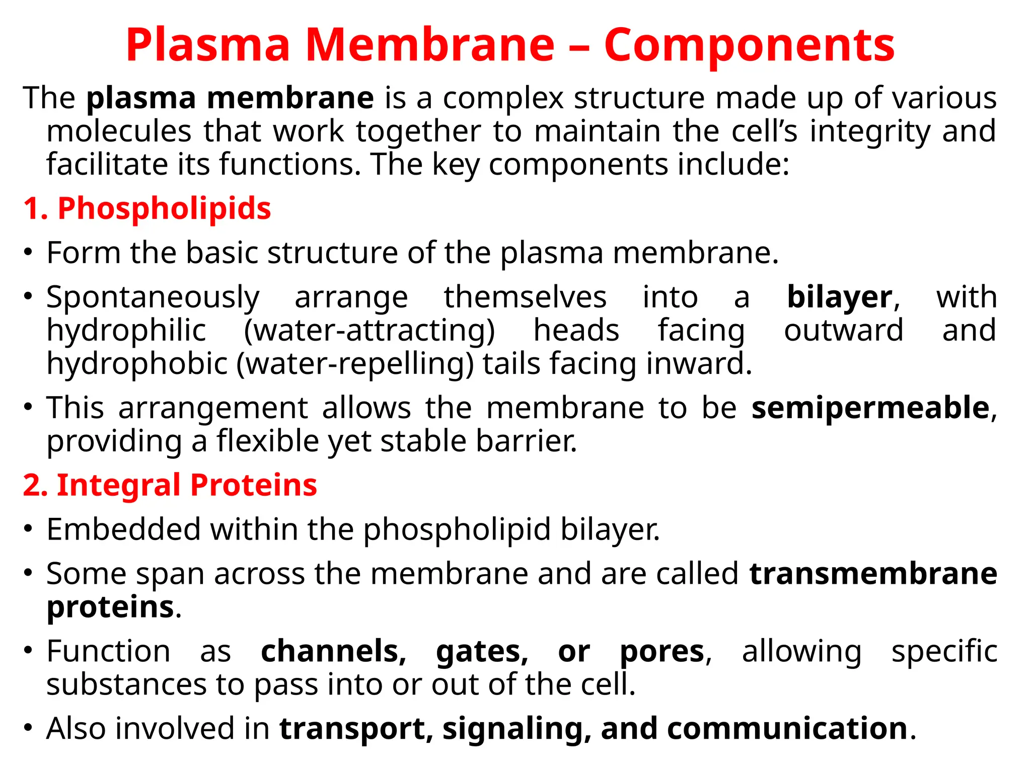
Plasma Membrane –Components
The plasma membrane is a complex structure made up of various
molecules that work together to maintain the cell’s integrity and
facilitate its functions. The key components include:
1. Phospholipids
• Form the basic structure of the plasma membrane.
• Spontaneously arrange themselves into a bilayer, with
hydrophilic (water-attracting) heads facing outward and
hydrophobic (water-repelling) tails facing inward.
• This arrangement allows the membrane to be semipermeable,
providing a flexible yet stable barrier.
2. Integral Proteins
• Embedded within the phospholipid bilayer.
• Some span across the membrane and are called transmembrane
proteins.
• Function as channels, gates, or pores, allowing specific
substances to pass into or out of the cell.
• Also involved in transport, signaling, and communication.
7.
3. Peripheral Proteins
•Attached to the outer or inner surfaces of the membrane.
• Not embedded in the lipid bilayer.
• Often act as enzymes, structural anchors, or part of
signaling pathways.
4. Cholesterol
• Interspersed between the phospholipid tails within the
bilayer.
• Helps to maintain membrane fluidity and stability,
especially across temperature changes.
5. Carbohydrates
• Attached to proteins or lipids on the extracellular surface
of the membrane, forming:
• Glycoproteins (carbohydrate + protein)
• Glycolipids (carbohydrate + lipid)
• Play a major role in cell recognition, signaling, and
adhesion.
8.
• Functions ofPlasma Membrane
• The plasma membrane acts as a physical barrier between the
cytoplasm and extracellular space and allows biochemical reactions
necessary for life to occur. The functions of plasma membrane are as
follows:
• Barrier: Separates cell contents from the external environment.
• Selective Permeability: Regulates the entry and exit of substances.
• Cell Communication: Contains receptor proteins for signal detection.
• Cell Cohesion: Adhesion proteins help cells stick together.
• Endocytosis & Exocytosis: Facilitates material transport in and out of
the cell.
• Homeostasis: Maintains internal balance by controlling molecule
movement.
• Environment Interaction: Governs interactions with surrounding
cells and molecules.
• Recognition: Displays unique patterns for cell identification.
• Flexibility: Allows cells to change shape and respond to the
environment.
• Supports Essential Functions: Essential for cell survival, growth, and
reproduction
9.
Structure of thePlasma Membrane (Biomembrane Structure)
The plasma membrane is described by the fluid mosaic model,
which means it is a flexible structure composed of lipids,
proteins, and carbohydrates that move fluidly within the layer.
Key Features:
• Selective Barrier: It is impermeable to ions and water-
soluble molecules, allowing only specific substances to pass
through via carrier proteins, transmembrane channels, or
pumps.
• Protein Function: Transmembrane proteins:
• Allow the passage of nutrients.
• Regulate ion concentration.
• Create an electrical potential across the membrane.
• Medical Relevance: A single amino acid mutation in a chloride
(Cl⁻) channel or plasma membrane pump can lead to disorders
such as cystic fibrosis.
• Lipid Content: Depending on location and function, lipids can
make up 20–80% of the membrane, with the remainder being
proteins.
10.
• Phospholipid BilayerStructure
• Composed of two layers of phospholipids arranged back-
to-back.
• Each phospholipid has:
• A hydrophilic (water-attracting) head faces outward
towards water inside and outside the cell.
• Two hydrophobic (water-repelling) tails face inward,
shielded from water.
• This unique arrangement forms a semipermeable
membrane that controls what enters and exits the cell.
• Phospholipids of the Plasma Membrane Phospholipids
are the most abundant lipids in the membrane and serve
both structural and signaling roles.
• Major Types in Animal Cells:
• Phosphatidylethanolamine (PE)Phosphatidylcholine (PC),
SphingomyelinPhosphatidylserine (PS)
11.
• Distribution inthe Bilayer:
• Inner Layer (cytoplasmic side): Rich in
phosphatidylserine (PS) and phosphatidylcholine (PE).
Outer Layer (extracellular side):
• Rich in sphingomyelin and phosphatidylcholine (PC).
This asymmetry of phospholipids is important for cell
function, signaling, and recognition.
• CholesterolInterspersed within the bilayer. Stabilizes
the membrane and maintains fluidity across
temperature changes.
• Conclusion plasma membrane is a dynamic and
essential structure, enabling the cell to maintain
homeostasis, interact with its environment, and
regulate internal conditions.
• Its unique composition of phospholipids, proteins,
carbohydrates, and cholesterol allows it to perform
these vital functions efficiently.
13.
• Molecular modelsof the Plasma membrane
• Various authors have given various models and concepts for the presence
and structure of the plasma membrane.
• But the Fluid Mosaic model proposed by Singer and Nicolson is the most
accepted of them all. In 1902, Overton proposed a basic model for the
transport of small neutral solutes.
• He experimented on numerous cells, both from plants and animals and
concluded that these special osmotic properties of living protoplasts are
due to the selective solubility mechanism of the membrane. He also
rightly guessed that the outer layer of the membrane contained fats and
sterols.
• Overton postulated that the plasma membrane is composed of a thin
layer of lipid.
• In 1926, Groter and Grendel conducted experiments on hemolyzed RBC
and concluded that the RBCs were covered by two layers of lipid molecules
over the entire cell surface.
• In 1931, Danielle and Harvey studied the surface tension of the cells.
Generally surface tension at the water-oil interface is around 0.01-0.015
newton/meter on the other hand the surface tension for cells is nil.
• This low surface tension for the cell is attributed to the presence of
proteins in the plasma membrane.
• The following are the molecular models of the plasma membrane
proposed by various biologists,
14.
• Sandwich model
•In 1935, Davison and Danielli proposed the sandwich or trilamellar model
for plasma membrane structure. According to this model, the plasma
membrane is a sheath-like structure composed of two lipid layers
sandwiched between continuous layers of proteins. The stability of the
membrane was maintained by the mutual attraction between
hydrocarbon chains of lipids and electrostatic forces between proteins
and lipid molecules.
• They also predicted the thickness of the lipid layer to be about 6.0 nm
and protein layer to be 1.0 nm. The total thickness was said to be around
8.0 nm. Finally electron micrograph studies also supported this model
proposed by Davison and Danielli
15.
• Unit membranemodel
• Later in 1959, Robertson proposed the unit membrane hypothesis,
which states that all cellular membranes have an identical
membrane structure.
• They named this identical membrane structure as unit membrane.
• According to this model, the unit membrane consists of a
bimolecular lipid leaflet packed in between the outer and inner
layers of protein.
16.
Fluid Mosaic Model
Finally,in 1972, Singer and Nicolson proposed the well-accepted Fluid Mosaic
model. As per this model, both lipids and proteins are distributed in a kind of
mosaic arrangement.
All the biological membranes are quasi-fluid structures in which lipids and
proteins can move.
In other words, the proteins are embedded in the lipid bilayer in such a way that
the proteins float in lipid sea. The surface of the lipid layers is interrupted by
randomly distributed protein molecules.
These proteins may either attach to the polar surface of the lipids or partially
penetrate the lipid bilayer. Some proteins are also found to be associated with the
sugar chains of glycoproteins.
17.
• Micellar modelof the Plasma membrane
• In 1963, Hilleir and Hoffman suggested that
biological membranes can have a non-lamellar
pattern. As per them, the plasma membrane has a
mosaic of globular subunits referred to as micelles
that are densely packed with a central core of lipid
molecules with a hydrophilic polar end.
• As lipid micelles tend spontaneously link, they are
probably building blocks for membranes. The
protein components of the membrane in this model
can establish a monolayer on either side of the
plane of lipid micelles.
• It is suggested that the gaps between the globular
micelles form water-filled pores, which are partially
lined by polar groups of micelles and partially by
polar groups of associate protein molecules.
19.
Model Name
Proposed By&
Year
Structure
Description
Key Features Limitations
1. Lipid and Lipid
Bilayer Model
Overton (1902),
Gorter & Grendel
(1925)
Suggested a
bilayer of lipids
based on
erythrocyte
membrane studies
- Double layer of
phospholipids
- Lacked explanation of
protein role
- Hydrophilic heads
face outward
- Did not explain
membrane permeability
and functionality
- Hydrophobic tails
inward
2. Davson–
Danielli Model
James Danielli &
Hugh Davson
(1935)
"Sandwich model" -
lipid bilayer
covered by protein
layers
- Lipid bilayer core
- Assumed static protein
layer
- Proteins coat
outer surfaces
- Could not explain
selective permeability
and protein movement
- First molecular-
level model of
membrane
3. Unit
Membrane
Model
J. David
Robertson (1953)
Trilaminar (three-
layered) structure
observed in
electron
micrographs
- Common
structure for all
membranes
- Assumed uniform
membrane thickness
- 2 dense protein
layers and 1 light
lipid layer
- Failed to explain
asymmetry and variety
among membranes
4. Fluid Mosaic S.J. Singer & G.L.
Dynamic model
with proteins
embedded in fluid
- Phospholipid
bilayer with
embedded proteins
- Most accepted and
updated model
- Proteins move
- Dynamic, accounts for
20.
Fill in theBlanks – Plasma Membrane Models (With
Answers)
1.The fluid mosaic model was proposed by __________ and __________
in 1972.
Answer: Singer, Nicolson
2.The plasma membrane is primarily composed of __________ and
__________.
Answer: phospholipids, proteins
3.The __________ heads of phospholipids face the aqueous
environment.
Answer: hydrophilic
4.The __________ tails of phospholipids face inward, away from water.
Answer: hydrophobic
5.The first suggestion of a lipid-based membrane came from
Overton in the year __________.
Answer: 1902
6.Gorter and Grendler proposed the lipid bilayer model in __________.
Answer: 1925
21.
•The Davson–Danielli modelis also known as the __________
model. Answer: sandwich
•In the Davson–Danielli model, the lipid bilayer is sandwiched
between two layers of __________. Answer: protein
•The unit membrane model was proposed by __________ in
1953. Answer: J. David Robertson
•The unit membrane shows a trilaminar structure, which
includes __________ dark bands and __________ light band.
Answer: two, one
•In the unit membrane model, the total thickness of the
membrane is about __________ Å. Answer: 75
•In the fluid mosaic model, membrane proteins can move
__________ within the lipid bilayer. Answer: lateral
22.
•The plasma membraneis __________ permeable, allowing
selective transport. Answer: selectively (or semi-)
•The lipid bilayer provides the membrane's basic __________
structure. Answer: fluid
•The outer protein layer in the unit membrane model is made of
__________ protein. Answer: mucoprotein
•The red cell membrane studies used by Gorter and Grendel
were conducted on __________. Answer: erythrocytes
•In the fluid mosaic model, cholesterol helps to maintain
membrane __________. Answer: fluidity
•The main limitation of the Davson–Danielli model was that it
could not explain __________ proteins. Answer:
transmembrane
23.
•The concept ofa universal membrane structure across
organelles was proposed in the __________ model. Answer:
unit membrane
•The plasma membrane typically ranges from __________ to
__________ nm in thickness. Answer: 5, 10
•The fluid mosaic model includes proteins, lipids,
carbohydrates, and __________.
Answer: cholesterol
•The inner mitochondrial membrane has a higher proportion of
__________ than lipids. Answer: proteins
•According to the fluid mosaic model, the membrane
resembles a __________ of various molecules. Answer:
mosaic
•The tails of phospholipids are composed of __________ fatty
acids. Answer: non-polar
•The lipid and lipid bilayer model was important because it laid
the __________ for later models. Answer: foundation