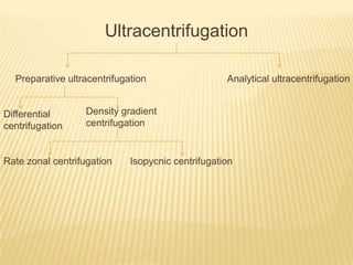
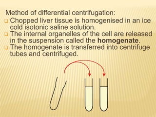
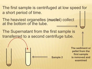
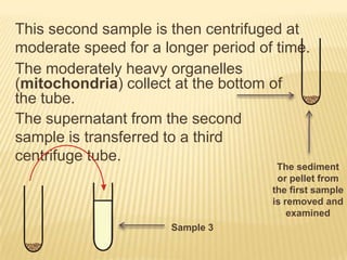
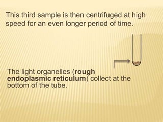
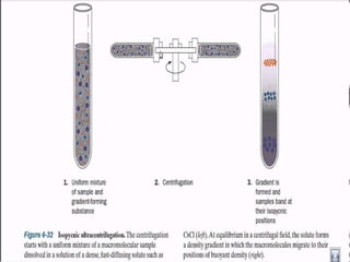
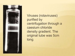
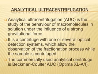
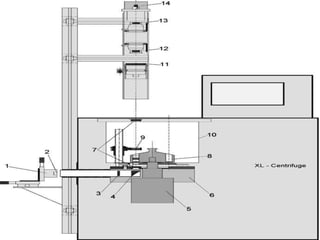
DIFFERENTIAL AND ULTRA
CENTRIFUGATION

By: Priya James
M.Pharm (Q.A.) 1ST Sem
School of Pharmaceutical Sciences,
RGPV, Bhopal.
CENTRIFUGATION
Centrifugation is a technique often
employed during isolation or analysis of
various cells, organelles and biopolymers,
dissolved or dispersed in biologically relevant
solvents.
 Biologists use the technique of centrifugation
to extract and isolate pure samples of
individual cell organelles for study.
 In this technique, the sample (comprising a
liquid phase and a solute) is placed in a
suitable vessel, and is spun in a centrifugal

THE FACTORS THAT DEFINE THE ENTIRE
SEDIMENTATION SYSTEM:
The rate at which the rotor spins.
 The length of time for which the centrifugal
force is applied.
 The solvent density.
 The system temperature.

Ultracentrifugation
Preparative ultracentrifugation

Differential
centrifugation

Analytical ultracentrifugation

Density gradient
centrifugation

Rate zonal centrifugation

Isopycnic centrifugation
PREPARATIVE CENTRIFUGATION
It aims to isolate and purify specific particles
such as sub. cellular organelles.
 It is mainly of two types:
 Differential centrifugation
 Density gradient centrifugation
 Molecules separate according to their
size, density and centrifugal force.

DIFFERENTIAL CENTRIFUGATION






Differential centrifugation, also termed
pelleting.
It fractionates particles according to their
weight.
The suspension of isolated organelles is
spun for different combination of speed and
time.
The lowest speed and shortest time
separates out the heaviest organelles and
high speed and longer time separating the
lightest organelles.
Method of differential centrifugation:
 Chopped liver tissue is homogenised in an ice
cold isotonic saline solution.
 The internal organelles of the cell are released
in the suspension called the homogenate.
 The homogenate is transferred into centrifuge
tubes and centrifuged.
The first sample is centrifuged at low speed for
a short period of time.
The heaviest organelles (nuclei) collect
at the bottom of the tube.
The Supernatant from the first sample is
transferred to a second centrifuge tube.

Sample 2

The sediment or
pellet from the
first sample
is removed and
examined
This second sample is then centrifuged at
moderate speed for a longer period of time.
The moderately heavy organelles
(mitochondria) collect at the bottom of
the tube.
The supernatant from the second
sample is transferred to a third
centrifuge tube.

The sediment
or pellet from
the first sample
is removed and
examined

Sample 3
This third sample is then centrifuged at high
speed for an even longer period of time.

The light organelles (rough
endoplasmic reticulum) collect at the
bottom of the tube.
DENSITY GRADIENT CENTRIFUGATION
Isolated organelles are separated on the
basis of their density.
 Solutions of increasing density, such as
sucrose solutions, are layered into a test
tube with the most concentrated solution at
the bottom of the tube.
 The suspension of isolated organelles is
pipetted onto the top of the most dilute
solution.
 As the tubes are spun, the organelles collect
in the layer which corresponds to their own
density.

GRADIENT MATERIAL
The properties of an ‘ideal’ solute for gradients
to be used in rate zonal or isopycnic
centrifugation:
 The solute should be stable in solution.
 It should be totally inert towards biological
materials
 It should not be absorbed in the UV or visible
range.
 It should be easily and completely removable
from the purified particles.
 It should be inexpensive and readily available.
RATE ZONAL CENTRIFUGATION
Particles are separated in terms of their
molecular mass.
 The sample solution is layered on top of the
density gradient, and under centrifugal force
the particles will begin sedimenting through
the gradient in distinct zones, each zone
consisting of particles characterized by the
same sedimentation rate.
 Force must be terminated before any of the
separated zones reaches the bottom of the
tube.

Photograph of an actual
tissue separation: sub
fractionation of a
rat hepatic light
mitochondrial fraction
by rate zonal
centrifugation using a
vertical rotor.
ISOPYCNIC CENTRIFUGATION
Isopycnic means "of the same density.
 Isopycnic centrifugation takes advantage of
differences in the effective buoyant densities of
particles- it does not rely on their
sedimentation rates and molecular mass.
 Each particle sediments only to the position in
the gradient at which its own effective buoyant
density is equal to the actual gradient density.

Viruses (rotaviruses)
purified by
centrifugation through a
caesium chloride
density gradient. The
original tube was 5cm
long.
ANALYTICAL ULTRACENTRIFUGATION
Analytical ultracentrifugation (AUC) is the
study of the behaviour of macromolecules in
solution under the influence of a strong
gravitational force.
 It is a centrifuge with one or several optical
detection systems, which allow the
observation of the fractionation process while
the sample is centrifuged.
 The commercially used analytical centrifuge
is Beckman-Coulter AUC (Optima XL-A/I),

Rotors:
The analytical rotors are built from one piece
of titanium.
 Depending on the rotor type, four or eight
holes are drilled into the titanium
Measuring
Cells:
The part of an analytical ultracentrifugation
system that is in direct contact to the sample
is the measuring cell.
AUC cells must fulfill two criteria:
They should not leak or distort even at high
centrifugal fields.
They should allow passage of light through
the cell.
Detectors:
UV/Vis

absorption optics is integrated in
modern XL-A/I analytical ultracentrifuges.
Fluorescence detector, is being recently used
commercially. This class of detectors makes
use of the sample’s specific light absorption or
fluorescence.
Mechanism:

Double-sector centerpiece.
The sample solution is
placed in one sector and a
sample of the solvent is
placed in the reference
sector.
The reference sector is
usually filled slightly more
than the sample sector, so
that the reference
meniscus does not
obscure the sample profile.
Light

from the flash lamp passes to hit the slit
assembly, where it is converted to
monochromatic light.
The lens-slit assembly moves stepwise as a
unit driven by an electrical motor, and allows
one to scan the sector of the measuring cell.
While the rotor spins, the lamp is triggered to
flash when the cell of interest passes the
optical path.
1957992 635169139917595000

1957992 635169139917595000

  • 1.
    DIFFERENTIAL AND ULTRA CENTRIFUGATION By:Priya James M.Pharm (Q.A.) 1ST Sem School of Pharmaceutical Sciences, RGPV, Bhopal.
  • 2.
    CENTRIFUGATION Centrifugation is atechnique often employed during isolation or analysis of various cells, organelles and biopolymers, dissolved or dispersed in biologically relevant solvents.  Biologists use the technique of centrifugation to extract and isolate pure samples of individual cell organelles for study.  In this technique, the sample (comprising a liquid phase and a solute) is placed in a suitable vessel, and is spun in a centrifugal 
  • 3.
    THE FACTORS THATDEFINE THE ENTIRE SEDIMENTATION SYSTEM: The rate at which the rotor spins.  The length of time for which the centrifugal force is applied.  The solvent density.  The system temperature. 
  • 4.
  • 5.
    PREPARATIVE CENTRIFUGATION It aimsto isolate and purify specific particles such as sub. cellular organelles.  It is mainly of two types:  Differential centrifugation  Density gradient centrifugation  Molecules separate according to their size, density and centrifugal force. 
  • 6.
    DIFFERENTIAL CENTRIFUGATION     Differential centrifugation,also termed pelleting. It fractionates particles according to their weight. The suspension of isolated organelles is spun for different combination of speed and time. The lowest speed and shortest time separates out the heaviest organelles and high speed and longer time separating the lightest organelles.
  • 7.
    Method of differentialcentrifugation:  Chopped liver tissue is homogenised in an ice cold isotonic saline solution.  The internal organelles of the cell are released in the suspension called the homogenate.  The homogenate is transferred into centrifuge tubes and centrifuged.
  • 8.
    The first sampleis centrifuged at low speed for a short period of time. The heaviest organelles (nuclei) collect at the bottom of the tube. The Supernatant from the first sample is transferred to a second centrifuge tube. Sample 2 The sediment or pellet from the first sample is removed and examined
  • 9.
    This second sampleis then centrifuged at moderate speed for a longer period of time. The moderately heavy organelles (mitochondria) collect at the bottom of the tube. The supernatant from the second sample is transferred to a third centrifuge tube. The sediment or pellet from the first sample is removed and examined Sample 3
  • 10.
    This third sampleis then centrifuged at high speed for an even longer period of time. The light organelles (rough endoplasmic reticulum) collect at the bottom of the tube.
  • 11.
    DENSITY GRADIENT CENTRIFUGATION Isolatedorganelles are separated on the basis of their density.  Solutions of increasing density, such as sucrose solutions, are layered into a test tube with the most concentrated solution at the bottom of the tube.  The suspension of isolated organelles is pipetted onto the top of the most dilute solution.  As the tubes are spun, the organelles collect in the layer which corresponds to their own density. 
  • 12.
    GRADIENT MATERIAL The propertiesof an ‘ideal’ solute for gradients to be used in rate zonal or isopycnic centrifugation:  The solute should be stable in solution.  It should be totally inert towards biological materials  It should not be absorbed in the UV or visible range.  It should be easily and completely removable from the purified particles.  It should be inexpensive and readily available.
  • 13.
    RATE ZONAL CENTRIFUGATION Particlesare separated in terms of their molecular mass.  The sample solution is layered on top of the density gradient, and under centrifugal force the particles will begin sedimenting through the gradient in distinct zones, each zone consisting of particles characterized by the same sedimentation rate.  Force must be terminated before any of the separated zones reaches the bottom of the tube. 
  • 15.
    Photograph of anactual tissue separation: sub fractionation of a rat hepatic light mitochondrial fraction by rate zonal centrifugation using a vertical rotor.
  • 16.
    ISOPYCNIC CENTRIFUGATION Isopycnic means"of the same density.  Isopycnic centrifugation takes advantage of differences in the effective buoyant densities of particles- it does not rely on their sedimentation rates and molecular mass.  Each particle sediments only to the position in the gradient at which its own effective buoyant density is equal to the actual gradient density. 
  • 18.
    Viruses (rotaviruses) purified by centrifugationthrough a caesium chloride density gradient. The original tube was 5cm long.
  • 19.
    ANALYTICAL ULTRACENTRIFUGATION Analytical ultracentrifugation(AUC) is the study of the behaviour of macromolecules in solution under the influence of a strong gravitational force.  It is a centrifuge with one or several optical detection systems, which allow the observation of the fractionation process while the sample is centrifuged.  The commercially used analytical centrifuge is Beckman-Coulter AUC (Optima XL-A/I), 
  • 21.
    Rotors: The analytical rotorsare built from one piece of titanium.  Depending on the rotor type, four or eight holes are drilled into the titanium
  • 22.
    Measuring Cells: The part ofan analytical ultracentrifugation system that is in direct contact to the sample is the measuring cell. AUC cells must fulfill two criteria: They should not leak or distort even at high centrifugal fields. They should allow passage of light through the cell.
  • 23.
    Detectors: UV/Vis absorption optics isintegrated in modern XL-A/I analytical ultracentrifuges. Fluorescence detector, is being recently used commercially. This class of detectors makes use of the sample’s specific light absorption or fluorescence.
  • 24.
    Mechanism: Double-sector centerpiece. The samplesolution is placed in one sector and a sample of the solvent is placed in the reference sector. The reference sector is usually filled slightly more than the sample sector, so that the reference meniscus does not obscure the sample profile.
  • 25.
    Light from the flashlamp passes to hit the slit assembly, where it is converted to monochromatic light. The lens-slit assembly moves stepwise as a unit driven by an electrical motor, and allows one to scan the sector of the measuring cell. While the rotor spins, the lamp is triggered to flash when the cell of interest passes the optical path.