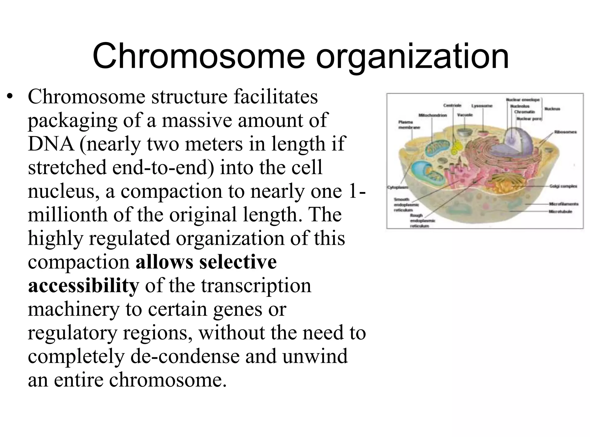
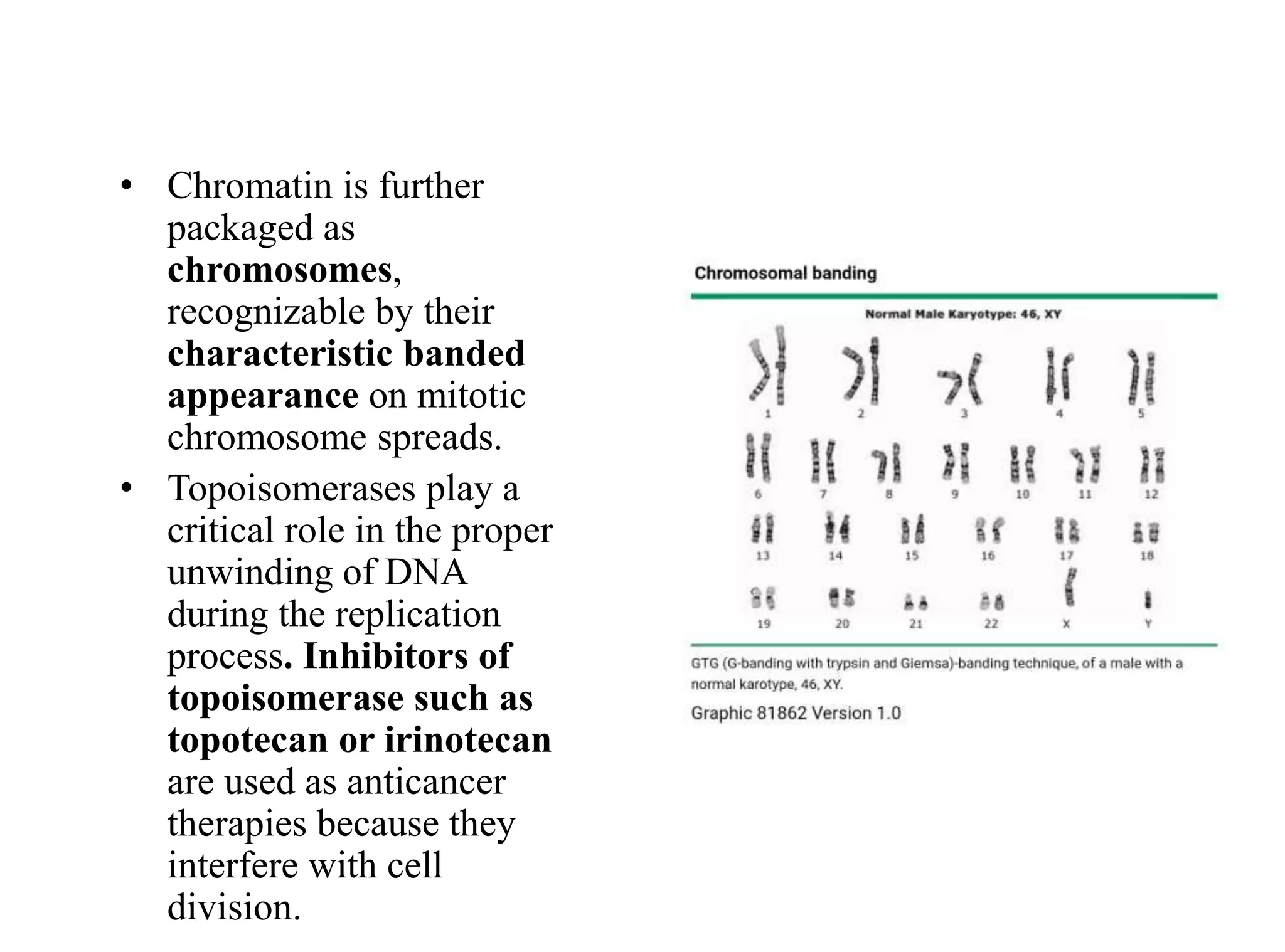
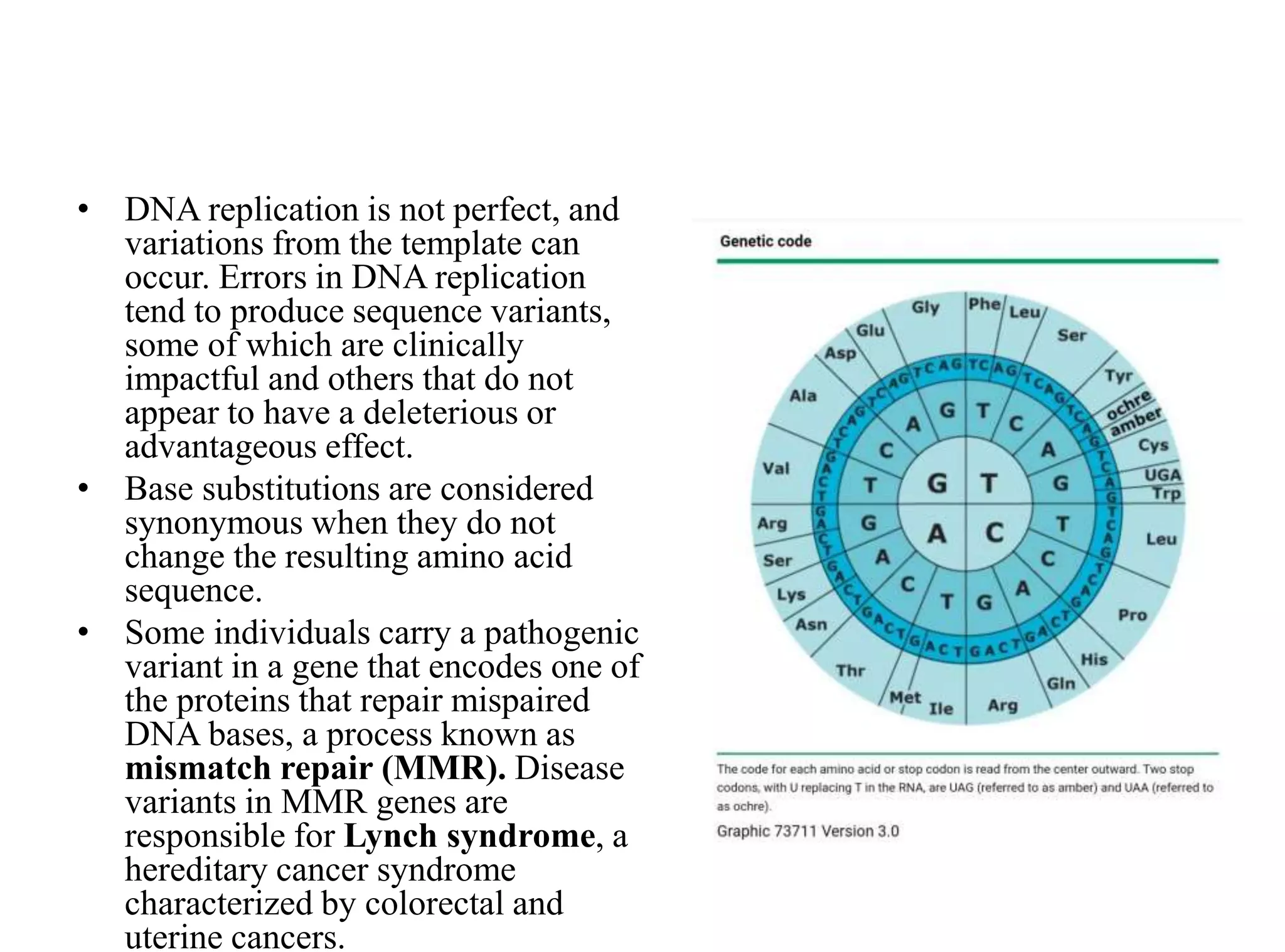
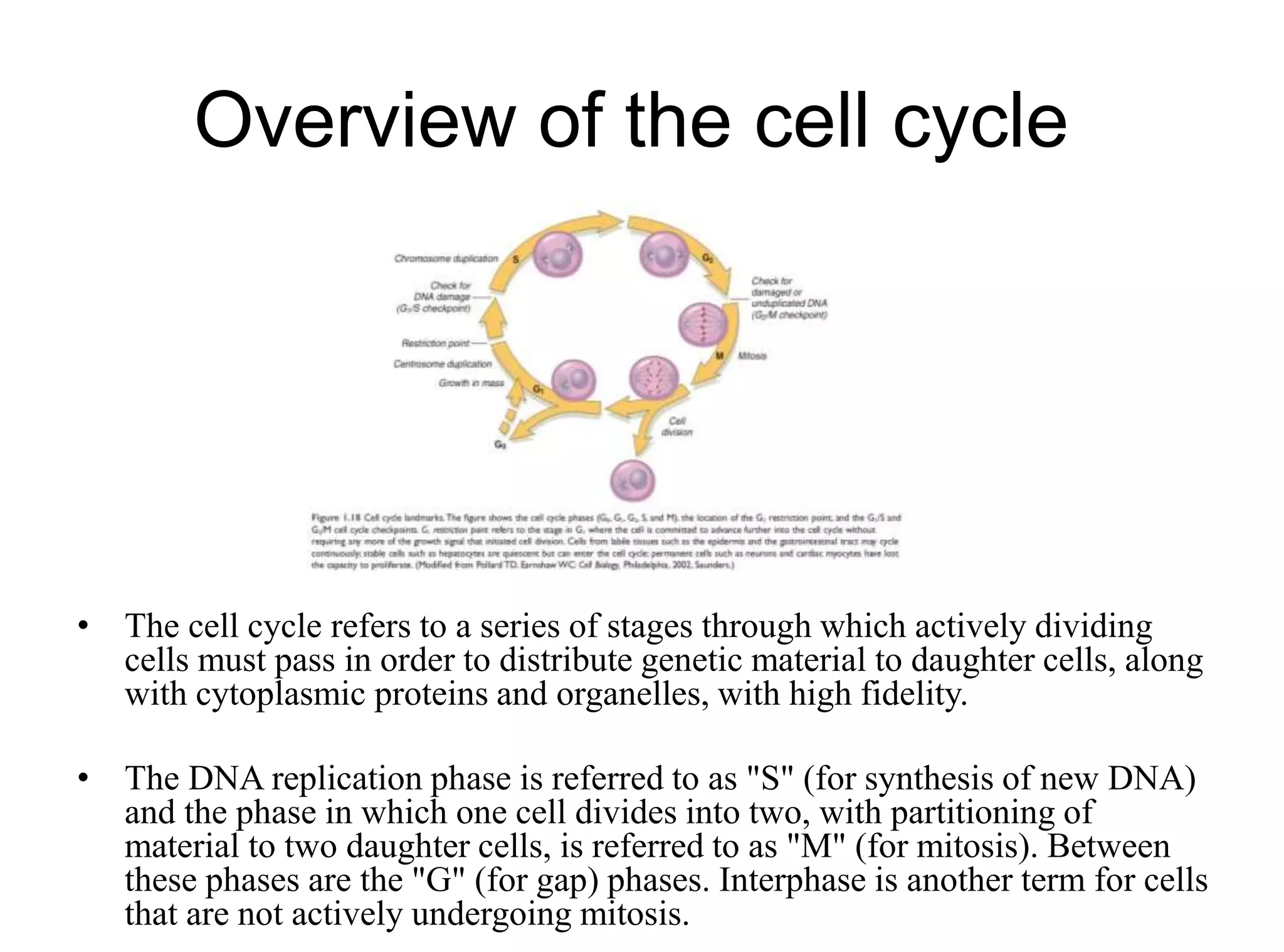
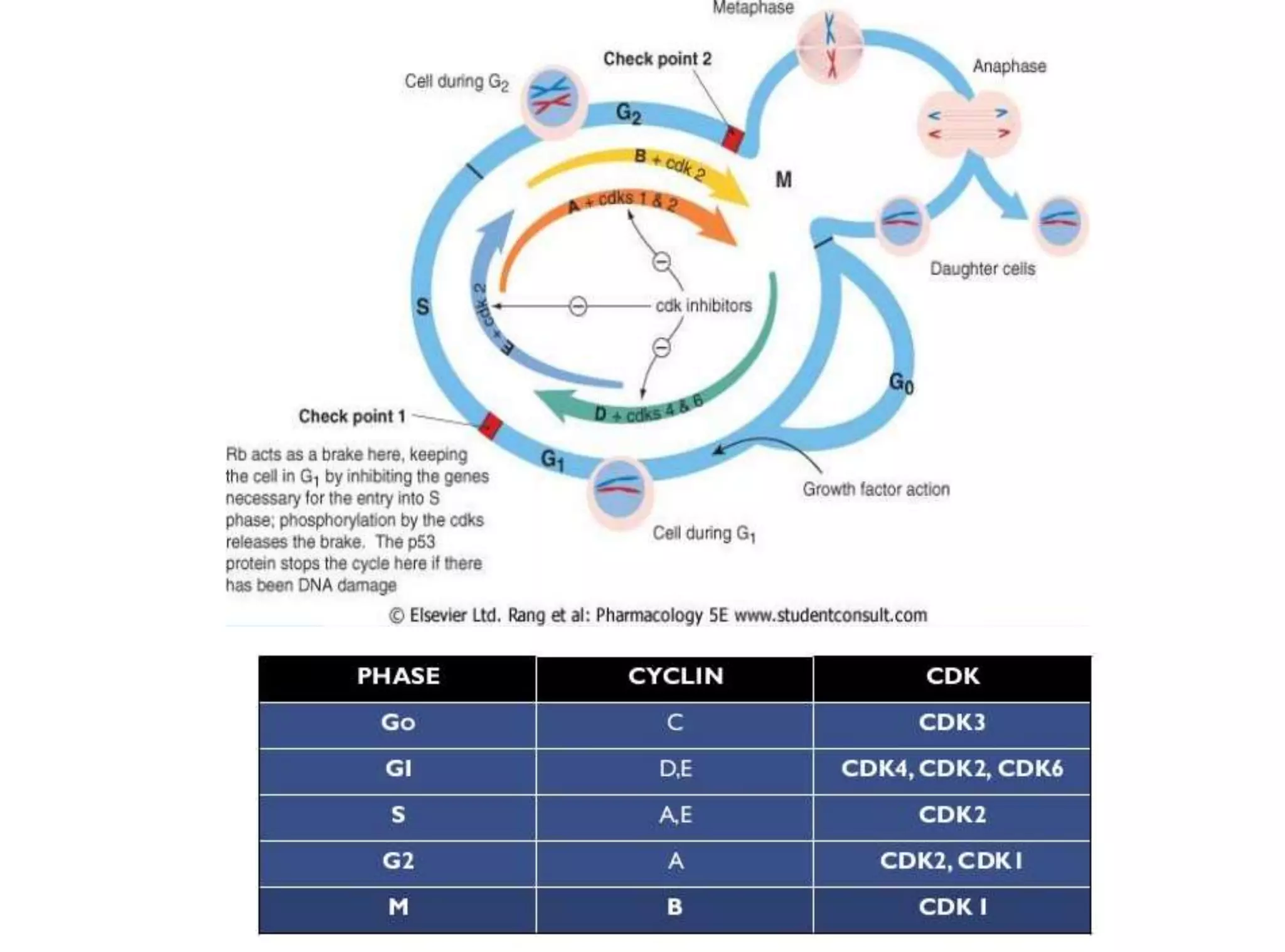
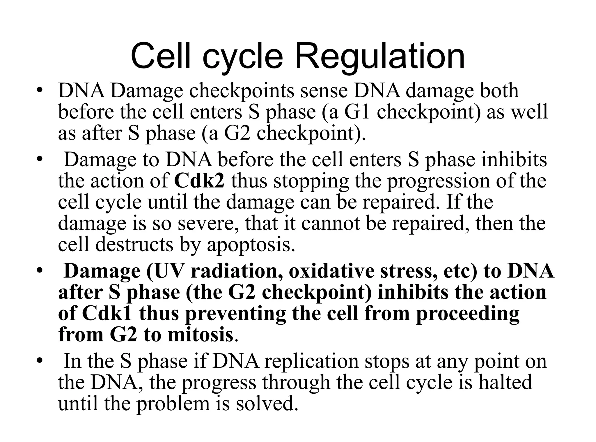
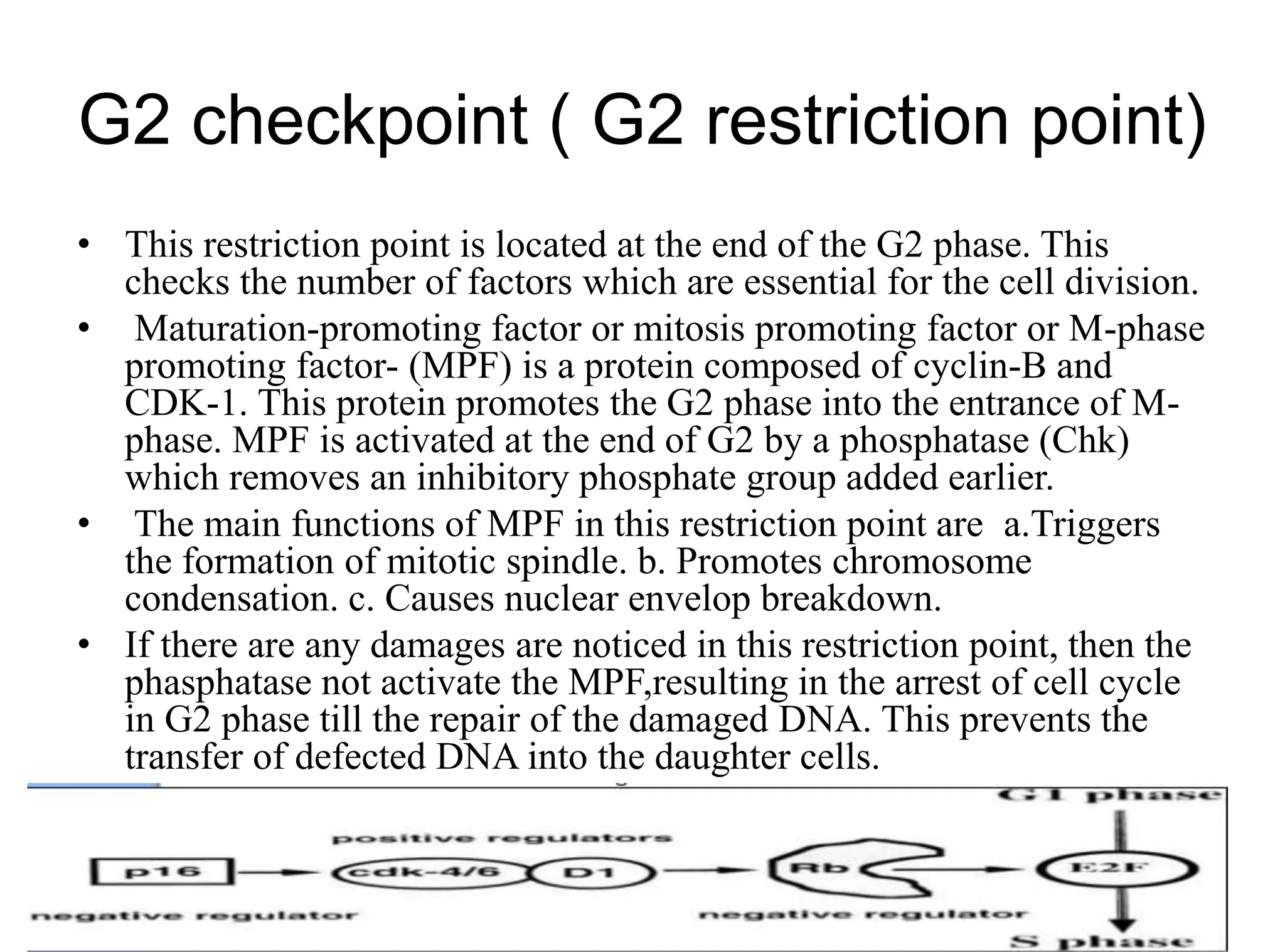
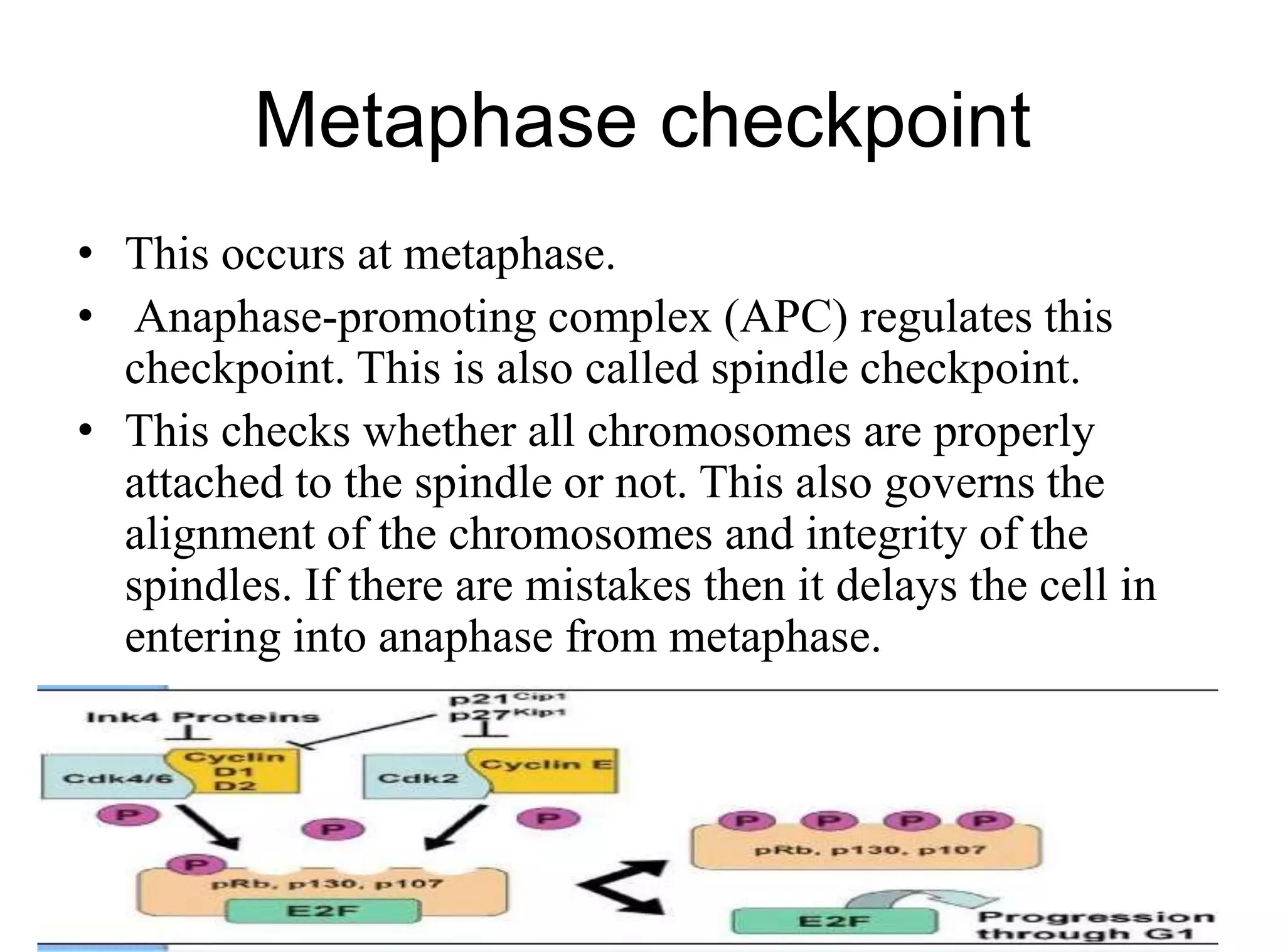
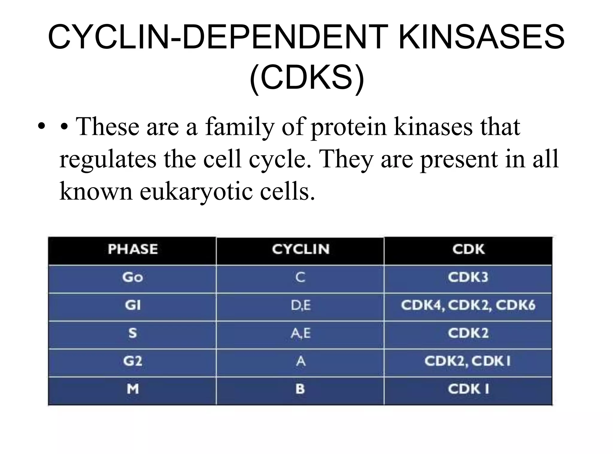
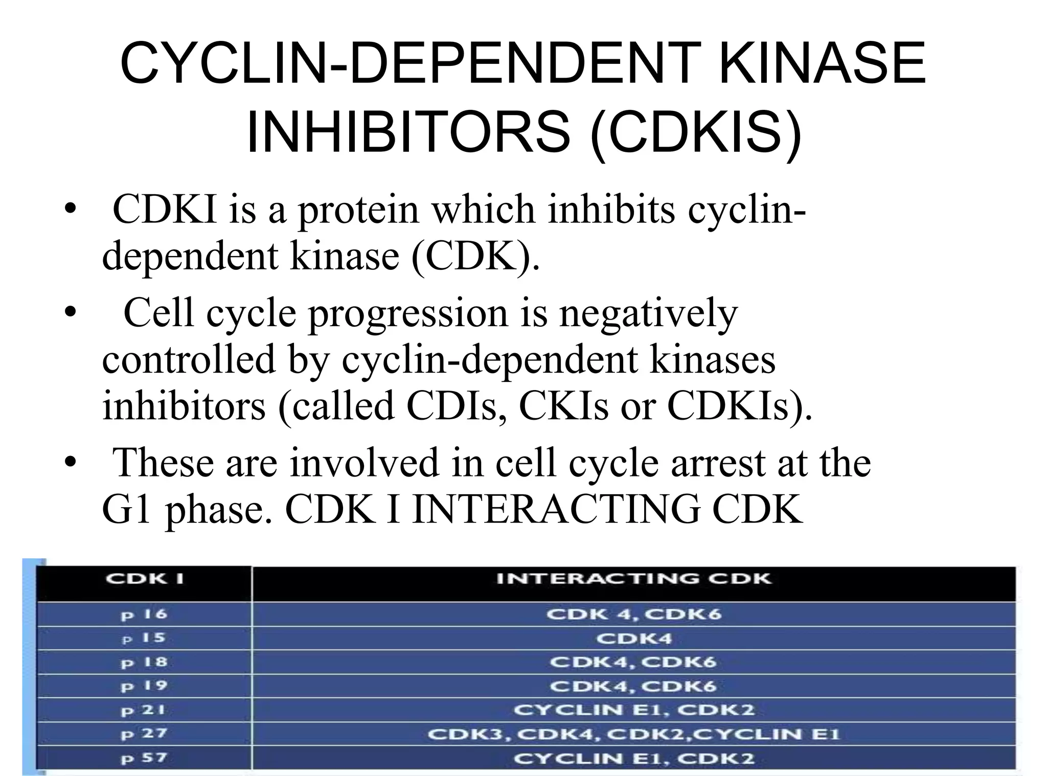
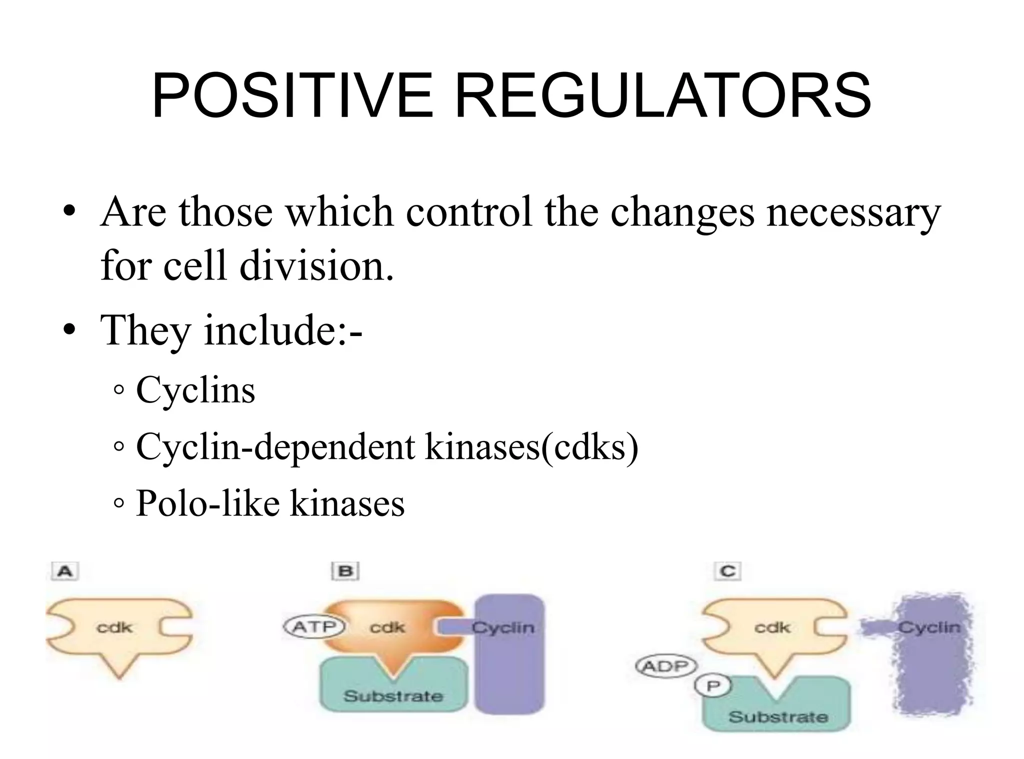
Cell cycle and its Regulation
Chromosome organization
• Chromosome structure facilitates
packaging of a massive amount of
DNA (nearly two meters in length if
stretched end-to-end) into the cell
nucleus, a compaction to nearly one 1-
millionth of the original length. The
highly regulated organization of this
compaction allows selective
accessibility of the transcription
machinery to certain genes or
regulatory regions, without the need to
completely de-condense and unwind
an entire chromosome.
Parts of the chromosome
Genomic DNA is organized into higher-order
structures, beginning with nucleosomes, which
consist of 147 base-pair stretches of double-
stranded DNA wrapped around a core of eight
histones. These nucleosomes in turn are further
organized into chromatin, which contains
additional regulatory proteins that facilitate tighter
packaging or unwinding, to allow access to
transcription factors and other protein complexes.
• Chromatin is further
packaged as
chromosomes,
recognizable by their
characteristic banded
appearance on mitotic
chromosome spreads.
• Topoisomerases play a
critical role in the proper
unwinding of DNA
during the replication
process. Inhibitors of
topoisomerase such as
topotecan or irinotecan
are used as anticancer
therapies because they
interfere with cell
division.
• DNA replication is not perfect, and
variations from the template can
occur. Errors in DNA replication
tend to produce sequence variants,
some of which are clinically
impactful and others that do not
appear to have a deleterious or
advantageous effect.
• Base substitutions are considered
synonymous when they do not
change the resulting amino acid
sequence.
• Some individuals carry a pathogenic
variant in a gene that encodes one of
the proteins that repair mispaired
DNA bases, a process known as
mismatch repair (MMR). Disease
variants in MMR genes are
responsible for Lynch syndrome, a
hereditary cancer syndrome
characterized by colorectal and
uterine cancers.
• Some individuals carry a pathogenic variant in a gene
that encodes one of the proteins involved in homologous
recombination, such as BRCA1 or BRCA2. Disease
variants in these homologous recombination repair
(HRR) genes compromise the ability of cells to repair
double-strand DNA breaks, increasing risks for
hereditary breast and ovarian cancer. In individuals with
HRR-deficient cancer, therapies that block the base
excision repair pathway can be useful therapeutically
because they can prevent correction of DNA damage,
leading to cell death.
Cell cycle
Overview of the cell cycle
• The cell cycle refers to a series of stages through which actively dividing
cells must pass in order to distribute genetic material to daughter cells, along
with cytoplasmic proteins and organelles, with high fidelity.
• The DNA replication phase is referred to as "S" (for synthesis of new DNA)
and the phase in which one cell divides into two, with partitioning of
material to two daughter cells, is referred to as "M" (for mitosis). Between
these phases are the "G" (for gap) phases. Interphase is another term for cells
that are not actively undergoing mitosis.
G0 Phase
• It is the resting phase.
• In these cells cyclin D is in decreased concentration.
• Rb protein is in hypo-phosphorylated (active form).
• Hence, holds the cell cycle at check point 1 by inhibiting the
expression of several transcription proteins(E2F) that codes
cyclins A and E necessary for cycle progression.
• Growth factor stimulation takes the G0 cells to G1 phase.
• In interphase the cell prepares itself to cycle. The term post-
mitotic is sometimes used to refer both quiescent and
senescent cells.
• Non-proliferative cells in eukaryotes generally enter the
quiescent Go state from G1 and may remain quiescent for
longer period of time or indefinitely (e.g.cardiac cells and
neurons).
• In multicellular eukaryotes, cells enter the Go phase from
the G1 phase and stop dividing. Some cells enter the Go
phase semi-permanently, e.g.some liver and kidney cells.
G1 Phase
• G stands for gap. It is the first phase within the interphase.
• It is also called the growth phase.
• This phase is the gap period between a mitotic phase and the S phase
of the cycle. Cell is preparing for S phase.
• This period starts immediately after division. The daughter cells
grow and increase in size during this phase.
• It is a longer phase. It lasts for even years. The nerve cells remain
permanently in G1 phase. Generally, this stage lasts for 25 to 50% of
the total interphase.
• During this phase 20 amino acids are formed, from which millions of
proteins and enzymes are formed, which are required in S phase.
During this phase mRNA, rRNA and tRNAs are formed. During this
phase new cell organelles are formed.
• Concentration of cyclin D increases, resulting in phosphorylation
and activation of necessary transcription proteins resulting in
synthesis of DNA polymerase DHFR etc which is required for
DNA replication.
• Formation of cyclin E complex is necessary for the transition
from G1 to S phase (check point 1)
• G1 phase consists of four sub-phases:
– Competence (g1a)
– Entry (g1b)
– Progression (g1c)
– Assembly (g1d)
• These sub-phases may be affected by limiting growth factors,
nutrient supply, temperature, and additional inhibiting factors.
• A rapidly dividing human cell which divides every 24h spends 9h in
G1 phase. The DNA in a G1 diploid eukaryotic cell is 2n, meaning
there are two sets of chromosomes present in the cell.The genetic
material exists as chromatin.
• There is a restriction point present at the end of G1 phase.
• Signals from extracellular growth factors are transducer in a typical
manner. Growth factors bind to receptors on the surface.
Accumulation of cyclin D is essential. Cyclins are a family of
proteins that control the progression of cells through the cell
cycle by activating cyclin-dependent kinases (Cdk) enzymes.
Cyclin D acts as a mitogenic signal sensor.
S PHASE
• Cyclin E/cdk and cyclin A/cdk regulate the processes in
phase S.
• By phosphorylating and activating proteins and enzymes
that are involved in DNA synthesis.
• S stands for synthesis. During this phase DNA synthesis
occurs. The DNA molecule duplicates. All the chromosomes
have been replicated. This period lasts for 35 to 40% of
interphase.
• During this phase, synthesis is completed as quickly as
possible due to the exposed base pairs may be destroyed
by the external proteins (drugs) or any mutagens (such as
nicotine).
• Cyclins, when bound with the dependent kinases such as
Cdk1 proteins form the maturation- promoting factor (MPF).
MPFs activate other proteins through phosphorylation. These
phosphorylated proteins, in turn, are responsible for specific
events during cell division such as microtubule formation
and chromatin remodeling.
G2 Phase
• Pre-mitotic phase.
• The G2 phase is the gap period between S-phase and mitotic
(M) phase of a cell cycle. It is the second growth phase. It is
a period of rapid cell growth and protein synthesis which the
cell ready itself for mitosis.
• The nucleus increases in volume. Metabolic activities
essential for cell division, occur during this phase. mRNA,
tRNA and rRNA synthesis also occur. It is not a necessary
part of the cell cycle.
• This phase has double the number of chromosomes.
• All the other cellular components are duplicated for the 2
daughter cells.
• Cyclin A/cdk and cyclin B/cdk complexes are
active which are necessary for the cell to enter
into M phase (check point 2)
M Phase (mitosis)
• G2 cells are divided into two daughter cells which may
enter the cycle again at G1 phase or come out of the cycle
to G0 phase.
• Mitosis is the distribution of the two sets of chromosomes
into two separate and equal nuclei.
• This is the division phase. During this phase the cell
divides. This phase has a short duration. A typical human
cell cycle has duration of 90h. Of these the M phase has
duration of 45 to 60min. This phase has two sub-phases
called karyokinesis and cytokinesis.
• Karyokinesis refers to the cell division of nucleus into two
daughter nuclei. It has 4 sub-stages, namely prophase,
metaphase, anaphase and telophase.
• Cytokinesis refers to the cell division of the cytoplasm
resulting in two daughter cells.
1) Pro phase- mitotic spindle formation
2) Meta phase- metaphase plate
3) Ana phase- mitotic apparatus
4) Telo phase- cytokinesis
Cell Cycle Regulation
Cell cycle Regulation
• DNA Damage checkpoints sense DNA damage both
before the cell enters S phase (a G1 checkpoint) as well
as after S phase (a G2 checkpoint).
• Damage to DNA before the cell enters S phase inhibits
the action of Cdk2 thus stopping the progression of the
cell cycle until the damage can be repaired. If the
damage is so severe, that it cannot be repaired, then the
cell destructs by apoptosis.
• Damage (UV radiation, oxidative stress, etc) to DNA
after S phase (the G2 checkpoint) inhibits the action
of Cdk1 thus preventing the cell from proceeding
from G2 to mitosis.
• In the S phase if DNA replication stops at any point on
the DNA, the progress through the cell cycle is halted
until the problem is solved.
• Ca breast: Abemaciclib is a potent small molecule cyclin-
dependent kinase (CDK) inhibitor which is selective for
CDK 4 and 6; it blocks retinoblastoma tumor suppressor
protein phosphorylation and prevents progression through
the cell cycle, resulting in arrest at the G1 phase (Sledge
2017). Abemaciclib either alone or in combination with
endocrine therapy has resulted in decreased tumor size.
• Palbociclib is a reversible small molecule cyclin-dependent
kinase (CDK) inhibitor which is selective for CDK 4 and 6.
CDKs have a role in regulating progression through the cell
cycle at the G1/S phase by blocking retinoblastoma (Rb)
hyperphosphorylation (Finn 2015). Palbociclib reduces
proliferation of breast cancer cell lines by preventing
progression from the G1 to the S cell cycle phase. The
combination of palbociclib with an antiestrogen provides for
increased inhibition of Rb phosphorylation, downstream
signaling, and tumor growth compared with each agent
alone.
•
• Cyclin dependent kinase (CDK) 4/6 inhibitors such as
abemaciclib and palbociclib have demonstrated activity in patients
with metastatic hormone receptor positive, HER2-negative breast
cancer, leading to interest in their use in the adjuvant setting as well.
In the monarchE trial, among over 5600 patients with high risk,
hormone receptor positive, HER2-negative early breast cancer, the
addition of abemaciclib to endocrine therapy resulted in absolute
improvements in invasive disease-free and distant relapse-free
survival by approximately 3 to 4 percent at two years. However, in
preliminary results of the PALLAS trial, the addition of palbociclib
to adjuvant endocrine therapy failed to improve outcomes. Pending
further data, use of CDK 4/6 inhibitors in the adjuvant setting
remains investigational.
• Ribociclib is a small molecule cyclin-dependent kinase (CDK)
inhibitor which is selective for CDK 4 and 6; it blocks retinoblastoma
protein phosphorylation and prevents progression through the cell
cycle, resulting in arrest at the G1 phase (Hortobagyi 2016). The
combination of ribociclib and an aromatase inhibitor causes
increased inhibition of tumor growth compared with each agent
alone. The combination of ribociclib and fulvestrant resulted in
tumor growth inhibition in estrogen receptor-positive breast cancer
models.
G1 checkpoint ( G1 restriction point)
• This checkpoint is present at the end of the G1 phase and before S phase.
• This checkpoint helps in taking the decision of whether the cell should divide, delay
division, or enter a resting stage (Go phase). If there are unfavourable conditions for
the cell division, then this restriction point restrict the progression to the next phase
by passing the cell to Go phase for an extended period of time.
• This restriction point is mainly controlled by the action of the CKI-p16 (CDK
inhibitor p16).The inhibited CDK not bind with cyclin D1, hence there is no cell
progression.
• Active cyclin D-cdk complexes phosphorylate retinoblastoma protein (pRb) in the
nucleus.
• Un-phosphorylated pRb acts as an inhibitor of G1 by preventing E2F-mediated
transcription.
• •Once pRb gets phosphorylated, E2F activates the transcription of cyclins E and A,
which then interacts with CDK2 to allow for G1 -S phase transition.
• This brings the cell to the end of the first checkpoint (unphosphorylated Rb inhibits
the E2F).
G2 checkpoint ( G2 restriction point)
• This restriction point is located at the end of the G2 phase. This
checks the number of factors which are essential for the cell division.
• Maturation-promoting factor or mitosis promoting factor or M-phase
promoting factor- (MPF) is a protein composed of cyclin-B and
CDK-1. This protein promotes the G2 phase into the entrance of M-
phase. MPF is activated at the end of G2 by a phosphatase (Chk)
which removes an inhibitory phosphate group added earlier.
• The main functions of MPF in this restriction point are a.Triggers
the formation of mitotic spindle. b. Promotes chromosome
condensation. c. Causes nuclear envelop breakdown.
• If there are any damages are noticed in this restriction point, then the
phasphatase not activate the MPF,resulting in the arrest of cell cycle
in G2 phase till the repair of the damaged DNA. This prevents the
transfer of defected DNA into the daughter cells.
Metaphase checkpoint
• This occurs at metaphase.
• Anaphase-promoting complex (APC) regulates this
checkpoint. This is also called spindle checkpoint.
• This checks whether all chromosomes are properly
attached to the spindle or not. This also governs the
alignment of the chromosomes and integrity of the
spindles. If there are mistakes then it delays the cell in
entering into anaphase from metaphase.
• Checkpoint controls function to ensure that chromosomes are intact and that
critical stages of the cell cycle are completed before the following stage is
initiated.
• The unreplicated-DNA checkpoint operates during S and G2 to prevent the
activation of MPF before DNA synthesis is complete by inhibiting the
activation of CDK1 by Cdc25C .
• The spindle-assembly checkpoint, which prevents premature initiation of
anaphase, utilizes Mad2 and other proteins to regulate theAPC specificity
factor Cdc20 that targets securin for polyubiquitination .
• The chromosome-segregation checkpoint prevents telophase and
cytokinesis until daughter chromosomes have been properly segregated,so
that the daughter cell has a full set of chromosomes.
• In the chromosome-segregation checkpoint, the small GTPase Tem1
controls the availability of Cdc14 phosphatase, which in turn activates the
APC specificity factor Cdh1 that targets B-type cyclins for degradation,
causing inactivation of MPF .
• The DNA-damage checkpoint arrests the cell cycle in response to DNA
damage until the damage is repaired. Three types of tumor-suppressor
proteins (ATM/ATR, Chk1/2, and p53) are critical to this checkpoint.
• Activation of the ATM or ATR protein kinases in response to DNA
damage due to UV light or –irradiation leads to arrest in G1 and the S
phase via a pathway that leads to loss of Cdc25A phosphatase
activity. A second pathway from activated ATM/R stabilizes p53,
which stimulates expression of p21CIP . Subsequent inhibition of
multiple CDK-cyclin complexes by p21CIP causes prolonged arrest
in G1 and G2 .
• In response to extensive DNA damage, p53 also activates genes that
induce apoptosis.
• The cell cycle is regulated by
• . 1. cyclin-dependent kinases (CDKs
• 2. cyclin-dependent kinase inhibitors (CDKIs).
• CYCLINS:
• Their concentration varies during the cell cycle. Cyclins are the
family of proteins which regulates the cell cycle.
• There are several types of cyclins that are active in different parts of
the cell cycle and causes phosphorylation of CDK.
• There are also several “orphan” cyclins for which no CDK partner
has been identified.
• • For example, cyclin F is an orphan cyclin that is essential for G2/M
transition.
• There are two main groups of cyclins.
• A. G1/S CYCLINS:
• Examples- Cyclin A,D and E.
• These cyclins are essential for the control of the cell cycle at the G1
/S transition.
• Cyclin A / CDK 2- active in S phase. Cyclin A binds to S phase CDK
2 and is required to progress through the S Phase. Cyclin A/CDK 2 is
inhibited by the complex p 21 CIP .
• The un-phophorylated form of Rb binds with E2F family of
transcription factors which controls expression of several genes
involved in cell cycle progression (example-cyclin-E).
• Rb acts as a repression, so in complex with E2F it prevents the
expression of E2F genes, and this inhibits the cell progression from
G1 to S phase.
• The binding of cyclin D/CDK4 and cyclin D/CDK 6 lead to partial
phosphorylation of Rb, by reducing its binding to E2F. The E2F gene
activates the expression of cyclin E bind with CDK 2 and causes
complete phosphorylation of Rb.This progresses the cell cycle from
G1 to S phase.
• G2/M CYCLINS:
• Example; cyclin B/ CDK1.
• These are essential for the control of the cell cycle at the G2
/M transition.
• G2/M cyclins accumulate steadily during G2 and are
abruptly destroyed as cells exit from mitosis (at the end of
the M-Phase).
• Cyclin B/ CDK1- regulates progression from G2 to M
Phase. Cyclin B is a mitotic cyclin. The amount of cyclin B
(which binds to CDK1) and the activity of cyclin B-CDK
complex rise through the cell cycle until mitosis.The
cyclinB-CDK1 complex is called maturation promoting
factor (MPF).
• There are two types of cyclin B.
• 1. CYCLIN B1/CDK1
This complex is involved in the early events of mitosis
such as chromosome condensation, nuclear envelop
breakdown. This complex is localized in the
microtubules.
• 2. CYCLIN B2/CDK1
 This complex is localized in the Golgi apparatus.
Cyclin B2 is also binds to a gene-transforming growth
factor (TGF beta receptor 2). This gene complex
transcribes several genes required for the cell
proliferation.
CYCLIN-DEPENDENT KINSASES
(CDKS)
• • These are a family of protein kinases that
regulates the cell cycle. They are present in all
known eukaryotic cells.
CYCLIN-DEPENDENT KINASE
INHIBITORS (CDKIS)
• CDKI is a protein which inhibits cyclin-
dependent kinase (CDK).
• Cell cycle progression is negatively
controlled by cyclin-dependent kinases
inhibitors (called CDIs, CKIs or CDKIs).
• These are involved in cell cycle arrest at the
G1 phase. CDK I INTERACTING CDK
POSITIVE REGULATORS
• Are those which control the changes necessary
for cell division.
• They include:-
◦ Cyclins
◦ Cyclin-dependent kinases(cdks)
◦ Polo-like kinases
NEGATIVE REGULATORS
• Are those which control the positive regulators.
They include :-
◦ Rb proteins
◦ P53 gene
◦ Inhibitors of cdks - which are of 2 types
Ink family (Inhibitors of kinases) P19 ,P15
CIP family (cdks inhibitory proteins) P21, P57
Rb protein
• Rb protein was initially identified as the product of the prototype
tumorsuppressor gene, RB.
• The products of tumor-suppressor genes function in various ways to
inhibit progression through the cell cycle (loss-of-function mutations
in RB are associated with the disease hereditary retinoblastoma. )
• A child with this disease inherits one normal RB+ allele from one
parentand one mutant RB- allele from the other. If the RB + allele in
any of the trillions of cells that make up the human body becomes
mutated to a RB-allele, then no functional protein is expressed and
the cell or one of its descendants is likely to become cancerous. Also,
in most human cancer cells Rb function is inactivated, either by
mutations in both alleles of RB, or by abnormal regulation of Rb
phosphorylation.
• Rb protein is one of the most significant substrates of mammalian
G1 cyclinCDK complexes. Phosphorylation of Rb protein at multiple
sites prevents its association with E2Fs, thereby permitting E2Fs to
activate transcription of genes required for entry into S phase.
• E2F stimulates its own expression and that of cyclin E and
CDK2, positive cross-regulation of E2F and cyclin E-CDK2
produces a rapid rise of both activities in late G1 .
• They accumulate, S-phase cyclin-CDK and mitotic cyclin-
CDK complexes maintain Rb protein in the phosphorylated
state throughout the S, G2 , and early M phases.
• After cells complete anaphase and enter early G1 or G0 , the
fall in cyclin-CDK levels leads to dephosphorylation of Rb
by unopposed phosphatases. As a consequence,
hypophosphorylated Rb is available to inhibit E2F activity
during early G1 of the next cycle and in G0-arrested cells.
• Unphosphorylated Rb protein binds to E2Fs, converting
them into transcriptional repressors.
• Phosphorylation of Rb by cyclin D-CDK4/6 in mid G1
liberates E2Fs to activate transcription of genes encoding
cyclin E, CDK2, and other proteins required for the S phase.
p53 PROTEIN
• DNA damage leads to the activation of the
gene regulatory protein p53, which
stimulates the transcription of several
genes.
• One of these encodes a CKI proteins p21,
which binds to G1 /S-Cdk and S-Cdk and
inhibits their activites, thereby helping to
block entry into S phase.
• In a normal cell p53 is inactivated by its
negative regulator, mdm2. Upon DNA
damage or other stresses, various pathways
will lead to the dissociation of the p53 and
mdm2 complex. Once activated, p53 will
induce a cell cycle arrest to allow either
repair and survival of the cell or apoptosis
to discard the damaged cell. How p53
makes this choice is currently unknown.
INHIBITORS OF CDKS
• The activities of mammalian cyclin-CDK complexes also are regulated by
CDK inhibitors (CIPs), which bind to and inhibit each of the mammalian
cyclin-CDK complexes, and INK4 proteins, which block passage through
G1 by specifically inhibiting CDK4 and CDK6.
CIP FAMILY (CDKS INHIBITORY PROTEINS)
• Three related CIPs—p21CIP, p27KIP2 , and p57KIP2—inhibit cyclin A-
CDK2 activity and must be degraded before DNA replication can begin.
• p21CIP plays a role in the response of mammalian cells to DNA damage.
INK FAMILY (INHIBITORS OF KINASES)
• A second class of cyclin-CDK inhibitors called INK4s (inhibitors of kinase
4) includes several small, closely related proteins that interact only with
CDK4 and CDK6 and thus function specifically in controlling the mid-G1
phase. •
• Binding of INK4s to CDK4/6 blocks their interaction with cyclin D and
hence their protein kinase activity. The resulting decreased phosphorylation
of Rb protein prevents transcriptional activation by E2Fs and entry into the
S phase. One INK4 called p16 is a tumor suppressor.
Cell cycle regulation in
Radiotherapy
Radio-Biology:
DNA is target for radiation cell kill.
Cells which are actively proliferating and poorly
differentiated are radiosensitive.
Cells are sensitive at or close to mitosis.
Late S Phase –Radioresistant (Because repair
occurs)
Early G1 Phase – Radioresistant
Late G1 – Radiosensitive(The defective cell
attempts to multiply)
G2M Phase – Highly Radiosensitive
• The functioning of Molecular checkpoint genes of
the cell cycle determines the radiosensitivity of the
cell.
• In general normal cells, has good check point
gene function which is lacking in tumour cells.
• Hence when given sufficient time for the repair,
the cells repopulate. This is the reason radiation
treatment is fractionated and given over the period
(for normal cell repair process).
• But when abnormally interrupted, tumour cells
also gain the repair capability.
• 4R’s of Radiobiology:
1. Reoxygenation (of tumor cells),
2. Reassortment (of tumor cells into radiosensitive phase
of cell cycle),
3. Repair (of normal cells),
4. Repopulation (of normal cells)
• Radiosensitivity (of tumor cells) is considered to
be 5th R of the radiobiology.
• Fractionation in radiotherapy is based on the
principle of 4 R’s of Radiobiology.
Thank you

Cell cycle and its regulation

  • 1.
    Cell cycle andits Regulation
  • 2.
    Chromosome organization • Chromosomestructure facilitates packaging of a massive amount of DNA (nearly two meters in length if stretched end-to-end) into the cell nucleus, a compaction to nearly one 1- millionth of the original length. The highly regulated organization of this compaction allows selective accessibility of the transcription machinery to certain genes or regulatory regions, without the need to completely de-condense and unwind an entire chromosome.
  • 3.
    Parts of thechromosome Genomic DNA is organized into higher-order structures, beginning with nucleosomes, which consist of 147 base-pair stretches of double- stranded DNA wrapped around a core of eight histones. These nucleosomes in turn are further organized into chromatin, which contains additional regulatory proteins that facilitate tighter packaging or unwinding, to allow access to transcription factors and other protein complexes.
  • 5.
    • Chromatin isfurther packaged as chromosomes, recognizable by their characteristic banded appearance on mitotic chromosome spreads. • Topoisomerases play a critical role in the proper unwinding of DNA during the replication process. Inhibitors of topoisomerase such as topotecan or irinotecan are used as anticancer therapies because they interfere with cell division.
  • 6.
    • DNA replicationis not perfect, and variations from the template can occur. Errors in DNA replication tend to produce sequence variants, some of which are clinically impactful and others that do not appear to have a deleterious or advantageous effect. • Base substitutions are considered synonymous when they do not change the resulting amino acid sequence. • Some individuals carry a pathogenic variant in a gene that encodes one of the proteins that repair mispaired DNA bases, a process known as mismatch repair (MMR). Disease variants in MMR genes are responsible for Lynch syndrome, a hereditary cancer syndrome characterized by colorectal and uterine cancers.
  • 7.
    • Some individualscarry a pathogenic variant in a gene that encodes one of the proteins involved in homologous recombination, such as BRCA1 or BRCA2. Disease variants in these homologous recombination repair (HRR) genes compromise the ability of cells to repair double-strand DNA breaks, increasing risks for hereditary breast and ovarian cancer. In individuals with HRR-deficient cancer, therapies that block the base excision repair pathway can be useful therapeutically because they can prevent correction of DNA damage, leading to cell death.
  • 8.
  • 10.
    Overview of thecell cycle • The cell cycle refers to a series of stages through which actively dividing cells must pass in order to distribute genetic material to daughter cells, along with cytoplasmic proteins and organelles, with high fidelity. • The DNA replication phase is referred to as "S" (for synthesis of new DNA) and the phase in which one cell divides into two, with partitioning of material to two daughter cells, is referred to as "M" (for mitosis). Between these phases are the "G" (for gap) phases. Interphase is another term for cells that are not actively undergoing mitosis.
  • 11.
    G0 Phase • Itis the resting phase. • In these cells cyclin D is in decreased concentration. • Rb protein is in hypo-phosphorylated (active form). • Hence, holds the cell cycle at check point 1 by inhibiting the expression of several transcription proteins(E2F) that codes cyclins A and E necessary for cycle progression. • Growth factor stimulation takes the G0 cells to G1 phase. • In interphase the cell prepares itself to cycle. The term post- mitotic is sometimes used to refer both quiescent and senescent cells. • Non-proliferative cells in eukaryotes generally enter the quiescent Go state from G1 and may remain quiescent for longer period of time or indefinitely (e.g.cardiac cells and neurons). • In multicellular eukaryotes, cells enter the Go phase from the G1 phase and stop dividing. Some cells enter the Go phase semi-permanently, e.g.some liver and kidney cells.
  • 12.
    G1 Phase • Gstands for gap. It is the first phase within the interphase. • It is also called the growth phase. • This phase is the gap period between a mitotic phase and the S phase of the cycle. Cell is preparing for S phase. • This period starts immediately after division. The daughter cells grow and increase in size during this phase. • It is a longer phase. It lasts for even years. The nerve cells remain permanently in G1 phase. Generally, this stage lasts for 25 to 50% of the total interphase. • During this phase 20 amino acids are formed, from which millions of proteins and enzymes are formed, which are required in S phase. During this phase mRNA, rRNA and tRNAs are formed. During this phase new cell organelles are formed. • Concentration of cyclin D increases, resulting in phosphorylation and activation of necessary transcription proteins resulting in synthesis of DNA polymerase DHFR etc which is required for DNA replication. • Formation of cyclin E complex is necessary for the transition from G1 to S phase (check point 1)
  • 13.
    • G1 phaseconsists of four sub-phases: – Competence (g1a) – Entry (g1b) – Progression (g1c) – Assembly (g1d) • These sub-phases may be affected by limiting growth factors, nutrient supply, temperature, and additional inhibiting factors. • A rapidly dividing human cell which divides every 24h spends 9h in G1 phase. The DNA in a G1 diploid eukaryotic cell is 2n, meaning there are two sets of chromosomes present in the cell.The genetic material exists as chromatin. • There is a restriction point present at the end of G1 phase. • Signals from extracellular growth factors are transducer in a typical manner. Growth factors bind to receptors on the surface. Accumulation of cyclin D is essential. Cyclins are a family of proteins that control the progression of cells through the cell cycle by activating cyclin-dependent kinases (Cdk) enzymes. Cyclin D acts as a mitogenic signal sensor.
  • 15.
    S PHASE • CyclinE/cdk and cyclin A/cdk regulate the processes in phase S. • By phosphorylating and activating proteins and enzymes that are involved in DNA synthesis. • S stands for synthesis. During this phase DNA synthesis occurs. The DNA molecule duplicates. All the chromosomes have been replicated. This period lasts for 35 to 40% of interphase. • During this phase, synthesis is completed as quickly as possible due to the exposed base pairs may be destroyed by the external proteins (drugs) or any mutagens (such as nicotine). • Cyclins, when bound with the dependent kinases such as Cdk1 proteins form the maturation- promoting factor (MPF). MPFs activate other proteins through phosphorylation. These phosphorylated proteins, in turn, are responsible for specific events during cell division such as microtubule formation and chromatin remodeling.
  • 16.
    G2 Phase • Pre-mitoticphase. • The G2 phase is the gap period between S-phase and mitotic (M) phase of a cell cycle. It is the second growth phase. It is a period of rapid cell growth and protein synthesis which the cell ready itself for mitosis. • The nucleus increases in volume. Metabolic activities essential for cell division, occur during this phase. mRNA, tRNA and rRNA synthesis also occur. It is not a necessary part of the cell cycle. • This phase has double the number of chromosomes. • All the other cellular components are duplicated for the 2 daughter cells. • Cyclin A/cdk and cyclin B/cdk complexes are active which are necessary for the cell to enter into M phase (check point 2)
  • 17.
    M Phase (mitosis) •G2 cells are divided into two daughter cells which may enter the cycle again at G1 phase or come out of the cycle to G0 phase. • Mitosis is the distribution of the two sets of chromosomes into two separate and equal nuclei. • This is the division phase. During this phase the cell divides. This phase has a short duration. A typical human cell cycle has duration of 90h. Of these the M phase has duration of 45 to 60min. This phase has two sub-phases called karyokinesis and cytokinesis. • Karyokinesis refers to the cell division of nucleus into two daughter nuclei. It has 4 sub-stages, namely prophase, metaphase, anaphase and telophase. • Cytokinesis refers to the cell division of the cytoplasm resulting in two daughter cells. 1) Pro phase- mitotic spindle formation 2) Meta phase- metaphase plate 3) Ana phase- mitotic apparatus 4) Telo phase- cytokinesis
  • 18.
  • 20.
    Cell cycle Regulation •DNA Damage checkpoints sense DNA damage both before the cell enters S phase (a G1 checkpoint) as well as after S phase (a G2 checkpoint). • Damage to DNA before the cell enters S phase inhibits the action of Cdk2 thus stopping the progression of the cell cycle until the damage can be repaired. If the damage is so severe, that it cannot be repaired, then the cell destructs by apoptosis. • Damage (UV radiation, oxidative stress, etc) to DNA after S phase (the G2 checkpoint) inhibits the action of Cdk1 thus preventing the cell from proceeding from G2 to mitosis. • In the S phase if DNA replication stops at any point on the DNA, the progress through the cell cycle is halted until the problem is solved.
  • 21.
    • Ca breast:Abemaciclib is a potent small molecule cyclin- dependent kinase (CDK) inhibitor which is selective for CDK 4 and 6; it blocks retinoblastoma tumor suppressor protein phosphorylation and prevents progression through the cell cycle, resulting in arrest at the G1 phase (Sledge 2017). Abemaciclib either alone or in combination with endocrine therapy has resulted in decreased tumor size. • Palbociclib is a reversible small molecule cyclin-dependent kinase (CDK) inhibitor which is selective for CDK 4 and 6. CDKs have a role in regulating progression through the cell cycle at the G1/S phase by blocking retinoblastoma (Rb) hyperphosphorylation (Finn 2015). Palbociclib reduces proliferation of breast cancer cell lines by preventing progression from the G1 to the S cell cycle phase. The combination of palbociclib with an antiestrogen provides for increased inhibition of Rb phosphorylation, downstream signaling, and tumor growth compared with each agent alone. •
  • 22.
    • Cyclin dependentkinase (CDK) 4/6 inhibitors such as abemaciclib and palbociclib have demonstrated activity in patients with metastatic hormone receptor positive, HER2-negative breast cancer, leading to interest in their use in the adjuvant setting as well. In the monarchE trial, among over 5600 patients with high risk, hormone receptor positive, HER2-negative early breast cancer, the addition of abemaciclib to endocrine therapy resulted in absolute improvements in invasive disease-free and distant relapse-free survival by approximately 3 to 4 percent at two years. However, in preliminary results of the PALLAS trial, the addition of palbociclib to adjuvant endocrine therapy failed to improve outcomes. Pending further data, use of CDK 4/6 inhibitors in the adjuvant setting remains investigational. • Ribociclib is a small molecule cyclin-dependent kinase (CDK) inhibitor which is selective for CDK 4 and 6; it blocks retinoblastoma protein phosphorylation and prevents progression through the cell cycle, resulting in arrest at the G1 phase (Hortobagyi 2016). The combination of ribociclib and an aromatase inhibitor causes increased inhibition of tumor growth compared with each agent alone. The combination of ribociclib and fulvestrant resulted in tumor growth inhibition in estrogen receptor-positive breast cancer models.
  • 23.
    G1 checkpoint (G1 restriction point) • This checkpoint is present at the end of the G1 phase and before S phase. • This checkpoint helps in taking the decision of whether the cell should divide, delay division, or enter a resting stage (Go phase). If there are unfavourable conditions for the cell division, then this restriction point restrict the progression to the next phase by passing the cell to Go phase for an extended period of time. • This restriction point is mainly controlled by the action of the CKI-p16 (CDK inhibitor p16).The inhibited CDK not bind with cyclin D1, hence there is no cell progression. • Active cyclin D-cdk complexes phosphorylate retinoblastoma protein (pRb) in the nucleus. • Un-phosphorylated pRb acts as an inhibitor of G1 by preventing E2F-mediated transcription. • •Once pRb gets phosphorylated, E2F activates the transcription of cyclins E and A, which then interacts with CDK2 to allow for G1 -S phase transition. • This brings the cell to the end of the first checkpoint (unphosphorylated Rb inhibits the E2F).
  • 24.
    G2 checkpoint (G2 restriction point) • This restriction point is located at the end of the G2 phase. This checks the number of factors which are essential for the cell division. • Maturation-promoting factor or mitosis promoting factor or M-phase promoting factor- (MPF) is a protein composed of cyclin-B and CDK-1. This protein promotes the G2 phase into the entrance of M- phase. MPF is activated at the end of G2 by a phosphatase (Chk) which removes an inhibitory phosphate group added earlier. • The main functions of MPF in this restriction point are a.Triggers the formation of mitotic spindle. b. Promotes chromosome condensation. c. Causes nuclear envelop breakdown. • If there are any damages are noticed in this restriction point, then the phasphatase not activate the MPF,resulting in the arrest of cell cycle in G2 phase till the repair of the damaged DNA. This prevents the transfer of defected DNA into the daughter cells.
  • 25.
    Metaphase checkpoint • Thisoccurs at metaphase. • Anaphase-promoting complex (APC) regulates this checkpoint. This is also called spindle checkpoint. • This checks whether all chromosomes are properly attached to the spindle or not. This also governs the alignment of the chromosomes and integrity of the spindles. If there are mistakes then it delays the cell in entering into anaphase from metaphase.
  • 27.
    • Checkpoint controlsfunction to ensure that chromosomes are intact and that critical stages of the cell cycle are completed before the following stage is initiated. • The unreplicated-DNA checkpoint operates during S and G2 to prevent the activation of MPF before DNA synthesis is complete by inhibiting the activation of CDK1 by Cdc25C . • The spindle-assembly checkpoint, which prevents premature initiation of anaphase, utilizes Mad2 and other proteins to regulate theAPC specificity factor Cdc20 that targets securin for polyubiquitination . • The chromosome-segregation checkpoint prevents telophase and cytokinesis until daughter chromosomes have been properly segregated,so that the daughter cell has a full set of chromosomes. • In the chromosome-segregation checkpoint, the small GTPase Tem1 controls the availability of Cdc14 phosphatase, which in turn activates the APC specificity factor Cdh1 that targets B-type cyclins for degradation, causing inactivation of MPF . • The DNA-damage checkpoint arrests the cell cycle in response to DNA damage until the damage is repaired. Three types of tumor-suppressor proteins (ATM/ATR, Chk1/2, and p53) are critical to this checkpoint.
  • 28.
    • Activation ofthe ATM or ATR protein kinases in response to DNA damage due to UV light or –irradiation leads to arrest in G1 and the S phase via a pathway that leads to loss of Cdc25A phosphatase activity. A second pathway from activated ATM/R stabilizes p53, which stimulates expression of p21CIP . Subsequent inhibition of multiple CDK-cyclin complexes by p21CIP causes prolonged arrest in G1 and G2 . • In response to extensive DNA damage, p53 also activates genes that induce apoptosis.
  • 29.
    • The cellcycle is regulated by • . 1. cyclin-dependent kinases (CDKs • 2. cyclin-dependent kinase inhibitors (CDKIs). • CYCLINS: • Their concentration varies during the cell cycle. Cyclins are the family of proteins which regulates the cell cycle. • There are several types of cyclins that are active in different parts of the cell cycle and causes phosphorylation of CDK. • There are also several “orphan” cyclins for which no CDK partner has been identified. • • For example, cyclin F is an orphan cyclin that is essential for G2/M transition.
  • 30.
    • There aretwo main groups of cyclins. • A. G1/S CYCLINS: • Examples- Cyclin A,D and E. • These cyclins are essential for the control of the cell cycle at the G1 /S transition. • Cyclin A / CDK 2- active in S phase. Cyclin A binds to S phase CDK 2 and is required to progress through the S Phase. Cyclin A/CDK 2 is inhibited by the complex p 21 CIP . • The un-phophorylated form of Rb binds with E2F family of transcription factors which controls expression of several genes involved in cell cycle progression (example-cyclin-E). • Rb acts as a repression, so in complex with E2F it prevents the expression of E2F genes, and this inhibits the cell progression from G1 to S phase. • The binding of cyclin D/CDK4 and cyclin D/CDK 6 lead to partial phosphorylation of Rb, by reducing its binding to E2F. The E2F gene activates the expression of cyclin E bind with CDK 2 and causes complete phosphorylation of Rb.This progresses the cell cycle from G1 to S phase.
  • 31.
    • G2/M CYCLINS: •Example; cyclin B/ CDK1. • These are essential for the control of the cell cycle at the G2 /M transition. • G2/M cyclins accumulate steadily during G2 and are abruptly destroyed as cells exit from mitosis (at the end of the M-Phase). • Cyclin B/ CDK1- regulates progression from G2 to M Phase. Cyclin B is a mitotic cyclin. The amount of cyclin B (which binds to CDK1) and the activity of cyclin B-CDK complex rise through the cell cycle until mitosis.The cyclinB-CDK1 complex is called maturation promoting factor (MPF).
  • 32.
    • There aretwo types of cyclin B. • 1. CYCLIN B1/CDK1 This complex is involved in the early events of mitosis such as chromosome condensation, nuclear envelop breakdown. This complex is localized in the microtubules. • 2. CYCLIN B2/CDK1  This complex is localized in the Golgi apparatus. Cyclin B2 is also binds to a gene-transforming growth factor (TGF beta receptor 2). This gene complex transcribes several genes required for the cell proliferation.
  • 33.
    CYCLIN-DEPENDENT KINSASES (CDKS) • •These are a family of protein kinases that regulates the cell cycle. They are present in all known eukaryotic cells.
  • 34.
    CYCLIN-DEPENDENT KINASE INHIBITORS (CDKIS) •CDKI is a protein which inhibits cyclin- dependent kinase (CDK). • Cell cycle progression is negatively controlled by cyclin-dependent kinases inhibitors (called CDIs, CKIs or CDKIs). • These are involved in cell cycle arrest at the G1 phase. CDK I INTERACTING CDK
  • 35.
    POSITIVE REGULATORS • Arethose which control the changes necessary for cell division. • They include:- ◦ Cyclins ◦ Cyclin-dependent kinases(cdks) ◦ Polo-like kinases
  • 36.
    NEGATIVE REGULATORS • Arethose which control the positive regulators. They include :- ◦ Rb proteins ◦ P53 gene ◦ Inhibitors of cdks - which are of 2 types Ink family (Inhibitors of kinases) P19 ,P15 CIP family (cdks inhibitory proteins) P21, P57
  • 37.
    Rb protein • Rbprotein was initially identified as the product of the prototype tumorsuppressor gene, RB. • The products of tumor-suppressor genes function in various ways to inhibit progression through the cell cycle (loss-of-function mutations in RB are associated with the disease hereditary retinoblastoma. ) • A child with this disease inherits one normal RB+ allele from one parentand one mutant RB- allele from the other. If the RB + allele in any of the trillions of cells that make up the human body becomes mutated to a RB-allele, then no functional protein is expressed and the cell or one of its descendants is likely to become cancerous. Also, in most human cancer cells Rb function is inactivated, either by mutations in both alleles of RB, or by abnormal regulation of Rb phosphorylation. • Rb protein is one of the most significant substrates of mammalian G1 cyclinCDK complexes. Phosphorylation of Rb protein at multiple sites prevents its association with E2Fs, thereby permitting E2Fs to activate transcription of genes required for entry into S phase.
  • 39.
    • E2F stimulatesits own expression and that of cyclin E and CDK2, positive cross-regulation of E2F and cyclin E-CDK2 produces a rapid rise of both activities in late G1 . • They accumulate, S-phase cyclin-CDK and mitotic cyclin- CDK complexes maintain Rb protein in the phosphorylated state throughout the S, G2 , and early M phases. • After cells complete anaphase and enter early G1 or G0 , the fall in cyclin-CDK levels leads to dephosphorylation of Rb by unopposed phosphatases. As a consequence, hypophosphorylated Rb is available to inhibit E2F activity during early G1 of the next cycle and in G0-arrested cells. • Unphosphorylated Rb protein binds to E2Fs, converting them into transcriptional repressors. • Phosphorylation of Rb by cyclin D-CDK4/6 in mid G1 liberates E2Fs to activate transcription of genes encoding cyclin E, CDK2, and other proteins required for the S phase.
  • 40.
    p53 PROTEIN • DNAdamage leads to the activation of the gene regulatory protein p53, which stimulates the transcription of several genes. • One of these encodes a CKI proteins p21, which binds to G1 /S-Cdk and S-Cdk and inhibits their activites, thereby helping to block entry into S phase. • In a normal cell p53 is inactivated by its negative regulator, mdm2. Upon DNA damage or other stresses, various pathways will lead to the dissociation of the p53 and mdm2 complex. Once activated, p53 will induce a cell cycle arrest to allow either repair and survival of the cell or apoptosis to discard the damaged cell. How p53 makes this choice is currently unknown.
  • 41.
    INHIBITORS OF CDKS •The activities of mammalian cyclin-CDK complexes also are regulated by CDK inhibitors (CIPs), which bind to and inhibit each of the mammalian cyclin-CDK complexes, and INK4 proteins, which block passage through G1 by specifically inhibiting CDK4 and CDK6. CIP FAMILY (CDKS INHIBITORY PROTEINS) • Three related CIPs—p21CIP, p27KIP2 , and p57KIP2—inhibit cyclin A- CDK2 activity and must be degraded before DNA replication can begin. • p21CIP plays a role in the response of mammalian cells to DNA damage. INK FAMILY (INHIBITORS OF KINASES) • A second class of cyclin-CDK inhibitors called INK4s (inhibitors of kinase 4) includes several small, closely related proteins that interact only with CDK4 and CDK6 and thus function specifically in controlling the mid-G1 phase. • • Binding of INK4s to CDK4/6 blocks their interaction with cyclin D and hence their protein kinase activity. The resulting decreased phosphorylation of Rb protein prevents transcriptional activation by E2Fs and entry into the S phase. One INK4 called p16 is a tumor suppressor.
  • 43.
    Cell cycle regulationin Radiotherapy
  • 44.
    Radio-Biology: DNA is targetfor radiation cell kill. Cells which are actively proliferating and poorly differentiated are radiosensitive. Cells are sensitive at or close to mitosis. Late S Phase –Radioresistant (Because repair occurs) Early G1 Phase – Radioresistant Late G1 – Radiosensitive(The defective cell attempts to multiply) G2M Phase – Highly Radiosensitive
  • 45.
    • The functioningof Molecular checkpoint genes of the cell cycle determines the radiosensitivity of the cell. • In general normal cells, has good check point gene function which is lacking in tumour cells. • Hence when given sufficient time for the repair, the cells repopulate. This is the reason radiation treatment is fractionated and given over the period (for normal cell repair process). • But when abnormally interrupted, tumour cells also gain the repair capability.
  • 46.
    • 4R’s ofRadiobiology: 1. Reoxygenation (of tumor cells), 2. Reassortment (of tumor cells into radiosensitive phase of cell cycle), 3. Repair (of normal cells), 4. Repopulation (of normal cells) • Radiosensitivity (of tumor cells) is considered to be 5th R of the radiobiology. • Fractionation in radiotherapy is based on the principle of 4 R’s of Radiobiology.
  • 47.