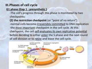
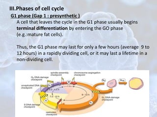
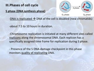
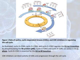
Cell Cycle
Maha Hammady Hemdan
MBBCh, Demonstrator at Medical Histology &
Cell Biology Department,
Faculty of Medicine Alexandria university
CELL CYCLE OVERVIEW
• The cell cycle : self-regulated sequence of events that
controls cell growth and division to produce two daughter
cells identical to those of the parental cell.
• It has 2 principal phases:
1.the interphase:
- representing continuous growth of the cell.
-subdivided into 3 phases:
G1 (gap1) phase, S ( synthesis) phase, and G2 (gap2) phase
2.the M phase (mitosis):
-characterized by the partition of the genome.
• The replication of cells is stimulated by growth factors or by
signaling from ECM
I.The checkpoints:
internal quality control mechanisms
• they are surveillance mechanisms that are sensing damage
to DNA and chromosomes
• When cells sense DNA damage, checkpoint activation
delays the cell cycle and triggers DNA repair mechanisms. If
DNA damage is too severe to be repaired, the cells are
eliminated by apoptosis, or enter a nonreplicative state called
senescence
• Mutations in genes regulating these checkpoints allow cells
with damaged DNA to divide, producing daughter cells
carrying mutations.
II.Regulation of the Cell Cycle
• the orderly progression through the cell cycle is regulated by :
1-proteins called cyclins, so called because of the cyclic nature
of their production and degradation,
2-associated enzymes, the cyclin dependent kinases (CDKs)
• forming CDK–cyclin complexes  phosphorylate crucial
target proteins that drive the cell through the cell cycle 
completion of this task, cyclin levels decline rapidly.
•More than 15 cyclins have been identified; cyclins D, E,A, and
B appear sequentially during the cell cycle and bind to one or
more CDKs (Fig 1).
• The activity of CDK–cyclin complexes is regulated by CDK
inhibitors (CDKIs), which enforce cell cycle checkpoints.
As illustrated, cyclin D–CDK4, cyclin D–CDK6, and cyclin E–CDK2 regulate the G1-to-S transition
by phosphorylating the Rb protein (pRb). Cyclin A–CDK2 and cyclin A–CDK1 are active in the S
phase. Cyclin B–CDK1 is essential for the G2-to-M transition.
CDK inhibitors can block activity of CDKs and progression through the cell cycle.
Figure 1 Role of cyclins, cyclin-dependent kinases (CDKs), and CDK inhibitors in regulating
the cell cycle.
Figure 1 Role of cyclins, cyclin-dependent kinases (CDKs), and CDK inhibitors in regulating
the cell cycle.
G1/s checkpoint mediated by interactions between the retinoblastoma susceptibility
protein (pRb) which has antiproliferative effects and a family of essential transcription factors
(E2F This interaction prevents transcription of genes like cyclin E that are needed for DNA
replication, and so the cells are arrested in G1(block cell-cycle progression).
The dephosphorylated form is the active form which prevent cell cycle progression
G1 phase (Gap 1 : presynthetic )
-it is the longest and the most variable phase of the cell cycle
-begins at the end of M phase of the previous cycle.
-During the G1 phase, the cell gathers nutrients and
synthesizes RNA and proteins necessary for DNA synthesis
III.Phases of cell cycle
G1 phase (Gap 1 : presynthetic )
-The cell’s progress through this phase is monitored by two
checkpoints:
(1) the G1 DNA-damage checkpoint, which monitors the
integrity of newly replicated DNA. e.g
DNA irreparable damage accumulation of protein p53
(central monitor of stress G1 DNA-damage checkpoint
detection  it does not allow the cell to enter the S
phase(apoptosis).
III.Phases of cell cycle
G1 phase (Gap 1 : presynthetic )
-The cell’s progress through this phase is monitored by two
checkpoints:
(2) the restriction checkpoint (or “point of no return”)
-normal cells become irreversibly committed to DNA replication
-the most important checkpoint in the cell cycle. At this
checkpoint, the cell self-evaluates its own replicative potential
before deciding to either enter the S phase and the next round
of cell division or to retire and leave the cell cycle.
A cell that leaves the cycle in the G1 phase usually begins
terminal differentiation by entering the GO phase (“O” stands
for “ outside”the cycle). Thus, the G1 phase may last for only a
few hours (average 9 to 12 hours) in a rapidly dividing cell, or it
may last a lifetime in a nondividing cell.
III.Phases of cell cycle
G1 phase (Gap 1 : presynthetic )
A cell that leaves the cycle in the G1 phase usually begins
terminal differentiation by entering the GO phase
(e.g.:mature fat cells).
Thus, the G1 phase may last for only a few hours (average 9 to
12 hours) in a rapidly dividing cell, or it may last a lifetime in a
non-dividing cell.
III.Phases of cell cycle
CELL RENEWAL
Somatic cells in the adult organism may be classified according
to their mitotic activity into :
1- Static cell populations
-no longer divide (postmitotic cells)
-central nervous system and skeletal or cardiac muscle cells.
2-Stable cell populations
-divide episodically and slowly to maintain normal tissue or
organ structure . (stimulated by injury).
-Periosteal and perichondrial cells, smooth muscle cells,
endothelial cells of blood vessels
3- Renewing cell populations
-regular mitotic activity.either slowly or rabidly
a)Slowly renewing populations : smooth muscle cells of most
hollow organs
b)Rapidly renewing populations : blood cells, epithelial cells
and dermal fibroblasts of the skin, and the epithelial cells of
S phase (DNA synthesis phase)
-DNA is replicated  DNA of the cell is doubled (new chromatids)
-about 7.5 to 10 hours in duration.
-Chromosome replication is initiated at many different sites called
replicons along the chromosomal DNA. Each replicon has a
specifically assigned time frame for replication during S phase.
- Presence of the S DNA-damage checkpoint in this phase
monitors quality of replicating DNA.
III.Phases of cell cycle
S phase (DNA synthesis phase)
III.Phases of cell cycle
G2 phase (Gap2 : premitotic )
-In the G2 phase, the cell prepares for cell division
by examining its replicated DNA , and
reorganization of cytoplasmic organelles
-The G2 phase may be as short as 1 hour in rapidly dividing
cells or of nearly indefinite duration in as the primary oocyte
that are arrested in G2 for extended periods.
- Two checkpoints monitor DNA quality:
1) G2 DNA-damage checkpoint
2)unreplicated-DNA checkpoint. The latter checkpoint
prevents the progression of the cell into the M phase before
DNA synthesis is complete.
III.Phases of cell cycle
M phase (Mitosis phase)
-Mitosis occurs in the M phase.
-Mitosis nearly always includes :
karyokinesis ( division of the nucleus)
cytokinesis (division of the cell)
-lasts about 1 hour.
-Mitosis takes place in several stages
-The M phase possesses two checkpoints:
1)spindle-assembly checkpoint:prevents premature entry into anaphase
2)chromosome-segregation checkpoint: prevents the process of
cytokinesis until all of the chromosomes have been correctly separated.
III.Phases of cell cycle
Regulation of the cell cycle by cyclin–Cdk
complexes. This diagram shows the changing
pattern of cyclin–Cdk
activities during diff erent phases of the cell
cycle.
III.Phases of cell cycle
IV.malfunction of cell cycle checkpoints
Cells that fail to execute the apoptotic cycle in response to
DNA or mitotic spindle damage are likely to divide
asymmetrically in the next round of cell division. This leads to
the generation of aneuploid cells (cells containing abnormal
chromosome numbers). Thus, a mitotic catastrophe may be
regarded as one of the mechanisms contributing to
oncogenesis (tumor cell development).
III.malfunction of cell cycle checkpoints
• The malfunction of the restriction checkpoint may be
facilitated by the viral proteins of several cancer-causing
viruses,
• all cancers appear to have genetic lesions that disable the
G1-S checkpoint, causing cells to continually reenter the S
phase.
• some tumer types shows overexpression of cyclins
• The CDKIs frequently are disabled by mutation or gene
silencing in many human malignancies.
BIBLIOGRAPHY
Ross, Michael H, and Wojciech Pawlina. Histology: A Text and Atlas :
with Correlated Cell and Molecular Biology. Baltimore, MD:
Lippincott Wiliams & Wilkins, 2006
Kumar, Abbas, Aster: Robbins Basic Pathology. 9th edition. Elsevier
Inc. 2013
B.OVERVIEW OF CELL INJURY AND CELL DEATH

The Cell cycle - maha hammady

  • 1.
    Cell Cycle Maha HammadyHemdan MBBCh, Demonstrator at Medical Histology & Cell Biology Department, Faculty of Medicine Alexandria university
  • 2.
    CELL CYCLE OVERVIEW •The cell cycle : self-regulated sequence of events that controls cell growth and division to produce two daughter cells identical to those of the parental cell. • It has 2 principal phases: 1.the interphase: - representing continuous growth of the cell. -subdivided into 3 phases: G1 (gap1) phase, S ( synthesis) phase, and G2 (gap2) phase 2.the M phase (mitosis): -characterized by the partition of the genome. • The replication of cells is stimulated by growth factors or by signaling from ECM
  • 3.
    I.The checkpoints: internal qualitycontrol mechanisms • they are surveillance mechanisms that are sensing damage to DNA and chromosomes • When cells sense DNA damage, checkpoint activation delays the cell cycle and triggers DNA repair mechanisms. If DNA damage is too severe to be repaired, the cells are eliminated by apoptosis, or enter a nonreplicative state called senescence • Mutations in genes regulating these checkpoints allow cells with damaged DNA to divide, producing daughter cells carrying mutations.
  • 4.
    II.Regulation of theCell Cycle • the orderly progression through the cell cycle is regulated by : 1-proteins called cyclins, so called because of the cyclic nature of their production and degradation, 2-associated enzymes, the cyclin dependent kinases (CDKs) • forming CDK–cyclin complexes  phosphorylate crucial target proteins that drive the cell through the cell cycle  completion of this task, cyclin levels decline rapidly. •More than 15 cyclins have been identified; cyclins D, E,A, and B appear sequentially during the cell cycle and bind to one or more CDKs (Fig 1). • The activity of CDK–cyclin complexes is regulated by CDK inhibitors (CDKIs), which enforce cell cycle checkpoints.
  • 5.
    As illustrated, cyclinD–CDK4, cyclin D–CDK6, and cyclin E–CDK2 regulate the G1-to-S transition by phosphorylating the Rb protein (pRb). Cyclin A–CDK2 and cyclin A–CDK1 are active in the S phase. Cyclin B–CDK1 is essential for the G2-to-M transition. CDK inhibitors can block activity of CDKs and progression through the cell cycle. Figure 1 Role of cyclins, cyclin-dependent kinases (CDKs), and CDK inhibitors in regulating the cell cycle.
  • 6.
    Figure 1 Roleof cyclins, cyclin-dependent kinases (CDKs), and CDK inhibitors in regulating the cell cycle. G1/s checkpoint mediated by interactions between the retinoblastoma susceptibility protein (pRb) which has antiproliferative effects and a family of essential transcription factors (E2F This interaction prevents transcription of genes like cyclin E that are needed for DNA replication, and so the cells are arrested in G1(block cell-cycle progression). The dephosphorylated form is the active form which prevent cell cycle progression
  • 7.
    G1 phase (Gap1 : presynthetic ) -it is the longest and the most variable phase of the cell cycle -begins at the end of M phase of the previous cycle. -During the G1 phase, the cell gathers nutrients and synthesizes RNA and proteins necessary for DNA synthesis III.Phases of cell cycle
  • 8.
    G1 phase (Gap1 : presynthetic ) -The cell’s progress through this phase is monitored by two checkpoints: (1) the G1 DNA-damage checkpoint, which monitors the integrity of newly replicated DNA. e.g DNA irreparable damage accumulation of protein p53 (central monitor of stress G1 DNA-damage checkpoint detection  it does not allow the cell to enter the S phase(apoptosis). III.Phases of cell cycle
  • 9.
    G1 phase (Gap1 : presynthetic ) -The cell’s progress through this phase is monitored by two checkpoints: (2) the restriction checkpoint (or “point of no return”) -normal cells become irreversibly committed to DNA replication -the most important checkpoint in the cell cycle. At this checkpoint, the cell self-evaluates its own replicative potential before deciding to either enter the S phase and the next round of cell division or to retire and leave the cell cycle. A cell that leaves the cycle in the G1 phase usually begins terminal differentiation by entering the GO phase (“O” stands for “ outside”the cycle). Thus, the G1 phase may last for only a few hours (average 9 to 12 hours) in a rapidly dividing cell, or it may last a lifetime in a nondividing cell. III.Phases of cell cycle
  • 10.
    G1 phase (Gap1 : presynthetic ) A cell that leaves the cycle in the G1 phase usually begins terminal differentiation by entering the GO phase (e.g.:mature fat cells). Thus, the G1 phase may last for only a few hours (average 9 to 12 hours) in a rapidly dividing cell, or it may last a lifetime in a non-dividing cell. III.Phases of cell cycle
  • 11.
    CELL RENEWAL Somatic cellsin the adult organism may be classified according to their mitotic activity into : 1- Static cell populations -no longer divide (postmitotic cells) -central nervous system and skeletal or cardiac muscle cells. 2-Stable cell populations -divide episodically and slowly to maintain normal tissue or organ structure . (stimulated by injury). -Periosteal and perichondrial cells, smooth muscle cells, endothelial cells of blood vessels 3- Renewing cell populations -regular mitotic activity.either slowly or rabidly a)Slowly renewing populations : smooth muscle cells of most hollow organs b)Rapidly renewing populations : blood cells, epithelial cells and dermal fibroblasts of the skin, and the epithelial cells of
  • 12.
    S phase (DNAsynthesis phase) -DNA is replicated  DNA of the cell is doubled (new chromatids) -about 7.5 to 10 hours in duration. -Chromosome replication is initiated at many different sites called replicons along the chromosomal DNA. Each replicon has a specifically assigned time frame for replication during S phase. - Presence of the S DNA-damage checkpoint in this phase monitors quality of replicating DNA. III.Phases of cell cycle
  • 13.
    S phase (DNAsynthesis phase) III.Phases of cell cycle
  • 14.
    G2 phase (Gap2: premitotic ) -In the G2 phase, the cell prepares for cell division by examining its replicated DNA , and reorganization of cytoplasmic organelles -The G2 phase may be as short as 1 hour in rapidly dividing cells or of nearly indefinite duration in as the primary oocyte that are arrested in G2 for extended periods. - Two checkpoints monitor DNA quality: 1) G2 DNA-damage checkpoint 2)unreplicated-DNA checkpoint. The latter checkpoint prevents the progression of the cell into the M phase before DNA synthesis is complete. III.Phases of cell cycle
  • 15.
    M phase (Mitosisphase) -Mitosis occurs in the M phase. -Mitosis nearly always includes : karyokinesis ( division of the nucleus) cytokinesis (division of the cell) -lasts about 1 hour. -Mitosis takes place in several stages -The M phase possesses two checkpoints: 1)spindle-assembly checkpoint:prevents premature entry into anaphase 2)chromosome-segregation checkpoint: prevents the process of cytokinesis until all of the chromosomes have been correctly separated. III.Phases of cell cycle
  • 16.
    Regulation of thecell cycle by cyclin–Cdk complexes. This diagram shows the changing pattern of cyclin–Cdk activities during diff erent phases of the cell cycle. III.Phases of cell cycle
  • 17.
    IV.malfunction of cellcycle checkpoints Cells that fail to execute the apoptotic cycle in response to DNA or mitotic spindle damage are likely to divide asymmetrically in the next round of cell division. This leads to the generation of aneuploid cells (cells containing abnormal chromosome numbers). Thus, a mitotic catastrophe may be regarded as one of the mechanisms contributing to oncogenesis (tumor cell development).
  • 18.
    III.malfunction of cellcycle checkpoints • The malfunction of the restriction checkpoint may be facilitated by the viral proteins of several cancer-causing viruses, • all cancers appear to have genetic lesions that disable the G1-S checkpoint, causing cells to continually reenter the S phase. • some tumer types shows overexpression of cyclins • The CDKIs frequently are disabled by mutation or gene silencing in many human malignancies.
  • 19.
    BIBLIOGRAPHY Ross, Michael H,and Wojciech Pawlina. Histology: A Text and Atlas : with Correlated Cell and Molecular Biology. Baltimore, MD: Lippincott Wiliams & Wilkins, 2006 Kumar, Abbas, Aster: Robbins Basic Pathology. 9th edition. Elsevier Inc. 2013
  • 20.
    B.OVERVIEW OF CELLINJURY AND CELL DEATH

Editor's Notes

  • #9 -the half-life of the p53 protein is increased drastically, leading to a quick accumulation of p53 in stressed cells -------------- • The p53 protein is the central monitor of stress in the cell and can be activated by anoxia, inappropriate oncogene signaling, or DNA damage. Activated p53 controls the expression and activity of genes involved in cell cycle arrest, DNA repair, cellular senescence, and apoptosis. • DNA damage leads to activation of p53 by phosphorylation. Activated p53 drives transcription of CDKN1A (p21), which prevents Rb phosphorylation, thereby causing a G1-S block in the cell cycle. This pause allows the cells to repair DNA damage. • If DNA damage cannot be repaired, p53 induces cellular senescence or apoptosis.
  • #10 self-evaluates its own replicative potential(size of the cell, the state of the cell’s physiologic processes, and its interactions with extracellular matrix)