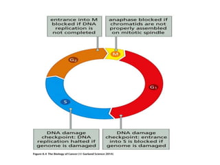
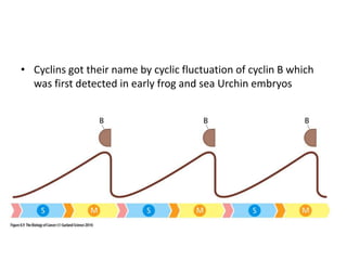
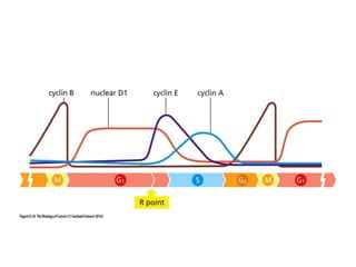
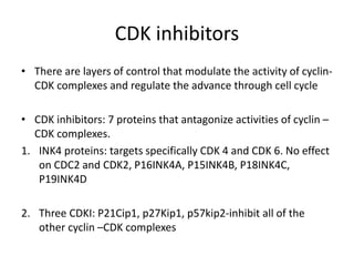
CELL CYCLE AND THERAPEUTIC
IMPLICATIONS
By
Dr Priyanka
Dept of surgical oncology
Points…..
1. Cell cycle
2. Cell cycle checkpoints
3. Regulation of cell cycle
4. Therapeutic implications
Cell Cycle clock
• The cell cycle clock is a network of interacting proteins- a
signal processing circuit that recieves signals from various
sources both outside and inside the cell, integrates them &
then decides cells fate
 It should decide in favour of proliferation.
 It should proceed to orchastrate complex transition: cell
growth and division.
 Should it decide in favour of quiescence.
• The proliferative behaviour of cancer cells indicates the
master governer of the cells fate is influenced not only by
normal proteins but also by oncogene proteins that inserts
themselves into various signaling pathway and disrupts
normal control mechanism.
• Similarly the deletion of tumour suppressor proteins acts by
disrupting the normal control mechanism.
Cell cycle
• Mammalian cell exhibit a complex cycle of growth & division
that is usually referred to as CELL CYCLE
• Two processes:
1. Mitosis
2. Cytokinesis
The cell recently formed by these processes must decide
whether it will once again initiate a new round of active growth
& division or retreat into non growing state: - G0
• Some cells never leave the G0 phase, or have completely lost
the ability to divide e.g neurons
• Some cells remain largely in G0 but can re-enter the cell cycle
when required – e.g kidney, liver, stomach cells
• Some cell have a sole functions of replenishing mucosal or
skin surfaces these cells never enter G0 phase – epithelial
cells
• This process is strongly influenced by mitogenic growth
factors and absence of these factors triggers the default
decision to proceed from mitosis into G0 quiescent state.
• This process is reversible if quiescent cell comes in contact
with mitogenic growth factors which induce cell to reenter
active growth and division.
• Once this cell division remains in active growth and division
cycle –it begins to prepare for next division which requires
macromolecular constituents for doubling of cells
• These constituents drives increase in cell size – cell growth –to
distinguish it from cell division which occurs via mitosis and
cytokinesis
• Cell growth implies both accumulation of cell constituents and
subsequent cell division :- cell proliferation
Growth Vs proliferation
Alteration of certain signaling protiens such as
One encoded by TSC 1 tumor suppressor gene,
Allow the processes of cell growth and division
To be uncoupled from one another.
Scanning electron micrograph of Drosophila eye,
Ommatidial cells in the upper portion of eye have
Been deprived of the fly ortholog of TSC 1,these
Cells are physically larger than the wild type cells
Because they have grown more during the cell cycles
That led to their formation.
Tuberous sclerosis: loss of TSC 1 TSG through germ
Line mutation and LOH
Giant cells present in a benign growth
• Prokaryotes: DNA replication – begins immediately after
daughter cells are formed by cell division
• Eukaryotes:
1. Accumulation of RNA and proteins & its synthesis is initiated
immediately after cell division and proceeds continuously
until next cell division
2. Task of replicating DNA is deferred for a number of hours as
many as 12 to 15 hrs after emergence of new daughter cells
from mitosis and cytokinesis.
• G1 ( first Gap): Period between birth of daughter cell and
subsequent onset of DNA synthesis. Cells make critical
decisions about growth Vs quiescence and whether as
quiescent cells they will differentiate
• S phase: period of DNA synthesis which requires 6-8 hrs for
completion , it can be shorter in rapidly dividing cells
embryonic cells and lymphocytes.
• G2 ( second Gap) : mammalian cells spend 3-5 hrs in second
Gap, preparing themselves In some still poorly understood
fashion for entrance into M phase and cell division.
• M:
1. It begins with two recently duplicated DNA helices within
each chromosome, these are carried in sister chromatids of
chromosome.
2. These are aligned adjacent to one another in nucleus, during
mitosis each daughter cell receive exactly one diploid
complement of chromatids.
• Distincts subphases of mitosis:
1. Prophase
2. Prometaphse
3. Metaphase
4. Anaphase
5. Telophase
Prophase: chromosome begins to condense & become visible, while
Centrosomes at the poles of the cells begin to assemble
Metaphase: chromosomes align along a plane that bisect the cell &
Become attached to microtubule fibres of mitotic spindle, nuclear
membrane has disappeared.
Anaphase: two halves of each chromosome – chromatids-pulled apart
By mitotic spindle to the two opposite poles of the cell.
Telophase: chromatids de condense and a new nuclear membrane
forms Around each set of chromatids ( now called chromosomes)
At the same time during process of cytokinesis the cytoplasm of
mother Cell divides, yielding two daughter cells.
Decisions about growth or quiescence
in G1 phase…????
• Cell consults their extracellular environment & its growth
regulating signals during a discrete window of time in active
cell cycle, namely, from the onset of G1 phase until an hour or
two before the G1 to S transition.
• This total dependence on extracellular signals followed by
entrance in late G1 into state of relative independence
indicates that decisions must be made toward the end of G1.
• So at this point cell must make up its mind whether it will
remain in G1, retreat from active cycle into G0 or advance into
late G1 and thereafter into remaining phases of cell cycle
• This critical decision is made at a transition that has been
called the restriction point or R point and it occurs several
hours before G1/S phase.
Cell cycle checkpoints
• Checkpoints impose quality control to ensure that a cell has
properly completed all the requisite steps of one phase of cell
cycle before it is allowed to advance into next phase.
• A cell will not be permitted to entre S phase until all steps of
G1 have been completed.
• It will be blocked from entering G2 until all of its chromosomal
DNA has been properly replicated.
• Similarly cell is not permitted to entre anaphase until all of its
chromosome are properly assembled on the mitotic spindle
during metaphase
• In addition cell is not allowed to advance into S or M if its DNA
has been damaged and not yet repaired
• Other controls ensure that once a specific step in the cell cycle
has been completed, it is not repeated until the next cell
cycle.
Consequences of loss of checkpoint controls
Always manifested as an altered karyotype
The normal human karyotype Is contrasted with that
Of a cell that has been deprived of rad 17 checkpoints
Protein, which Is responsible for preventing inadvertent re-
Replication of already –replicated chromosomal DNA,
Resulting in endoreduplication and increases in ploidy of
Cell.
The Bub 1 protein normally prevents seperation of
Chromosome in the event that one or more
Chromosomal pairs are not properly aligned on the
Metaphase plate.
In its absence cell gain or lose chromosomes, as seen
In this spectral karyotyping analysis, which indicates
That this human cell has chromosome 1( yellow arrow)
& one chromosome 6 ( red arrow)
ATR ( ataxia telangiectasia and Rad 3 related) protein kinase is responsible for halting further
DNA replication until stalled replication forks are repaired.
In its absence fragile sites –sites in chromosome prone to breakage –become visible upon
Karyotyping analysis
Fragile sites in chr 3 & 6 in above pic
Cell cycle regulation
• Cell cycle clock uses group of enzymes called protein kinases
to execute the various steps of cell cycle progression.
• Example: phosphorylation of centrosome associated proteins
at G1/S boundry, prior to S phase, histone proteins in
anticipation of S & M phase places chromatin In configuration.
Phosphorylation of proteins forming nuclear membrane.
• Kinases deployed by cell cycle machinery are called – cyclin
dependent kinsases (CDKs): never act on their own
• Cyclins :- regulatory subunits
• CDK+ Cyclin complex responsible for sending out signals from
cell cycle clock to dozens if not hundreds of responder
molecules that carry actual work of moving cell through its
growth –division cycle.
• Cyclins got their name by cyclic fluctuation of cyclin B which
was first detected in early frog and sea Urchin embryos
CDK inhibitors
• There are layers of control that modulate the activity of cyclin-
CDK complexes and regulate the advance through cell cycle
• CDK inhibitors: 7 proteins that antagonize activities of cyclin –
CDK complexes.
1. INK4 proteins: targets specifically CDK 4 and CDK 6. No effect
on CDC2 and CDK2, P16INK4A, P15INK4B, P18INK4C,
P19INK4D
2. Three CDKI: P21Cip1, p27Kip1, p57kip2-inhibit all of the
other cyclin –CDK complexes
• 3 families of genes -
– Cip/Kip (CDK interacting protein/ Kinase inhibitory protein)
family
– INK4a/ARF (Inhibitor of Kinase 4/Alternative Reading
Frame) family
– pRb protein family
1. Cip/Kip family includes the genes p21, p27 and p57. They
halt cell cycle in G1 phase
– p21 is activated by p53 (which, in turn, is triggered by
DNA damage e.g. due to radiation).
– p27 is activated by Transforming Growth Factor of β (TGF
β), a growth inhibitor.
INK4a/ARF family includes p15, p16, p18 & p19.
– p16INK4a, which binds to CDK4 & CDK 6
– and arrests the cell cycle in G1 phase,
3. pRb protein family – p107 and p130.
– Well characterised functions as transcriptional inhibitors
– They are also potent cyclin E/A – CDK2 inhibitors
pRb and viral oncoprotein
• 3 different DNA tumor virus oncoproteins adenovirus E1A,
SV40 large T, HPV E7: target common cellular proteins
• Viruses can subotage activities of pRb by dispatching viral
oncoproteins that seek out & bind pRb, sequestering and
apparently inactivating it and removes pRb from regulatory
circuitry of cell
• Target hypophosphorylated pRb in early and mid G1.
Cell Cycle and Oncogenesis
• Cancer is partly a disease of uncontrolled proliferation
• Cell cycle and checkpoint genes are misregulated or mutated
in cancer
• Cancerous cell division:
– Ignores the cell cycle checkpoints
• Caused by DNA mutations
• Cells grow and divide out of control
• Cancerous cells do not perform designated purpose.
• Crowd out normal cells that do perform designated
purpose.
• To achieve uncontrolled cell division – 2 basic requirements
must be met:-
1. Strong constitutive proliferation signal capable of overriding
restraints on division
2. Barrier of senescence to render tumour cells immortal.
Cancer Genes
• An oncogene is a gene that when mutated gains a function or
is expressed at abnormally-high levels and as a result
contributes to converting a normal cell into a cancer cell.
Often cause increase in transcription of genes through
kinases. Proteins that act in this way are called transcription
factors.
• A tumor suppressor gene encodes for a protein that is
involved in suppressing cell division (p53, or other checkpoint
proteins). When mutated it is no longer functional.
Proto Oncogenes
• They are crucial in normal cell function and converted to
oncogene by gain –of –function mutations.
• Only one allele needs to be triggered for uncontrolled growth
unlike tumour suppressor gene which requires both genes to
be altered.
• Genes for Cyclin Dependent Kinases especially CDK4 & 6.
Ras Proto-oncogenes
Ras family genes mutated in 40% of all cancers.
– Involved in signal transduction pathway from growth factor
receptor to nucleus
– Mutant form lacks GTPase activity and remains active
Tumor Suppressor Genes
• 1. Cell Cycle Regulators:
– Cdkis (cyclin-depndent kinase inhibitors)
– Rb (Retinoblastoma protein)
– P53
• 2. Signaling proteins: APC (adenomatous polyposis coli)
• 3. DNA damage repair proteins: BRCA1 (Breast cancer gene)
• 4. Enzymes: PTEN (phosphatase and tensin homolog deleted
in chromosome ten)
p53 Tumor Suppressor Gene
• Mutated (inactivated) in more than 50% of all cancers
• p53 regulates (activates or represses) transcription of more
than 50 different genes
• p53 regulated by Mdm2 (prevents the phosphorylations and
acetylations that activate inactive p53)
• Activated p53 levels rise rapidly if DNA is damaged or repair
intermediates accumulate
RB1 Tumor Suppressor Gene
• Retinoblastoma 1 gene
• Involved in breast, bone, lung, bladder and retinal cancers
(among others)
• Tumor suppressor protein that controls the G1/S checkpoint
Cell cycle & Cancer Therapy
SURGERY
ALL CELLS!
Chemotherapy Radiation
Dividing
Cells
R+ resection (Tumour
Debulking)
Residual cells pushed into cell
cycle
Chemotherapy
• Few chemotherapy drugs acts at more than one phase of cell
cycle & few are phase speciific
• G0 phase: Glucocorticoids for mature lymphocytes
• G1 phase : L- asparginase for ALL
• S phase : Procarbazine & antimetabolites
• G2 phase : Bleomycin and plant alkaloids
• M phase : Mitotic spindle agents (taxanes)
Radiation
• DNA is the main target of radiation action in cells
• Base damages, cross links, single and double stranded breaks
– remains unrepaired and leads to cell death – “Mitotic
death”
• In general, cells are most radiosensitive in late M and G2
phases and most resistant in late S
CDK 4 inhibitors in breast cancer
• neoMONARCH: Change in Ki67 Expression and Ki67 Response
• The neoMONARCH study is a presurgical study looking at
abemaciclib in combination with anastrozole vs anastrozole
alone vs abemaciclib alone
• This is in a short leading phase at which time biopsies are
compared at baseline and then after the relative treatments
• As expected, there is a very potent decrease in Ki67 with the
abemaciclib containing regimens
• That is also seen with anastrozole alone, but consistent with
preclinical data there seems to be a greater effect when the 2
drugs are used in combination
• MONARCH 1:
• Abemaciclib also has been studied as a single agent in a large
phase 1 and phase 2 study called the MONARCH 1 study
• All of these patients were HRpositive, human epidermal
growth factor receptor 2 (HER2)negative, and had prior
chemotherapy for metastatic disease
• The response rate was a modest 19.7% with PFS about 6
months, as well as an overall survival of just under 18 months
This was a single agent
• Abemaciclib did get a breakthrough therapy designation in
October of 2016 as a single agent based on some of the
phase 1 data with this compound
THANK YOU

Cell cycle and therapeutic implications

  • 1.
    CELL CYCLE ANDTHERAPEUTIC IMPLICATIONS By Dr Priyanka Dept of surgical oncology
  • 2.
    Points….. 1. Cell cycle 2.Cell cycle checkpoints 3. Regulation of cell cycle 4. Therapeutic implications
  • 3.
    Cell Cycle clock •The cell cycle clock is a network of interacting proteins- a signal processing circuit that recieves signals from various sources both outside and inside the cell, integrates them & then decides cells fate  It should decide in favour of proliferation.  It should proceed to orchastrate complex transition: cell growth and division.  Should it decide in favour of quiescence.
  • 4.
    • The proliferativebehaviour of cancer cells indicates the master governer of the cells fate is influenced not only by normal proteins but also by oncogene proteins that inserts themselves into various signaling pathway and disrupts normal control mechanism. • Similarly the deletion of tumour suppressor proteins acts by disrupting the normal control mechanism.
  • 6.
    Cell cycle • Mammaliancell exhibit a complex cycle of growth & division that is usually referred to as CELL CYCLE • Two processes: 1. Mitosis 2. Cytokinesis The cell recently formed by these processes must decide whether it will once again initiate a new round of active growth & division or retreat into non growing state: - G0
  • 7.
    • Some cellsnever leave the G0 phase, or have completely lost the ability to divide e.g neurons • Some cells remain largely in G0 but can re-enter the cell cycle when required – e.g kidney, liver, stomach cells • Some cell have a sole functions of replenishing mucosal or skin surfaces these cells never enter G0 phase – epithelial cells
  • 8.
    • This processis strongly influenced by mitogenic growth factors and absence of these factors triggers the default decision to proceed from mitosis into G0 quiescent state. • This process is reversible if quiescent cell comes in contact with mitogenic growth factors which induce cell to reenter active growth and division.
  • 9.
    • Once thiscell division remains in active growth and division cycle –it begins to prepare for next division which requires macromolecular constituents for doubling of cells • These constituents drives increase in cell size – cell growth –to distinguish it from cell division which occurs via mitosis and cytokinesis • Cell growth implies both accumulation of cell constituents and subsequent cell division :- cell proliferation
  • 10.
    Growth Vs proliferation Alterationof certain signaling protiens such as One encoded by TSC 1 tumor suppressor gene, Allow the processes of cell growth and division To be uncoupled from one another. Scanning electron micrograph of Drosophila eye, Ommatidial cells in the upper portion of eye have Been deprived of the fly ortholog of TSC 1,these Cells are physically larger than the wild type cells Because they have grown more during the cell cycles That led to their formation.
  • 11.
    Tuberous sclerosis: lossof TSC 1 TSG through germ Line mutation and LOH Giant cells present in a benign growth
  • 12.
    • Prokaryotes: DNAreplication – begins immediately after daughter cells are formed by cell division • Eukaryotes: 1. Accumulation of RNA and proteins & its synthesis is initiated immediately after cell division and proceeds continuously until next cell division 2. Task of replicating DNA is deferred for a number of hours as many as 12 to 15 hrs after emergence of new daughter cells from mitosis and cytokinesis.
  • 13.
    • G1 (first Gap): Period between birth of daughter cell and subsequent onset of DNA synthesis. Cells make critical decisions about growth Vs quiescence and whether as quiescent cells they will differentiate • S phase: period of DNA synthesis which requires 6-8 hrs for completion , it can be shorter in rapidly dividing cells embryonic cells and lymphocytes.
  • 14.
    • G2 (second Gap) : mammalian cells spend 3-5 hrs in second Gap, preparing themselves In some still poorly understood fashion for entrance into M phase and cell division. • M: 1. It begins with two recently duplicated DNA helices within each chromosome, these are carried in sister chromatids of chromosome. 2. These are aligned adjacent to one another in nucleus, during mitosis each daughter cell receive exactly one diploid complement of chromatids.
  • 15.
    • Distincts subphasesof mitosis: 1. Prophase 2. Prometaphse 3. Metaphase 4. Anaphase 5. Telophase
  • 16.
    Prophase: chromosome beginsto condense & become visible, while Centrosomes at the poles of the cells begin to assemble Metaphase: chromosomes align along a plane that bisect the cell & Become attached to microtubule fibres of mitotic spindle, nuclear membrane has disappeared. Anaphase: two halves of each chromosome – chromatids-pulled apart By mitotic spindle to the two opposite poles of the cell. Telophase: chromatids de condense and a new nuclear membrane forms Around each set of chromatids ( now called chromosomes) At the same time during process of cytokinesis the cytoplasm of mother Cell divides, yielding two daughter cells.
  • 18.
    Decisions about growthor quiescence in G1 phase…???? • Cell consults their extracellular environment & its growth regulating signals during a discrete window of time in active cell cycle, namely, from the onset of G1 phase until an hour or two before the G1 to S transition. • This total dependence on extracellular signals followed by entrance in late G1 into state of relative independence indicates that decisions must be made toward the end of G1.
  • 19.
    • So atthis point cell must make up its mind whether it will remain in G1, retreat from active cycle into G0 or advance into late G1 and thereafter into remaining phases of cell cycle • This critical decision is made at a transition that has been called the restriction point or R point and it occurs several hours before G1/S phase.
  • 21.
    Cell cycle checkpoints •Checkpoints impose quality control to ensure that a cell has properly completed all the requisite steps of one phase of cell cycle before it is allowed to advance into next phase. • A cell will not be permitted to entre S phase until all steps of G1 have been completed. • It will be blocked from entering G2 until all of its chromosomal DNA has been properly replicated.
  • 22.
    • Similarly cellis not permitted to entre anaphase until all of its chromosome are properly assembled on the mitotic spindle during metaphase • In addition cell is not allowed to advance into S or M if its DNA has been damaged and not yet repaired • Other controls ensure that once a specific step in the cell cycle has been completed, it is not repeated until the next cell cycle.
  • 24.
    Consequences of lossof checkpoint controls Always manifested as an altered karyotype The normal human karyotype Is contrasted with that Of a cell that has been deprived of rad 17 checkpoints Protein, which Is responsible for preventing inadvertent re- Replication of already –replicated chromosomal DNA, Resulting in endoreduplication and increases in ploidy of Cell.
  • 25.
    The Bub 1protein normally prevents seperation of Chromosome in the event that one or more Chromosomal pairs are not properly aligned on the Metaphase plate. In its absence cell gain or lose chromosomes, as seen In this spectral karyotyping analysis, which indicates That this human cell has chromosome 1( yellow arrow) & one chromosome 6 ( red arrow)
  • 26.
    ATR ( ataxiatelangiectasia and Rad 3 related) protein kinase is responsible for halting further DNA replication until stalled replication forks are repaired. In its absence fragile sites –sites in chromosome prone to breakage –become visible upon Karyotyping analysis Fragile sites in chr 3 & 6 in above pic
  • 27.
    Cell cycle regulation •Cell cycle clock uses group of enzymes called protein kinases to execute the various steps of cell cycle progression. • Example: phosphorylation of centrosome associated proteins at G1/S boundry, prior to S phase, histone proteins in anticipation of S & M phase places chromatin In configuration. Phosphorylation of proteins forming nuclear membrane.
  • 28.
    • Kinases deployedby cell cycle machinery are called – cyclin dependent kinsases (CDKs): never act on their own • Cyclins :- regulatory subunits • CDK+ Cyclin complex responsible for sending out signals from cell cycle clock to dozens if not hundreds of responder molecules that carry actual work of moving cell through its growth –division cycle.
  • 30.
    • Cyclins gottheir name by cyclic fluctuation of cyclin B which was first detected in early frog and sea Urchin embryos
  • 32.
    CDK inhibitors • Thereare layers of control that modulate the activity of cyclin- CDK complexes and regulate the advance through cell cycle • CDK inhibitors: 7 proteins that antagonize activities of cyclin – CDK complexes. 1. INK4 proteins: targets specifically CDK 4 and CDK 6. No effect on CDC2 and CDK2, P16INK4A, P15INK4B, P18INK4C, P19INK4D 2. Three CDKI: P21Cip1, p27Kip1, p57kip2-inhibit all of the other cyclin –CDK complexes
  • 33.
    • 3 familiesof genes - – Cip/Kip (CDK interacting protein/ Kinase inhibitory protein) family – INK4a/ARF (Inhibitor of Kinase 4/Alternative Reading Frame) family – pRb protein family 1. Cip/Kip family includes the genes p21, p27 and p57. They halt cell cycle in G1 phase – p21 is activated by p53 (which, in turn, is triggered by DNA damage e.g. due to radiation). – p27 is activated by Transforming Growth Factor of β (TGF β), a growth inhibitor.
  • 34.
    INK4a/ARF family includesp15, p16, p18 & p19. – p16INK4a, which binds to CDK4 & CDK 6 – and arrests the cell cycle in G1 phase, 3. pRb protein family – p107 and p130. – Well characterised functions as transcriptional inhibitors – They are also potent cyclin E/A – CDK2 inhibitors
  • 35.
    pRb and viraloncoprotein
  • 36.
    • 3 differentDNA tumor virus oncoproteins adenovirus E1A, SV40 large T, HPV E7: target common cellular proteins • Viruses can subotage activities of pRb by dispatching viral oncoproteins that seek out & bind pRb, sequestering and apparently inactivating it and removes pRb from regulatory circuitry of cell • Target hypophosphorylated pRb in early and mid G1.
  • 37.
    Cell Cycle andOncogenesis • Cancer is partly a disease of uncontrolled proliferation • Cell cycle and checkpoint genes are misregulated or mutated in cancer • Cancerous cell division: – Ignores the cell cycle checkpoints • Caused by DNA mutations • Cells grow and divide out of control • Cancerous cells do not perform designated purpose. • Crowd out normal cells that do perform designated purpose.
  • 38.
    • To achieveuncontrolled cell division – 2 basic requirements must be met:- 1. Strong constitutive proliferation signal capable of overriding restraints on division 2. Barrier of senescence to render tumour cells immortal.
  • 39.
    Cancer Genes • Anoncogene is a gene that when mutated gains a function or is expressed at abnormally-high levels and as a result contributes to converting a normal cell into a cancer cell. Often cause increase in transcription of genes through kinases. Proteins that act in this way are called transcription factors. • A tumor suppressor gene encodes for a protein that is involved in suppressing cell division (p53, or other checkpoint proteins). When mutated it is no longer functional.
  • 40.
    Proto Oncogenes • Theyare crucial in normal cell function and converted to oncogene by gain –of –function mutations. • Only one allele needs to be triggered for uncontrolled growth unlike tumour suppressor gene which requires both genes to be altered. • Genes for Cyclin Dependent Kinases especially CDK4 & 6.
  • 41.
    Ras Proto-oncogenes Ras familygenes mutated in 40% of all cancers. – Involved in signal transduction pathway from growth factor receptor to nucleus – Mutant form lacks GTPase activity and remains active
  • 43.
    Tumor Suppressor Genes •1. Cell Cycle Regulators: – Cdkis (cyclin-depndent kinase inhibitors) – Rb (Retinoblastoma protein) – P53 • 2. Signaling proteins: APC (adenomatous polyposis coli) • 3. DNA damage repair proteins: BRCA1 (Breast cancer gene) • 4. Enzymes: PTEN (phosphatase and tensin homolog deleted in chromosome ten)
  • 44.
    p53 Tumor SuppressorGene • Mutated (inactivated) in more than 50% of all cancers • p53 regulates (activates or represses) transcription of more than 50 different genes • p53 regulated by Mdm2 (prevents the phosphorylations and acetylations that activate inactive p53) • Activated p53 levels rise rapidly if DNA is damaged or repair intermediates accumulate
  • 46.
    RB1 Tumor SuppressorGene • Retinoblastoma 1 gene • Involved in breast, bone, lung, bladder and retinal cancers (among others) • Tumor suppressor protein that controls the G1/S checkpoint
  • 48.
    Cell cycle &Cancer Therapy SURGERY ALL CELLS! Chemotherapy Radiation Dividing Cells R+ resection (Tumour Debulking) Residual cells pushed into cell cycle
  • 49.
    Chemotherapy • Few chemotherapydrugs acts at more than one phase of cell cycle & few are phase speciific • G0 phase: Glucocorticoids for mature lymphocytes • G1 phase : L- asparginase for ALL • S phase : Procarbazine & antimetabolites • G2 phase : Bleomycin and plant alkaloids • M phase : Mitotic spindle agents (taxanes)
  • 50.
    Radiation • DNA isthe main target of radiation action in cells • Base damages, cross links, single and double stranded breaks – remains unrepaired and leads to cell death – “Mitotic death” • In general, cells are most radiosensitive in late M and G2 phases and most resistant in late S
  • 52.
    CDK 4 inhibitorsin breast cancer
  • 54.
    • neoMONARCH: Changein Ki67 Expression and Ki67 Response • The neoMONARCH study is a presurgical study looking at abemaciclib in combination with anastrozole vs anastrozole alone vs abemaciclib alone • This is in a short leading phase at which time biopsies are compared at baseline and then after the relative treatments • As expected, there is a very potent decrease in Ki67 with the abemaciclib containing regimens • That is also seen with anastrozole alone, but consistent with preclinical data there seems to be a greater effect when the 2 drugs are used in combination
  • 55.
    • MONARCH 1: •Abemaciclib also has been studied as a single agent in a large phase 1 and phase 2 study called the MONARCH 1 study • All of these patients were HRpositive, human epidermal growth factor receptor 2 (HER2)negative, and had prior chemotherapy for metastatic disease • The response rate was a modest 19.7% with PFS about 6 months, as well as an overall survival of just under 18 months This was a single agent • Abemaciclib did get a breakthrough therapy designation in October of 2016 as a single agent based on some of the phase 1 data with this compound
  • 56.