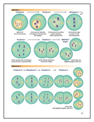
DEPARTMENT OF BIOTECHNOLOGY
CELL CYCLE
2
CELL CYCLE:
 Cell division is a very important processes in all living organisms
 During the cell division,DNA replication and cell growth takes place
 “The sequence of events by which a cell duplicates its genome,
synthesizes the other constituents of the cell and eventually divides
into two daughter cells is termed as cell cycle”
 “Rudolf Virchow” suggested “omniscellula e cellula” means every
cell is derived from pre- existing cells.
 The duration of the cell cycle varies greatly from one cell type to
another cell
 A single-celled yeast can divide every 90-120minutes
 While a mammalian liver cell divides on average less than once a
year
 Muscle cells,nerve cells, and RBC are the only cells in our body
which do not divide
 Before discussion about cell division first we should know the
following terms
CHROMATIN:
 DNA coiled around histone proteins
CHROMATID:
 one half of a duplicated chromosome
CHROMOSOME:
 are two chromatids together ,made of condensed chromatin
CENTROMERE:
 the constriction region that divide the chromosome into
twochromatids
 primary constriction of the condensed chromosome
 clearly visible with a light microscope
3
 made up of highly repetitive condensed AT rich, heterchromatic
DNA with non-histone proteins
 cannot bind with microtubules themselves
 it is the site for kinetochore assembly
 it ensures delivery of one copy of each chromosome to each
daughter at cell division
KINETOCHORE:
 Kinetochores are disc shaped protein complexes imtimately
associated with centromere
 Kinetochore can only be seen using electron microscope
 It is the site of assembly and disassembly of microtubules
 Made of multiple proteins
 It has a trilaminar structure the inner layer is in close contact with
condensed centromeric heterochromatin microtubules are attached
to the outer layer
 Made up of specialized types of histones like CENP-A
 Attachment site for spindle microtubule
 Involved in the movement of chromosomes during mitosis and
meiosis
4
CELL DIVISION IN PROKARYOTES:
 Binary fusion
5
CELL DIVISION IN EUKARYOTES:
 A typical eukaryotic cell cycle is classified into two phases
1. interphase
2. M phase
INTERPHASE:
 It is a long, metabolically active phase between two successive
mitotic cell division, it has three substages
 Approximately 95% of the cell cycle is spent in interphase
6
G1 PHASE:
 The period between the end of M phase and the start of DNA
replication
 Last for about 11 hours
 Cell is metabolically active and continuously grows
 Do not replicate its DNA
 Raw materials(enzymes,ATP) required for the S phase synthesized
 Increase in cell size
S PHASE:
 Also called synthesis phase
 Period during which DNA replication takes place
 The amount of DNA per cell divides from 2C to 4C
 But the number of chromosome remains same i.e., 2N
 In animals, centriole also divides in cytoplasm but not in plants
due to the absence of centriole
G2 PHASE:
 The gap period between the DNA replication and the initiation of
M phase
 The synthesis of proteins required for the synthesis of spindle
fibres takes place
 Cell growth continues
 ATP synthesis increases
 Synthesis of plasma membrane proteins takes place
7
G0 STAGE:
 Also called “quiescent stage or inactive stage”
 Cells that do not divide and exit G phase and enter an inactive
stage called Go stage
 Cells at this stage remain metabolically active but do not
proliferate
 Cells are able to enter reversible or irreversible Go stage
 Quiescent state represent a reversible resting stage, cells in this
stage remain metabolically active and do not proliferate unless
depending on the requirement
 Senescent cells are dysfunctional cells that have ceased
proliferation and are permanently withdraw from the cell cycle
 Terminally differentiated cells(muscle cells and nerve cell) are
those cells that in the course of acquiring specialized functions,
have irreversibly lost its ability to proliferate
M PHASE
 Also called as mitotic phase
 It is a short phase
 It includes two important processes that occur simultaneously
they are karyokinesis(division of the nucleus) and
cytokinesis(division of the cytoplasm) resulting in two daughter
cells
 After M phase the cell may enter interphase to repeat the cell
division or Go phase to arrest the cell cycle
 Two transient cytoskeletol structures that mediate M phase in
animal cells.
 Mitotic spindle assembles first to separate the chromosome
8
 Contractile ring ( actin and myosin filaments) assembles to divide
the cell in two
MITOSIS:
 It was first observed by strasburger in plant cells and Boveri and
Flemming in animal cells
 It is a type of cell division in which a parental cell produces two
similar daughter cells that resembles the parental cells.
 Also called equational cell division because there is no change in
chromosome number
 Occur in somatic cells of the body , so it is also called somatic cell
division
 Mitosis occurs in two stages i.e 1.karyokinesis 2. Cytokinesis
KARYOKINESIS:
 It is the division of nuclear material, it occurs in four stages as
follows
1.PROPHASE:
 It is the longest phase
 Chromatin begins to condense and becomes visible in light
microscope as chromosomes
 Centrioles move towards the opposite poles
 Spindle apparatus begin to appear
9
PROMETAPHASE:
 Starts with the breakdown of the nuclear membrane
 Chromosome can now attach to spindle microtubules via their
kinetochores, and undergo active movement
 Nucleolus disappear
 Chromosome are set free in the cytoplasm
PROMETAPHASE
10
 they are three classes of microtubules in a mitotic spindle
 1.kinetochore microtubule: which attach to the kinetochore
region of the chromosome
 2.polar microtubules: microtubules do not interact with the
chromosomes but overlap with polar microtubules from the
opposite pole
 3.unattached microtubules: which are unattached and are free
11
METAPHASE:
 Spindle fibres are completely formed
 The chromosome become short and thick with two distinct
chromatids each
 All the chromosome move towards the centre of the cell and
arrange in the equatorial plane to form metsphasic plate
 Chromosomes are attached to spindle fibres at their kinetochore
region
12
ANAPHASE:
 The paired chromatids separate to form two daughter
chromosomes
 Daughter chromosomes are pulled towards the pole
 Chromosome number changes from 2N to 4N (due to the
separation of chromatids)
13
TELOPHASE:
 The daughter chromosome reach the opposite poles
 Nucleolus and nuclear membrane reappears
 The spindle fibres disappear
CYTOKINESIS:
 It is the division of protoplasm into two daughter cells after
karyokinesis
 The contractile ring (actin and myosin filament) assembles to
divide the cell into two cells
 In plant cells, cell wall formation begins in the centre and grow
outwards
 In some organisms karyokinesis is not followed by cytokinesis
,leads to the formation of syncytium (liquid endosperm in
coconut)
 Membrane bound organelles are usully present in large number
and will be safely inherited , but other organelles like golgi
complex , ER break up into small fragments during mitosis
14
SIGNIFICANCE OF MITOSIS:
 It maintains genetic stability with in the population of cells
derived from same parental cell
 It helps the growth and tissue repair
 It helps in the replacement of dead and worn out cells
 It is a means of reproduction in lower animals
 Takes place in somatic cells
 There is no change in chromosome number so called equational
division
15
MEIOSIS:
 The term meiosis was coined by Former and Moore
 Type of cell division in which the daughter cells receive only half
of the original set of chromosome of the parental cell so called
reductional division
 Reduction of chromosome number is done by one round of DNA
replication being followed by two rounds of chromosome
segregation
 Occurs only in germinal cells
 At the end of meiosis four daughter cells are formed
 Each of the daughter cell has one half of the number of
chromosomes as the parent
 Male gamete (sperm) and female gamete (ovum) fertilizes to form
diploid zygote develops into a individual having diploid numbers
of chromosome, so meiosis helps in maintain the constant
number of chromosomes
INTERPHASE 1:
 As we already know during interphase the duplication of DNA,
centrioles and synthesis of RNA and proteins take place
MEIOSIS I:
KARYOKINESIS:
PROPHASE I: it is the longest phase of meiosis and divided into 5 sub
stages
LEPTOTENE:
 The chromatin condenses to form chromosome and visible under
light microscope
 Each chromosome has two chromatids that are not distinctly
visible
16
 Also called as bouquet stage because the chromosome ends are
attached to the inner nuclear envelop and helps in homologous
chromosome pairing and synapsis
 Centriole move towards outer pole
 Spindle apparatus begin to appear
ZYGOTENE:
 Pairing of homologous chromosome takes place called synapsis.
The pair is called bivalent
 Synaptonemal complex is a protein complex that forms between
homologous chromosomes
 The chromosome continues to undergo condensation
 Centrioles moving towards opposite poles
PACHYTENE:
 The chromosome become more short and thick
 Each bivalent shows four chromatids called tetrad
 In this stage the exchange of genetic material takes place
between the non sister chromatids of homologous chromosome
this process is called crossing over
 Results in genetic recombinations which is responsible for
variations
DIPLOTENE:
 The beginning of the diplotene is recognized by the dissolution of
the synaptonemal complex
 And the chromosome separated at each other except at the sites
of crossing over
 X – shaped structures are called chaismata
17
DIAKINESIS:
 Terminalisation of chaismata
 Chromosome is fully condensed
 At the end nucleolus and nuclear envelop disappears
18
METAPHASE:
 Spindle fibres are completely formed
 The chromosome become short and thick with two distinct
chromatids each
 All the chromosome move towards the centre of the cell and
arrange in the equatorial plane to form metsphasic plate
 Chromosomes are attached to spindle fibres at their kinetochore
region
ANAPHASE:
 The homologous chromosomes separate, while sister chromatids
remain associated at their centromeres
19
TELOPHASE:
 The homologous chromosome reach the opposite poles
 Nucleolus and nuclear membrane reappears
 The spindle fibres disappear
CYTOKINESIS:
 It is the division of protoplasm into two daughter cells after
karyokinesis
 The contractile ring (actin and myosin filament) assembles to
divide the cell into two cells
 In plant cells, cell wall formation begins in the centreand grow
outwards
 In some organisms karyokinesis is not followed by cytokinesis
,leads to the formation of syncytium (liquid endosperm in
coconut)
 Membrane bound organelles are usully present in large number
and will be safely inherited , but other organelles like golgi
complex , ER break up into small fragments during mitosis
20
INTERKINESIS:
 The interphase after the first meiotic division is called interkinesis
 Or the time gap between the meiosis I and meiosis II
 Generally it is short or may not occur at all
 No DNA replication occur during this stage
MEIOSIS II:
 Similar to the normal mitosis
 There is no S phase
 The chromatids of each chromosome are no longer identical
because of recombination during prophase I of meiosis I
KARYOKINESIS:
 It is the division of nuclear material, it occurs in four stages as
follows
21
1.PROPHASE II:
 It is the longest phase
 Chromatin begins to condense and becomes visible in light
microscope as chromosomes
 Centrioles move towards the opposite poles
 Spindle apparatus begin to appear
PROMETAPHASE II:
 Starts with the breakdown of the nuclear membrane
 Chromosome can now attach to spindle microtubules via their
kinetochores, and undergo active movement
 Nucleolus disappear
 Chromosome are set free in the cytoplasm
PROMETAPHASE
22
METAPHASE II:
 Spindle fibres are completely formed
 The chromosome become short and thick with two distinct
chromatids each
 All the chromosome move towards the centre of the cell and
arrange in the equatorial plane to form metaphasic plate
 Chromosomes are attached to spindle fibers at their kinetochore
region
ANAPHASE II:
 The paired chromatids separate to form two daughter
chromosomes
 Daughter chromosomes are pulled towards the pole
 Chromosome number changes from 2N to 4N (due to the
separation of chromatids)
23
TELOPHASE II:
 The daughter chromosome reach the opposite poles
 Nucleolus and nuclear membrane reappears
 The spindle fibres disappear
CYTOKINESIS II:
 It is the division of protoplasm into two daughter cells after
karyokinesis
 The contractile ring (actin and myosin filament) assembles to
divide the cell into two cells
 In plant cells, cell wall formation begins in the centre and grow
outward
24
SIGNIFICANCE OF MEIOSIS:
 It helps to restore diploidy and maintain the constant number of
chromosomes for a species
 Meiosis produce new combination of chromosomes and genes by
crossing over
 Increases the genetic variations ,important for the process of
evolution
 Reduction of chromosome number so called reductional division
25
26
DIFFERENCE BETWEEN MITOSIS AND MEIOSIS:
 MITOSIS  MEIOSIS
Take place in the somatic cells of the
body
Take place in the germ cells
Occurs in both sexually as well as
asexually reproducing organisms
Occurs only in sexual reproducing
organisms
The cell divide only once There are two cell divisions the first and
the second meiotic division
Interphase occur prior to each division Interphase preceeds only in meiosis 1. It
does not occur prior in meiosis II
DNA replication takes place during
interphase I
DNA replication takes place during
interphase I but not interphase II
The duration of prophase is short usually
of a few hours
Prophase is comparatively longer and
may take days
Prophase is comparatively simple Prophase is complicated and divided into
leptotene,zygotene, pachytene, diplotene,
and diakinesis.
The cell divides only once and the
chromosome also divide only once
There is no synapsis
There are two cell divisions but the
chromosome divide only once
Synapsis of homologous chromosome
take place during prophase
The two chromatids of a chromosome do
not exchange segments during prophase
Chromatids of two homologous
chromosome exchange segments during
crossing over
The arms of the prophase chromatids are
close to one another
The arms of the chromatids are separated
widely in prophase II
Chromosomes are already duplicated at
the beginning of prophase
When prophase I commences the
chromosomes appear single,(although
DNA replication taken place in interphase
I)
No bouguet stage is recorded Chromosome of animals and some plants
27
show convergence towards one side
during early prophase I it is known as
bouquet stage
A synaptionemal complex is absent Synapsed homologous chromosome
develop a synaptionemal complex
Crossing over is absent Crossing over or exchange of similar
segments between nonsister chromatids
of homologous chromosomes usually take
place during pachytene stage
Chiasmata are absent Chiasmata or visible connections between
homologous chromosomes of bivalents
are observed during diplotene,
diakinesis(prophaseI) and metaphase I
In the metaphase plate all the
centromeres line up in same plate
In metaphase I the centromers are lined
up in two planes which are parallel to one
other
The metaphase plate is made up of
chromosome pairs
The metaphase plate is made up of paired
chromosome pairs
Two chromatids of a chromosome are
genetically similar
The genetic constitution of the daughter
cells is identical to that of the parent cells
Two chromatids of chromosome are often
genetically different due to the
crossingover
The genetic constitution of the daughter
cells differs from that of the parent cells.
The chromosome of daughter cells usually
contain a mixture of maternal and
paternal genes
Division of the centromeres take place
during anaphase
There is no centromeric division during
anaphaseI centromeres divide only during
anaphase II
The chromosome seperatessimulta-
neously during anaphase
Short chromosomes seperates
early,separation of long chromosome is
delayed
Anaphase chromosome are single
stranded
Chromosomes are double stranded in
anaphase I. but single stranded in
anaphase II
Similar chromosome move towards the Dissimilar chromosomes move towards
28
opposite poles in anaphase the opposite poles both in anaphase I and
II
Spindle fibers disappear completely in
telophase
Spindle fibers do not disappear
completely in telophase I
nucleoli reappear at telophase nucleoli do not reappear at telophase
cytokinesis follows every mitosis. It
produces two new cells
cytokinesis often does not occur after the
first or reduction division. It is often
simultaneously after second division to
result in four cells
The chromosome number remains
constant at the end of mitosis
the chromosome number is reduced from
the diploid to the haploid
it helps in multiplication of cells multiplication of cells is not involved
Take part in healing and repair Take part in the formation of gametes and
maintainance of chromosome number of
the race

Cell cycle

  • 1.
  • 2.
    2 CELL CYCLE:  Celldivision is a very important processes in all living organisms  During the cell division,DNA replication and cell growth takes place  “The sequence of events by which a cell duplicates its genome, synthesizes the other constituents of the cell and eventually divides into two daughter cells is termed as cell cycle”  “Rudolf Virchow” suggested “omniscellula e cellula” means every cell is derived from pre- existing cells.  The duration of the cell cycle varies greatly from one cell type to another cell  A single-celled yeast can divide every 90-120minutes  While a mammalian liver cell divides on average less than once a year  Muscle cells,nerve cells, and RBC are the only cells in our body which do not divide  Before discussion about cell division first we should know the following terms CHROMATIN:  DNA coiled around histone proteins CHROMATID:  one half of a duplicated chromosome CHROMOSOME:  are two chromatids together ,made of condensed chromatin CENTROMERE:  the constriction region that divide the chromosome into twochromatids  primary constriction of the condensed chromosome  clearly visible with a light microscope
  • 3.
    3  made upof highly repetitive condensed AT rich, heterchromatic DNA with non-histone proteins  cannot bind with microtubules themselves  it is the site for kinetochore assembly  it ensures delivery of one copy of each chromosome to each daughter at cell division KINETOCHORE:  Kinetochores are disc shaped protein complexes imtimately associated with centromere  Kinetochore can only be seen using electron microscope  It is the site of assembly and disassembly of microtubules  Made of multiple proteins  It has a trilaminar structure the inner layer is in close contact with condensed centromeric heterochromatin microtubules are attached to the outer layer  Made up of specialized types of histones like CENP-A  Attachment site for spindle microtubule  Involved in the movement of chromosomes during mitosis and meiosis
  • 4.
    4 CELL DIVISION INPROKARYOTES:  Binary fusion
  • 5.
    5 CELL DIVISION INEUKARYOTES:  A typical eukaryotic cell cycle is classified into two phases 1. interphase 2. M phase INTERPHASE:  It is a long, metabolically active phase between two successive mitotic cell division, it has three substages  Approximately 95% of the cell cycle is spent in interphase
  • 6.
    6 G1 PHASE:  Theperiod between the end of M phase and the start of DNA replication  Last for about 11 hours  Cell is metabolically active and continuously grows  Do not replicate its DNA  Raw materials(enzymes,ATP) required for the S phase synthesized  Increase in cell size S PHASE:  Also called synthesis phase  Period during which DNA replication takes place  The amount of DNA per cell divides from 2C to 4C  But the number of chromosome remains same i.e., 2N  In animals, centriole also divides in cytoplasm but not in plants due to the absence of centriole G2 PHASE:  The gap period between the DNA replication and the initiation of M phase  The synthesis of proteins required for the synthesis of spindle fibres takes place  Cell growth continues  ATP synthesis increases  Synthesis of plasma membrane proteins takes place
  • 7.
    7 G0 STAGE:  Alsocalled “quiescent stage or inactive stage”  Cells that do not divide and exit G phase and enter an inactive stage called Go stage  Cells at this stage remain metabolically active but do not proliferate  Cells are able to enter reversible or irreversible Go stage  Quiescent state represent a reversible resting stage, cells in this stage remain metabolically active and do not proliferate unless depending on the requirement  Senescent cells are dysfunctional cells that have ceased proliferation and are permanently withdraw from the cell cycle  Terminally differentiated cells(muscle cells and nerve cell) are those cells that in the course of acquiring specialized functions, have irreversibly lost its ability to proliferate M PHASE  Also called as mitotic phase  It is a short phase  It includes two important processes that occur simultaneously they are karyokinesis(division of the nucleus) and cytokinesis(division of the cytoplasm) resulting in two daughter cells  After M phase the cell may enter interphase to repeat the cell division or Go phase to arrest the cell cycle  Two transient cytoskeletol structures that mediate M phase in animal cells.  Mitotic spindle assembles first to separate the chromosome
  • 8.
    8  Contractile ring( actin and myosin filaments) assembles to divide the cell in two MITOSIS:  It was first observed by strasburger in plant cells and Boveri and Flemming in animal cells  It is a type of cell division in which a parental cell produces two similar daughter cells that resembles the parental cells.  Also called equational cell division because there is no change in chromosome number  Occur in somatic cells of the body , so it is also called somatic cell division  Mitosis occurs in two stages i.e 1.karyokinesis 2. Cytokinesis KARYOKINESIS:  It is the division of nuclear material, it occurs in four stages as follows 1.PROPHASE:  It is the longest phase  Chromatin begins to condense and becomes visible in light microscope as chromosomes  Centrioles move towards the opposite poles  Spindle apparatus begin to appear
  • 9.
    9 PROMETAPHASE:  Starts withthe breakdown of the nuclear membrane  Chromosome can now attach to spindle microtubules via their kinetochores, and undergo active movement  Nucleolus disappear  Chromosome are set free in the cytoplasm PROMETAPHASE
  • 10.
    10  they arethree classes of microtubules in a mitotic spindle  1.kinetochore microtubule: which attach to the kinetochore region of the chromosome  2.polar microtubules: microtubules do not interact with the chromosomes but overlap with polar microtubules from the opposite pole  3.unattached microtubules: which are unattached and are free
  • 11.
    11 METAPHASE:  Spindle fibresare completely formed  The chromosome become short and thick with two distinct chromatids each  All the chromosome move towards the centre of the cell and arrange in the equatorial plane to form metsphasic plate  Chromosomes are attached to spindle fibres at their kinetochore region
  • 12.
    12 ANAPHASE:  The pairedchromatids separate to form two daughter chromosomes  Daughter chromosomes are pulled towards the pole  Chromosome number changes from 2N to 4N (due to the separation of chromatids)
  • 13.
    13 TELOPHASE:  The daughterchromosome reach the opposite poles  Nucleolus and nuclear membrane reappears  The spindle fibres disappear CYTOKINESIS:  It is the division of protoplasm into two daughter cells after karyokinesis  The contractile ring (actin and myosin filament) assembles to divide the cell into two cells  In plant cells, cell wall formation begins in the centre and grow outwards  In some organisms karyokinesis is not followed by cytokinesis ,leads to the formation of syncytium (liquid endosperm in coconut)  Membrane bound organelles are usully present in large number and will be safely inherited , but other organelles like golgi complex , ER break up into small fragments during mitosis
  • 14.
    14 SIGNIFICANCE OF MITOSIS: It maintains genetic stability with in the population of cells derived from same parental cell  It helps the growth and tissue repair  It helps in the replacement of dead and worn out cells  It is a means of reproduction in lower animals  Takes place in somatic cells  There is no change in chromosome number so called equational division
  • 15.
    15 MEIOSIS:  The termmeiosis was coined by Former and Moore  Type of cell division in which the daughter cells receive only half of the original set of chromosome of the parental cell so called reductional division  Reduction of chromosome number is done by one round of DNA replication being followed by two rounds of chromosome segregation  Occurs only in germinal cells  At the end of meiosis four daughter cells are formed  Each of the daughter cell has one half of the number of chromosomes as the parent  Male gamete (sperm) and female gamete (ovum) fertilizes to form diploid zygote develops into a individual having diploid numbers of chromosome, so meiosis helps in maintain the constant number of chromosomes INTERPHASE 1:  As we already know during interphase the duplication of DNA, centrioles and synthesis of RNA and proteins take place MEIOSIS I: KARYOKINESIS: PROPHASE I: it is the longest phase of meiosis and divided into 5 sub stages LEPTOTENE:  The chromatin condenses to form chromosome and visible under light microscope  Each chromosome has two chromatids that are not distinctly visible
  • 16.
    16  Also calledas bouquet stage because the chromosome ends are attached to the inner nuclear envelop and helps in homologous chromosome pairing and synapsis  Centriole move towards outer pole  Spindle apparatus begin to appear ZYGOTENE:  Pairing of homologous chromosome takes place called synapsis. The pair is called bivalent  Synaptonemal complex is a protein complex that forms between homologous chromosomes  The chromosome continues to undergo condensation  Centrioles moving towards opposite poles PACHYTENE:  The chromosome become more short and thick  Each bivalent shows four chromatids called tetrad  In this stage the exchange of genetic material takes place between the non sister chromatids of homologous chromosome this process is called crossing over  Results in genetic recombinations which is responsible for variations DIPLOTENE:  The beginning of the diplotene is recognized by the dissolution of the synaptonemal complex  And the chromosome separated at each other except at the sites of crossing over  X – shaped structures are called chaismata
  • 17.
    17 DIAKINESIS:  Terminalisation ofchaismata  Chromosome is fully condensed  At the end nucleolus and nuclear envelop disappears
  • 18.
    18 METAPHASE:  Spindle fibresare completely formed  The chromosome become short and thick with two distinct chromatids each  All the chromosome move towards the centre of the cell and arrange in the equatorial plane to form metsphasic plate  Chromosomes are attached to spindle fibres at their kinetochore region ANAPHASE:  The homologous chromosomes separate, while sister chromatids remain associated at their centromeres
  • 19.
    19 TELOPHASE:  The homologouschromosome reach the opposite poles  Nucleolus and nuclear membrane reappears  The spindle fibres disappear CYTOKINESIS:  It is the division of protoplasm into two daughter cells after karyokinesis  The contractile ring (actin and myosin filament) assembles to divide the cell into two cells  In plant cells, cell wall formation begins in the centreand grow outwards  In some organisms karyokinesis is not followed by cytokinesis ,leads to the formation of syncytium (liquid endosperm in coconut)  Membrane bound organelles are usully present in large number and will be safely inherited , but other organelles like golgi complex , ER break up into small fragments during mitosis
  • 20.
    20 INTERKINESIS:  The interphaseafter the first meiotic division is called interkinesis  Or the time gap between the meiosis I and meiosis II  Generally it is short or may not occur at all  No DNA replication occur during this stage MEIOSIS II:  Similar to the normal mitosis  There is no S phase  The chromatids of each chromosome are no longer identical because of recombination during prophase I of meiosis I KARYOKINESIS:  It is the division of nuclear material, it occurs in four stages as follows
  • 21.
    21 1.PROPHASE II:  Itis the longest phase  Chromatin begins to condense and becomes visible in light microscope as chromosomes  Centrioles move towards the opposite poles  Spindle apparatus begin to appear PROMETAPHASE II:  Starts with the breakdown of the nuclear membrane  Chromosome can now attach to spindle microtubules via their kinetochores, and undergo active movement  Nucleolus disappear  Chromosome are set free in the cytoplasm PROMETAPHASE
  • 22.
    22 METAPHASE II:  Spindlefibres are completely formed  The chromosome become short and thick with two distinct chromatids each  All the chromosome move towards the centre of the cell and arrange in the equatorial plane to form metaphasic plate  Chromosomes are attached to spindle fibers at their kinetochore region ANAPHASE II:  The paired chromatids separate to form two daughter chromosomes  Daughter chromosomes are pulled towards the pole  Chromosome number changes from 2N to 4N (due to the separation of chromatids)
  • 23.
    23 TELOPHASE II:  Thedaughter chromosome reach the opposite poles  Nucleolus and nuclear membrane reappears  The spindle fibres disappear CYTOKINESIS II:  It is the division of protoplasm into two daughter cells after karyokinesis  The contractile ring (actin and myosin filament) assembles to divide the cell into two cells  In plant cells, cell wall formation begins in the centre and grow outward
  • 24.
    24 SIGNIFICANCE OF MEIOSIS: It helps to restore diploidy and maintain the constant number of chromosomes for a species  Meiosis produce new combination of chromosomes and genes by crossing over  Increases the genetic variations ,important for the process of evolution  Reduction of chromosome number so called reductional division
  • 25.
  • 26.
    26 DIFFERENCE BETWEEN MITOSISAND MEIOSIS:  MITOSIS  MEIOSIS Take place in the somatic cells of the body Take place in the germ cells Occurs in both sexually as well as asexually reproducing organisms Occurs only in sexual reproducing organisms The cell divide only once There are two cell divisions the first and the second meiotic division Interphase occur prior to each division Interphase preceeds only in meiosis 1. It does not occur prior in meiosis II DNA replication takes place during interphase I DNA replication takes place during interphase I but not interphase II The duration of prophase is short usually of a few hours Prophase is comparatively longer and may take days Prophase is comparatively simple Prophase is complicated and divided into leptotene,zygotene, pachytene, diplotene, and diakinesis. The cell divides only once and the chromosome also divide only once There is no synapsis There are two cell divisions but the chromosome divide only once Synapsis of homologous chromosome take place during prophase The two chromatids of a chromosome do not exchange segments during prophase Chromatids of two homologous chromosome exchange segments during crossing over The arms of the prophase chromatids are close to one another The arms of the chromatids are separated widely in prophase II Chromosomes are already duplicated at the beginning of prophase When prophase I commences the chromosomes appear single,(although DNA replication taken place in interphase I) No bouguet stage is recorded Chromosome of animals and some plants
  • 27.
    27 show convergence towardsone side during early prophase I it is known as bouquet stage A synaptionemal complex is absent Synapsed homologous chromosome develop a synaptionemal complex Crossing over is absent Crossing over or exchange of similar segments between nonsister chromatids of homologous chromosomes usually take place during pachytene stage Chiasmata are absent Chiasmata or visible connections between homologous chromosomes of bivalents are observed during diplotene, diakinesis(prophaseI) and metaphase I In the metaphase plate all the centromeres line up in same plate In metaphase I the centromers are lined up in two planes which are parallel to one other The metaphase plate is made up of chromosome pairs The metaphase plate is made up of paired chromosome pairs Two chromatids of a chromosome are genetically similar The genetic constitution of the daughter cells is identical to that of the parent cells Two chromatids of chromosome are often genetically different due to the crossingover The genetic constitution of the daughter cells differs from that of the parent cells. The chromosome of daughter cells usually contain a mixture of maternal and paternal genes Division of the centromeres take place during anaphase There is no centromeric division during anaphaseI centromeres divide only during anaphase II The chromosome seperatessimulta- neously during anaphase Short chromosomes seperates early,separation of long chromosome is delayed Anaphase chromosome are single stranded Chromosomes are double stranded in anaphase I. but single stranded in anaphase II Similar chromosome move towards the Dissimilar chromosomes move towards
  • 28.
    28 opposite poles inanaphase the opposite poles both in anaphase I and II Spindle fibers disappear completely in telophase Spindle fibers do not disappear completely in telophase I nucleoli reappear at telophase nucleoli do not reappear at telophase cytokinesis follows every mitosis. It produces two new cells cytokinesis often does not occur after the first or reduction division. It is often simultaneously after second division to result in four cells The chromosome number remains constant at the end of mitosis the chromosome number is reduced from the diploid to the haploid it helps in multiplication of cells multiplication of cells is not involved Take part in healing and repair Take part in the formation of gametes and maintainance of chromosome number of the race