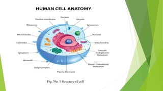
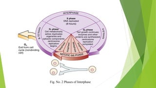
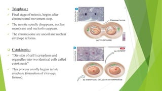
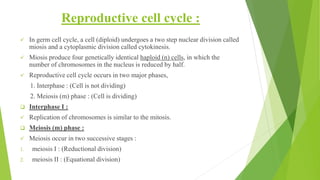
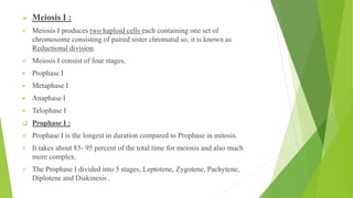
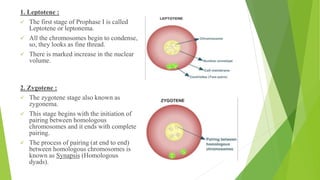
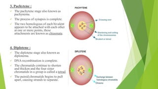
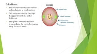
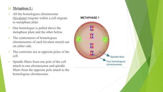
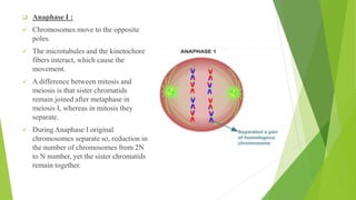
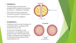
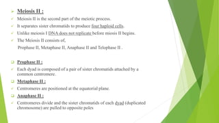
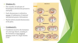
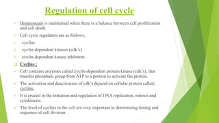
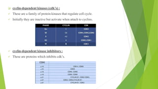
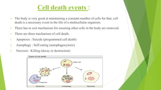
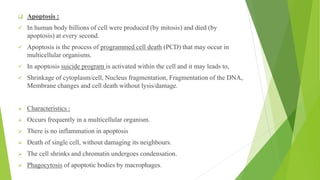
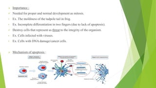
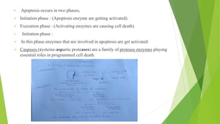
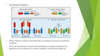
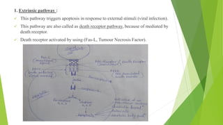
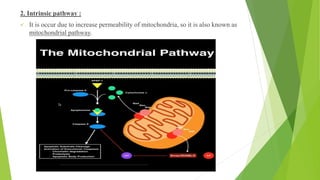
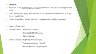
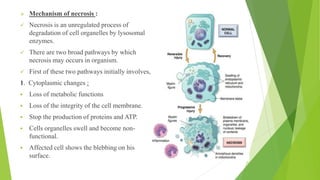
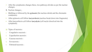
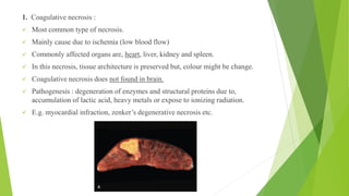
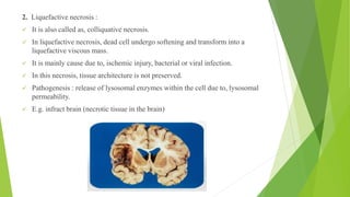
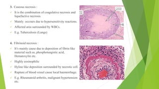
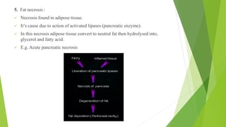
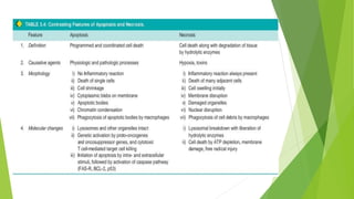
Cell Biology
Presented by,
Avinash Gunjal
F. Y. M-pharm (Pharmacology)
Dr. D Y Patil College of Pharmacy,
Akurdi.
Guided by,
Dr. Nikita Saraswat
Assistant Professor
Dept. of Pharmacology
Dr. D Y Patil College of Pharmacy,
Akurdi.
Fig. No. 1 Structure of cell
Cell cycle and it’s regulation :
 Cell cycle :
✓ “The cell cycle is an orderly sequence of events in which a cell
duplicates it’s content and divides into daughter cells”
✓ In somatic cell cycle, a cell undergoes a nuclear division called mitosis
and a cytoplasmic division called cytokinesis.
✓ Mitosis produce two genetically identical cells, each with the same
number of chromosomes as the parent cell.
✓ In germ cell cycle, a cell undergoes a two step nuclear division called
miosis and a cytoplasmic division called cytokinesis.
✓ Miosis produce four genetically identical cells, in which the number of
chromosomes in the nucleus is reduced by half.
Somatic cell cycle :
✓ “Somatic cell cycle is an orderly sequence of events in which a somatic cell
duplicates it’s content and divides to produce two identical diploid (2n) cells”.
✓ Somatic cell cycle consist of two major phases,
1) Interphase : (Cell is not dividing)
2) Mitotic (M) phase : (Cell is dividing)
1. Interphase :
✓ Interphase is the phase of the cell cycle in which a typical cell spends most of
its life.
✓ Interphase is the metabolic phase of the cell, in which the cell obtains nutrients
and metabolizes them, grows, replicates its DNA.
✓ Interphase consists of three phases, First gap phase (G1), Synthesis of DNA (S)
and Second gap phase (G2).
Fig. No. 2 Phases of Interphase
1. Mitotic (M) Phase :
✓ In this phase, a cell undergoes a nuclear division (karyokinesis) called mitosis
and a cytoplasmic division called cytokinesis to form two genetically identical
cells.
✓ Mitotic phase divide into four phases are as follow, Prophase, Metaphase,
Anaphase and Telophase.
➢ Prophase :
✓ First phase of mitosis, begins after interphase
✓ DNA has been replicated before prophase begins
✓ Prophase is longest phase of karyokinesis.
✓ Prophase divides into two phases,
▪ Early prophase
▪ Late Prophase
❑ Early prophase :
✓ Chromatin fibers condense and shorten into chromosomes (called chromatid at
this stage)
✓ Nucleolus disappears and nuclear membrane break down
❑ Late Prophase :
✓ Separation of centrosomes
✓ Centrosome start to form mitotic spindle..
✓ Cell organelles and nuclear membrane start to disappear.
➢ Metaphase :
✓ The microtubule of the mitotic spindle
align the centromeres of chromatid pair at
the centre of mitotic spindle.
✓ This plane alignment of the centromeres is
called metaphase plate.
➢ Anaphase :
✓ The centromeres are seprated, each pair of
sister chromatid migrate towards end of
spindle.
✓ Separated chromatids (called chromosome)
move to opposite pole of the cell.
✓ As microtubule pulled chromosome
towards pole they appear V-shaped.
➢ Telophase :
✓ Final stage of mitosis, begins after
chromosomal movement stop.
✓ The mitotic spindle disappears, nuclear
membrane and nucleoli reappears.
✓ The chromosome are uncoil and nuclear
envelope reforms.
❑ Cytokinesis :
✓ “Division of cell’s cytoplasm and
organelles into two identical cells called
cytokinesis”
✓ This process usually begins in late
anaphase (formation of cleavage
furrow).
Reproductive cell cycle :
✓ In germ cell cycle, a cell (diploid) undergoes a two step nuclear division called
miosis and a cytoplasmic division called cytokinesis.
✓ Miosis produce four genetically identical haploid (n) cells, in which the
number of chromosomes in the nucleus is reduced by half.
✓ Reproductive cell cycle occurs in two major phases,
1. Interphase : (Cell is not dividing)
2. Meiosis (m) phase : (Cell is dividing)
❑ Interphase I :
✓ Replication of chromosomes is similar to the mitosis.
❑ Meiosis (m) phase :
✓ Meiosis occur in two successive stages :
1. meiosis I : (Reductional division)
2. meiosis II : (Equational division)
➢ Meiosis I :
✓ Meiosis I produces two haploid cells each containing one set of
chromosome consisting of paired sister chromatid so, it is known as
Reductional division.
✓ Meiosis I consist of four stages,
▪ Prophase I
▪ Metaphase I
▪ Anaphase I
▪ Telophase I
❑ Prophase I :
✓ Prophase I is the longest in duration compared to Prophase in mitosis.
✓ It takes about 85- 95 percent of the total time for meiosis and also much
more complex.
✓ The Prophase I divided into 5 stages, Leptotene, Zygotene, Pachytene,
Diplotene and Diakinesis .
1. Leptotene :
✓ The first stage of Prophase I is called
Leptotene or leptonema.
✓ All the chromosomes begin to condense,
so, they looks as fine thread.
✓ There is marked increase in the nuclear
volume.
2. Zygotene :
✓ The zygotene stage also known as
zygonema.
✓ This stage begins with the initiation of
pairing between homologous
chromosomes and it ends with complete
pairing.
✓ The process of pairing (at end to end)
between homologous chromosomes is
known as Synapsis (Homologous
dyads).
3. Pachytene :
✓ The pachytene stage also known as
pachynema.
✓ The process of synapsis is complete.
✓ The two homologous of each bivalent
appears to be attached with each other
at one or more points, these
attachments are known as chiasmata.
4. Diplotene :
✓ The diplotene stage also known as
diplonema.
✓ DNA recombination is complete.
✓ The chromatids continue to shorten
and thicken and the four sister
chromatids in a group is called a tetrad.
✓ The paired chromatids begins to pull
apart, causing strands to separate.
5. Diakinesis :
✓ The chromosomes become shorter
and thicker due to condensation.
✓ Nucleolus and nuclear envelope
disappear towards the end of
diakinesis.
✓ The spindle apparatus becomes
organized and the centrioles migrate
away from one another.
❑ Metaphase I :
✓ All the homologous chromosome
(bivalents) migrate within a cell migrate
to metaphase plate.
✓ One homologue is pulled above the
metaphase plate and the other below.
✓ The centromeres of homologous
chromosomes of each bivalent stretch out
on either side.
✓ The centrioles are at opposite poles of the
cell.
✓ Spindle fibers from one pole of the cell
attach to one chromosome and spindle
fibers from the opposite pole attach to the
homologous chromosome.
❑ Anaphase I :
✓ Chromosomes move to the opposite
poles.
✓ The microtubules and the kinetochore
fibers interact, which cause the
movement.
✓ A difference between mitosis and
meiosis is that sister chromatids
remain joined after metaphase in
meiosis I, whereas in mitosis they
separate.
✓ During Anaphase I original
chromosomes separate so, reduction in
the number of chromosomes from 2N
to N number, yet the sister chromatids
remain together.
❑ Telophase I :
✓ The homologous chromosome
complete their migration towards the
two poles i.e. shortning of spindles.
✓ The nuclear envelope organized
around two groups of chromosomes.
✓ The nucleolus also reappears.
➢ Cytokinesis I :
➢ Cytokinesis involves the formation of
a cleavage furrow, resulting in the
formation of two cells.
➢ At the end of Telophase I and
Cytokinesis, two daughter cells are
produced, each with one half of the
number of chromosomes (haploid set
of replicated chromosomes) of the
original parent cell.
➢ Meiosis II :
✓ Meiosis II is the second part of the meiotic process.
✓ It separates sister chromatids to produce four haploid cells.
✓ Unlike meiosis I DNA does not replicate before miosis II begins.
✓ The Meiosis II consists of,
Prophase II, Metaphase II, Anaphase II and Telophase II .
❑ Prophase II :
✓ Each dyad is composed of a pair of sister chromatids attached by a
common centromere.
❑ Metaphase II :
✓ Centromeres are positioned at the equatorial plane.
❑ Anaphase II :
✓ Centromeres divide and the sister chromatids of each dyad (duplicated
chromosome) are pulled to opposite poles
❑ Telophase II :
✓ One member of each pair of
homologous chromosome present in
each pole.
✓ Each chromosome is referred as
monad. (Combination of maternal
and paternal genetic information).
✓ Nuclei reform around chromosomes
at the poles.
➢ Cytokinesis II :
✓ Cytokinesis involves the formation
of a cleavage furrow, resulting in
the formation of four haploid
gametes result of a single meiotic
event.
Regulation of cell cycle
✓ Homeostasis is maintained when there is a balance between cell proliferation
and cell death.
✓ Cell cycle regulators are as follows,
1. cyclins
2. cyclin-dependent kinases (cdk’s)
3. cyclin-dependent kinase inhibitors
❑ Cyclins :
✓ Cell contains enzymes called cyclin-dependent protein kinase (cdk’s), that
transfer phosphate group from ATP to a protein to activate the protein.
✓ The activation and deactivation of cdk’s depend on cellular protein called,
cyclins.
✓ It is crucial in the initiation and regulation of DNA replication, mitosis and
cytokinesis.
✓ The level of cyclins in the cell are very important in determining timing and
sequence of cell division.
❑ cyclin-dependent kinases (cdk’s) :
✓ These are a family of protein kinases that regulate cell cycle.
✓ Initially they are inactive but activate when attach to cyclins.
✓ cyclin-dependent kinase inhibitors :
✓ These are proteins which inhibits cdk’s.
Cell death events :
✓ The body is very good at maintaining a constant number of cells for that, cell
death is a necessary event in the life of a multicellular organism.
✓ There has to exit mechanism for ensuring other cells in the body are removed.
✓ There are three mechanism of cell death,
1. Apoptosis : Suicide (programmed cell death)
2. Autophagy : Self-eating (autophagocytosis)
3. Necrosis : Killing (decay or destruction)
❑ Apoptosis :
✓ In human body billions of cell were produced (by mitosis) and died (by
apoptosis) at every second.
✓ Apoptosis is the process of programmed cell death (PCD) that may occur in
multicellular organisms.
✓ In apoptosis suicide program is activated within the cell and it may leads to,
✓ Shrinkage of cytoplasm/cell, Nucleus fragmentation, Fragmentation of the DNA,
Membrane changes and cell death without lysis/damage.
➢ Characteristics :
➢ Occurs frequently in a multicellular organism.
➢ There is no inflammation in apoptosis
➢ Death of single cell, without damaging its neighbours.
➢ The cell shrinks and chromatin undergoes condensation.
➢ Phagocytosis of apoptotic bodies by macrophages.
➢ Importance :
✓ Needed for proper and normal development as mitosis.
• Ex. The moldiness of the tadpole tail in frog.
• Ex. Incomplete differentiation in two fingers (due to lack of apoptosis).
✓ Destroy cells that represent as threat to the integrity of the organism.
• Ex. Cells infected with viruses.
• Ex. Cells with DNA damage/cancer cells.
➢ Mechanism of apoptosis :
▪ Apoptosis occurs in two phases,
✓ Initiation phase : (Apoptosis enzyme are getting activated).
✓ Execution phase : (Activating enzymes are causing cell death).
1. Initiation phase :
✓ In this phase enzymes that are involved in apoptosis are get activated.
✓ Caspases (cysteine-aspartic proteases) are a family of protease enzymes playing
essential roles in programmed cell death.
▪ Activation of caspases :
✓ When initiator caspases activated, they are going to activate executioner
caspases.
✓ Once the executioner caspases activated they are going to perform the
apoptosis by two pathways, extrinsic pathway and intrinsic pathway.
1. Extrinsic pathway :
✓ This pathway triggers apoptosis in response to external stimuli (viral infection).
✓ This pathway are also called as death receptor pathway, because of mediated by
death receptor.
✓ Death receptor activated by using (Fas-L, Tumour Necrosis Factor).
2. Intrinsic pathway :
✓ It is occur due to increase permeability of mitochondria, so it is also known as
mitochondrial pathway.
❑ Necrosis :
✓ Necrosis, is the morphological changes that follow cell death in living tissue or
organ.
✓ It is a form of cell injury which results in the premature death of cells in living
tissue by autolysis.
✓ It is an unregulated digestion of cell components by hydrolytic enzymes.
➢ Causes of necrosis :
✓ External causes : Mechanical trauma,
Damage to blood vessel,
Thermal effect.
✓ Internal causes : Trophoneurotic disorder,
Pancreatic enzymes (lipase),
Bacterial toxins and pathogens.
➢ Mechanism of necrosis :
✓ Necrosis is an unregulated process of
degradation of cell organelles by lysosomal
enzymes.
✓ There are two broad pathways by which
necrosis may occurs in organism.
✓ First of these two pathways initially involves,
1. Cytoplasmic changes :
▪ Loss of metabolic functions
▪ Loss of the integrity of the cell membrane.
▪ Stop the production of proteins and ATP.
▪ Cells organelles swell and become non-
functional.
▪ Affected cell shows the blebbing on his
surface.
✓ After the cytoplasmic changes these, two pathways divides as per the nuclear
changes.
2. Nuclear changes :
▪ Blebbing is followed by the pyknosis (the nucleus shrink and the chromatin
condenses).
▪ After pyknosis cell follow karyorrhexis (nucleus break down into fragments)
▪ After karyorrhexis cell follow karyolysis (cell nuclei dissolved into the
cytoplasm).
❑ Types of necrosis :
1. Coagulative necrosis :
2. Liquefaction necrosis :
3. Caseous necrosis :
4. Fat necrosis :
5. Fibrinoid necrosis :
1. Coagulative necrosis :
✓ Most common type of necrosis.
✓ Mainly cause due to ischemia (low blood flow)
✓ Commonly affected organs are, heart, liver, kidney and spleen.
✓ In this necrosis, tissue architecture is preserved but, colour might be change.
✓ Coagulative necrosis does not found in brain.
✓ Pathogenesis : degeneration of enzymes and structural proteins due to,
accumulation of lactic acid, heavy metals or expose to ionizing radiation.
✓ E.g. myocardial infraction, zenker’s degenerative necrosis etc.
2. Liquefactive necrosis :
✓ It is also called as, colliquative necrosis.
✓ In liquefactive necrosis, dead cell undergo softening and transform into a
liquefactive viscous mass.
✓ It is mainly cause due to, ischemic injury, bacterial or viral infection.
✓ In this necrosis, tissue architecture is not preserved.
✓ Pathogenesis : release of lysosomal enzymes within the cell due to, lysosomal
permeability.
✓ E.g. infract brain (necrotic tissue in the brain)
3. Caseous necrosis :
✓ It is the combination of coagulative necrosis and
liquefactive necrosis.
✓ Mainly occours due to hypersensitivity reactions.
✓ Affected area surrounded by WBCs.
✓ E.g. Tuberculosis (Lungs)
4. Fibrinoid necrosis :
✓ It’s mainly cause due to deposition of fibrin like
material such as, phosphotungistic acid,
Hematoxylin etc.
✓ Highly eosinophilic
✓ Hyline like deposition surrounded by necrotic cell.
✓ Rupture of blood vessel cause local haemorrhage.
✓ E.g. Rheumatoid arthritis, malignant hypertension
etc.
5. Fat necrosis :
✓ Necrosis found in adipose tissue.
✓ It’s cause due to action of activated lipases (pancreatic enzyme).
✓ In this necrosis adipose tissue convert to neutral fat then hydrolysed into,
glycerol and fatty acid.
✓ E.g. Acute pancreatic necrosis
❑ Autophagy :
✓ Degradation of cytoplasmic components with the help of auto-phagosomes.
✓ It is an intracellular process of cellular cleaning, cytoprotection and self-
degredation
✓ Mainly seen due to, physiological and pathological condition.
✓ Autophagy is survival mechanism under the stressful condition.
✓ It maintain integrity of cell by recycling metabolites and clear intracellular
debris.
➢ Autophagy Pathway :
✓ It is occurs into three steps are as follows,
1. Initiation
2. Elongation
3. Fusion and degradation
➢ Mechanism of autophagy :
➢ References :
1. Gerard J. Tortora, Bryan Derrickson, “Principles of Anatomy and Physiology”
15th Edition 2017.
2. Anne Waugh, Allison Grant, “Ross and Wilson Anatomy & Physiology” in
health and illness, 12th Edition, 2014.
3. Gerald karp, “Cell and Molecular Biology Concept And Experiment” 7th
Edition, 2016
4. www.Wikipedia.org
5. www.slideshare.net

Cell Biology

  • 1.
    Cell Biology Presented by, AvinashGunjal F. Y. M-pharm (Pharmacology) Dr. D Y Patil College of Pharmacy, Akurdi. Guided by, Dr. Nikita Saraswat Assistant Professor Dept. of Pharmacology Dr. D Y Patil College of Pharmacy, Akurdi.
  • 2.
    Fig. No. 1Structure of cell
  • 3.
    Cell cycle andit’s regulation :  Cell cycle : ✓ “The cell cycle is an orderly sequence of events in which a cell duplicates it’s content and divides into daughter cells” ✓ In somatic cell cycle, a cell undergoes a nuclear division called mitosis and a cytoplasmic division called cytokinesis. ✓ Mitosis produce two genetically identical cells, each with the same number of chromosomes as the parent cell. ✓ In germ cell cycle, a cell undergoes a two step nuclear division called miosis and a cytoplasmic division called cytokinesis. ✓ Miosis produce four genetically identical cells, in which the number of chromosomes in the nucleus is reduced by half.
  • 4.
    Somatic cell cycle: ✓ “Somatic cell cycle is an orderly sequence of events in which a somatic cell duplicates it’s content and divides to produce two identical diploid (2n) cells”. ✓ Somatic cell cycle consist of two major phases, 1) Interphase : (Cell is not dividing) 2) Mitotic (M) phase : (Cell is dividing) 1. Interphase : ✓ Interphase is the phase of the cell cycle in which a typical cell spends most of its life. ✓ Interphase is the metabolic phase of the cell, in which the cell obtains nutrients and metabolizes them, grows, replicates its DNA. ✓ Interphase consists of three phases, First gap phase (G1), Synthesis of DNA (S) and Second gap phase (G2).
  • 5.
    Fig. No. 2Phases of Interphase
  • 6.
    1. Mitotic (M)Phase : ✓ In this phase, a cell undergoes a nuclear division (karyokinesis) called mitosis and a cytoplasmic division called cytokinesis to form two genetically identical cells. ✓ Mitotic phase divide into four phases are as follow, Prophase, Metaphase, Anaphase and Telophase. ➢ Prophase : ✓ First phase of mitosis, begins after interphase ✓ DNA has been replicated before prophase begins ✓ Prophase is longest phase of karyokinesis. ✓ Prophase divides into two phases, ▪ Early prophase ▪ Late Prophase
  • 7.
    ❑ Early prophase: ✓ Chromatin fibers condense and shorten into chromosomes (called chromatid at this stage) ✓ Nucleolus disappears and nuclear membrane break down ❑ Late Prophase : ✓ Separation of centrosomes ✓ Centrosome start to form mitotic spindle.. ✓ Cell organelles and nuclear membrane start to disappear.
  • 8.
    ➢ Metaphase : ✓The microtubule of the mitotic spindle align the centromeres of chromatid pair at the centre of mitotic spindle. ✓ This plane alignment of the centromeres is called metaphase plate. ➢ Anaphase : ✓ The centromeres are seprated, each pair of sister chromatid migrate towards end of spindle. ✓ Separated chromatids (called chromosome) move to opposite pole of the cell. ✓ As microtubule pulled chromosome towards pole they appear V-shaped.
  • 9.
    ➢ Telophase : ✓Final stage of mitosis, begins after chromosomal movement stop. ✓ The mitotic spindle disappears, nuclear membrane and nucleoli reappears. ✓ The chromosome are uncoil and nuclear envelope reforms. ❑ Cytokinesis : ✓ “Division of cell’s cytoplasm and organelles into two identical cells called cytokinesis” ✓ This process usually begins in late anaphase (formation of cleavage furrow).
  • 10.
    Reproductive cell cycle: ✓ In germ cell cycle, a cell (diploid) undergoes a two step nuclear division called miosis and a cytoplasmic division called cytokinesis. ✓ Miosis produce four genetically identical haploid (n) cells, in which the number of chromosomes in the nucleus is reduced by half. ✓ Reproductive cell cycle occurs in two major phases, 1. Interphase : (Cell is not dividing) 2. Meiosis (m) phase : (Cell is dividing) ❑ Interphase I : ✓ Replication of chromosomes is similar to the mitosis. ❑ Meiosis (m) phase : ✓ Meiosis occur in two successive stages : 1. meiosis I : (Reductional division) 2. meiosis II : (Equational division)
  • 11.
    ➢ Meiosis I: ✓ Meiosis I produces two haploid cells each containing one set of chromosome consisting of paired sister chromatid so, it is known as Reductional division. ✓ Meiosis I consist of four stages, ▪ Prophase I ▪ Metaphase I ▪ Anaphase I ▪ Telophase I ❑ Prophase I : ✓ Prophase I is the longest in duration compared to Prophase in mitosis. ✓ It takes about 85- 95 percent of the total time for meiosis and also much more complex. ✓ The Prophase I divided into 5 stages, Leptotene, Zygotene, Pachytene, Diplotene and Diakinesis .
  • 12.
    1. Leptotene : ✓The first stage of Prophase I is called Leptotene or leptonema. ✓ All the chromosomes begin to condense, so, they looks as fine thread. ✓ There is marked increase in the nuclear volume. 2. Zygotene : ✓ The zygotene stage also known as zygonema. ✓ This stage begins with the initiation of pairing between homologous chromosomes and it ends with complete pairing. ✓ The process of pairing (at end to end) between homologous chromosomes is known as Synapsis (Homologous dyads).
  • 13.
    3. Pachytene : ✓The pachytene stage also known as pachynema. ✓ The process of synapsis is complete. ✓ The two homologous of each bivalent appears to be attached with each other at one or more points, these attachments are known as chiasmata. 4. Diplotene : ✓ The diplotene stage also known as diplonema. ✓ DNA recombination is complete. ✓ The chromatids continue to shorten and thicken and the four sister chromatids in a group is called a tetrad. ✓ The paired chromatids begins to pull apart, causing strands to separate.
  • 14.
    5. Diakinesis : ✓The chromosomes become shorter and thicker due to condensation. ✓ Nucleolus and nuclear envelope disappear towards the end of diakinesis. ✓ The spindle apparatus becomes organized and the centrioles migrate away from one another.
  • 15.
    ❑ Metaphase I: ✓ All the homologous chromosome (bivalents) migrate within a cell migrate to metaphase plate. ✓ One homologue is pulled above the metaphase plate and the other below. ✓ The centromeres of homologous chromosomes of each bivalent stretch out on either side. ✓ The centrioles are at opposite poles of the cell. ✓ Spindle fibers from one pole of the cell attach to one chromosome and spindle fibers from the opposite pole attach to the homologous chromosome.
  • 16.
    ❑ Anaphase I: ✓ Chromosomes move to the opposite poles. ✓ The microtubules and the kinetochore fibers interact, which cause the movement. ✓ A difference between mitosis and meiosis is that sister chromatids remain joined after metaphase in meiosis I, whereas in mitosis they separate. ✓ During Anaphase I original chromosomes separate so, reduction in the number of chromosomes from 2N to N number, yet the sister chromatids remain together.
  • 17.
    ❑ Telophase I: ✓ The homologous chromosome complete their migration towards the two poles i.e. shortning of spindles. ✓ The nuclear envelope organized around two groups of chromosomes. ✓ The nucleolus also reappears. ➢ Cytokinesis I : ➢ Cytokinesis involves the formation of a cleavage furrow, resulting in the formation of two cells. ➢ At the end of Telophase I and Cytokinesis, two daughter cells are produced, each with one half of the number of chromosomes (haploid set of replicated chromosomes) of the original parent cell.
  • 18.
    ➢ Meiosis II: ✓ Meiosis II is the second part of the meiotic process. ✓ It separates sister chromatids to produce four haploid cells. ✓ Unlike meiosis I DNA does not replicate before miosis II begins. ✓ The Meiosis II consists of, Prophase II, Metaphase II, Anaphase II and Telophase II . ❑ Prophase II : ✓ Each dyad is composed of a pair of sister chromatids attached by a common centromere. ❑ Metaphase II : ✓ Centromeres are positioned at the equatorial plane. ❑ Anaphase II : ✓ Centromeres divide and the sister chromatids of each dyad (duplicated chromosome) are pulled to opposite poles
  • 19.
    ❑ Telophase II: ✓ One member of each pair of homologous chromosome present in each pole. ✓ Each chromosome is referred as monad. (Combination of maternal and paternal genetic information). ✓ Nuclei reform around chromosomes at the poles. ➢ Cytokinesis II : ✓ Cytokinesis involves the formation of a cleavage furrow, resulting in the formation of four haploid gametes result of a single meiotic event.
  • 20.
    Regulation of cellcycle ✓ Homeostasis is maintained when there is a balance between cell proliferation and cell death. ✓ Cell cycle regulators are as follows, 1. cyclins 2. cyclin-dependent kinases (cdk’s) 3. cyclin-dependent kinase inhibitors ❑ Cyclins : ✓ Cell contains enzymes called cyclin-dependent protein kinase (cdk’s), that transfer phosphate group from ATP to a protein to activate the protein. ✓ The activation and deactivation of cdk’s depend on cellular protein called, cyclins. ✓ It is crucial in the initiation and regulation of DNA replication, mitosis and cytokinesis. ✓ The level of cyclins in the cell are very important in determining timing and sequence of cell division.
  • 21.
    ❑ cyclin-dependent kinases(cdk’s) : ✓ These are a family of protein kinases that regulate cell cycle. ✓ Initially they are inactive but activate when attach to cyclins. ✓ cyclin-dependent kinase inhibitors : ✓ These are proteins which inhibits cdk’s.
  • 22.
    Cell death events: ✓ The body is very good at maintaining a constant number of cells for that, cell death is a necessary event in the life of a multicellular organism. ✓ There has to exit mechanism for ensuring other cells in the body are removed. ✓ There are three mechanism of cell death, 1. Apoptosis : Suicide (programmed cell death) 2. Autophagy : Self-eating (autophagocytosis) 3. Necrosis : Killing (decay or destruction)
  • 23.
    ❑ Apoptosis : ✓In human body billions of cell were produced (by mitosis) and died (by apoptosis) at every second. ✓ Apoptosis is the process of programmed cell death (PCD) that may occur in multicellular organisms. ✓ In apoptosis suicide program is activated within the cell and it may leads to, ✓ Shrinkage of cytoplasm/cell, Nucleus fragmentation, Fragmentation of the DNA, Membrane changes and cell death without lysis/damage. ➢ Characteristics : ➢ Occurs frequently in a multicellular organism. ➢ There is no inflammation in apoptosis ➢ Death of single cell, without damaging its neighbours. ➢ The cell shrinks and chromatin undergoes condensation. ➢ Phagocytosis of apoptotic bodies by macrophages.
  • 24.
    ➢ Importance : ✓Needed for proper and normal development as mitosis. • Ex. The moldiness of the tadpole tail in frog. • Ex. Incomplete differentiation in two fingers (due to lack of apoptosis). ✓ Destroy cells that represent as threat to the integrity of the organism. • Ex. Cells infected with viruses. • Ex. Cells with DNA damage/cancer cells. ➢ Mechanism of apoptosis :
  • 25.
    ▪ Apoptosis occursin two phases, ✓ Initiation phase : (Apoptosis enzyme are getting activated). ✓ Execution phase : (Activating enzymes are causing cell death). 1. Initiation phase : ✓ In this phase enzymes that are involved in apoptosis are get activated. ✓ Caspases (cysteine-aspartic proteases) are a family of protease enzymes playing essential roles in programmed cell death.
  • 26.
    ▪ Activation ofcaspases : ✓ When initiator caspases activated, they are going to activate executioner caspases. ✓ Once the executioner caspases activated they are going to perform the apoptosis by two pathways, extrinsic pathway and intrinsic pathway.
  • 27.
    1. Extrinsic pathway: ✓ This pathway triggers apoptosis in response to external stimuli (viral infection). ✓ This pathway are also called as death receptor pathway, because of mediated by death receptor. ✓ Death receptor activated by using (Fas-L, Tumour Necrosis Factor).
  • 28.
    2. Intrinsic pathway: ✓ It is occur due to increase permeability of mitochondria, so it is also known as mitochondrial pathway.
  • 30.
    ❑ Necrosis : ✓Necrosis, is the morphological changes that follow cell death in living tissue or organ. ✓ It is a form of cell injury which results in the premature death of cells in living tissue by autolysis. ✓ It is an unregulated digestion of cell components by hydrolytic enzymes. ➢ Causes of necrosis : ✓ External causes : Mechanical trauma, Damage to blood vessel, Thermal effect. ✓ Internal causes : Trophoneurotic disorder, Pancreatic enzymes (lipase), Bacterial toxins and pathogens.
  • 31.
    ➢ Mechanism ofnecrosis : ✓ Necrosis is an unregulated process of degradation of cell organelles by lysosomal enzymes. ✓ There are two broad pathways by which necrosis may occurs in organism. ✓ First of these two pathways initially involves, 1. Cytoplasmic changes : ▪ Loss of metabolic functions ▪ Loss of the integrity of the cell membrane. ▪ Stop the production of proteins and ATP. ▪ Cells organelles swell and become non- functional. ▪ Affected cell shows the blebbing on his surface.
  • 32.
    ✓ After thecytoplasmic changes these, two pathways divides as per the nuclear changes. 2. Nuclear changes : ▪ Blebbing is followed by the pyknosis (the nucleus shrink and the chromatin condenses). ▪ After pyknosis cell follow karyorrhexis (nucleus break down into fragments) ▪ After karyorrhexis cell follow karyolysis (cell nuclei dissolved into the cytoplasm). ❑ Types of necrosis : 1. Coagulative necrosis : 2. Liquefaction necrosis : 3. Caseous necrosis : 4. Fat necrosis : 5. Fibrinoid necrosis :
  • 33.
    1. Coagulative necrosis: ✓ Most common type of necrosis. ✓ Mainly cause due to ischemia (low blood flow) ✓ Commonly affected organs are, heart, liver, kidney and spleen. ✓ In this necrosis, tissue architecture is preserved but, colour might be change. ✓ Coagulative necrosis does not found in brain. ✓ Pathogenesis : degeneration of enzymes and structural proteins due to, accumulation of lactic acid, heavy metals or expose to ionizing radiation. ✓ E.g. myocardial infraction, zenker’s degenerative necrosis etc.
  • 34.
    2. Liquefactive necrosis: ✓ It is also called as, colliquative necrosis. ✓ In liquefactive necrosis, dead cell undergo softening and transform into a liquefactive viscous mass. ✓ It is mainly cause due to, ischemic injury, bacterial or viral infection. ✓ In this necrosis, tissue architecture is not preserved. ✓ Pathogenesis : release of lysosomal enzymes within the cell due to, lysosomal permeability. ✓ E.g. infract brain (necrotic tissue in the brain)
  • 35.
    3. Caseous necrosis: ✓ It is the combination of coagulative necrosis and liquefactive necrosis. ✓ Mainly occours due to hypersensitivity reactions. ✓ Affected area surrounded by WBCs. ✓ E.g. Tuberculosis (Lungs) 4. Fibrinoid necrosis : ✓ It’s mainly cause due to deposition of fibrin like material such as, phosphotungistic acid, Hematoxylin etc. ✓ Highly eosinophilic ✓ Hyline like deposition surrounded by necrotic cell. ✓ Rupture of blood vessel cause local haemorrhage. ✓ E.g. Rheumatoid arthritis, malignant hypertension etc.
  • 36.
    5. Fat necrosis: ✓ Necrosis found in adipose tissue. ✓ It’s cause due to action of activated lipases (pancreatic enzyme). ✓ In this necrosis adipose tissue convert to neutral fat then hydrolysed into, glycerol and fatty acid. ✓ E.g. Acute pancreatic necrosis
  • 38.
    ❑ Autophagy : ✓Degradation of cytoplasmic components with the help of auto-phagosomes. ✓ It is an intracellular process of cellular cleaning, cytoprotection and self- degredation ✓ Mainly seen due to, physiological and pathological condition. ✓ Autophagy is survival mechanism under the stressful condition. ✓ It maintain integrity of cell by recycling metabolites and clear intracellular debris. ➢ Autophagy Pathway : ✓ It is occurs into three steps are as follows, 1. Initiation 2. Elongation 3. Fusion and degradation
  • 39.
    ➢ Mechanism ofautophagy :
  • 40.
    ➢ References : 1.Gerard J. Tortora, Bryan Derrickson, “Principles of Anatomy and Physiology” 15th Edition 2017. 2. Anne Waugh, Allison Grant, “Ross and Wilson Anatomy & Physiology” in health and illness, 12th Edition, 2014. 3. Gerald karp, “Cell and Molecular Biology Concept And Experiment” 7th Edition, 2016 4. www.Wikipedia.org 5. www.slideshare.net