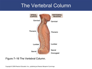
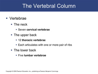
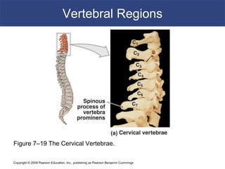
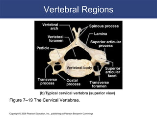
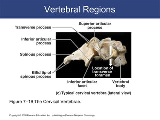
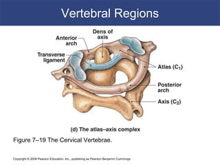
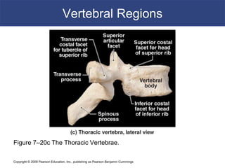
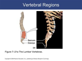
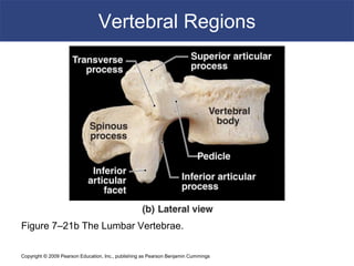
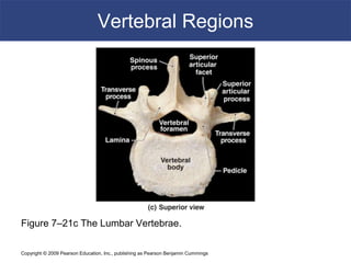
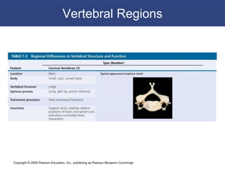
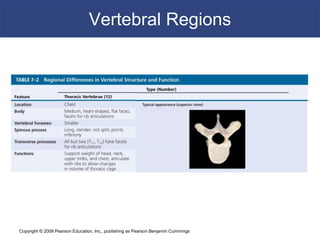
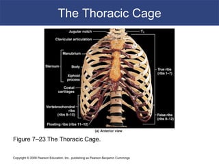
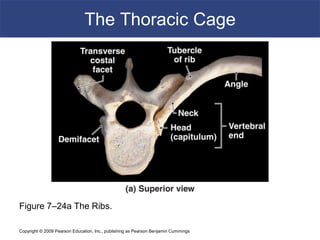
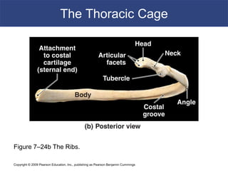
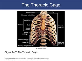
Copyright © 2009 Pearson Education, Inc., publishing as Pearson Benjamin Cummings
C h a p t e r
7
The Axial
Skeleton - Spine
PowerPoint® Lecture Slides
prepared by Jason LaPres
Lone Star College - North Harris
Modified by CHB 11/09
Copyright © 2009 Pearson Education, Inc.,
publishing as Pearson Benjamin Cummings
Copyright © 2009 Pearson Education, Inc., publishing as Pearson Benjamin Cummings
The Vertebral Column
 The spine or vertebral column
 Protects the spinal cord
 Supports the head and body
 26 bones
 24 vertebrae, the sacrum, and the coccyx
The Vertebral Column
Copyright © 2009 Pearson Education, Inc., publishing as Pearson Benjamin Cummings
The Vertebral Column
Figure 7–16 The Vertebral Column.
Copyright © 2009 Pearson Education, Inc., publishing as Pearson Benjamin Cummings
The Vertebral Column
 Vertebrae
 The neck
 Seven cervical vertebrae
 The upper back
 12 thoracic vertebrae
 Each articulates with one or more pair of ribs
 The lower back
 Five lumbar vertebrae
Copyright © 2009 Pearson Education, Inc., publishing as Pearson Benjamin Cummings
The Vertebral Column
 The Sacrum and Coccyx
 The fifth lumbar vertebra articulates with the
sacrum
 The sacrum articulates with the coccyx
Copyright © 2009 Pearson Education, Inc., publishing as Pearson Benjamin Cummings
The Vertebral Column
 Four Curvatures of the Vertebral Column
 Cervical curve
 Thoracic curve
 Lumbar curve
 Sacral curve
Copyright © 2009 Pearson Education, Inc., publishing as Pearson Benjamin Cummings
The Vertebral Column
 Thoracic and sacral curves
 Are called primary curves (present during fetal
development)
 Or accommodation curves (accommodate internal
organs)
 Lumbar and cervical curves
 Are called secondary curves (appear after birth)
 Or compensation curves (shift body weight for
upright posture)
Copyright © 2009 Pearson Education, Inc., publishing as Pearson Benjamin Cummings
The Vertebral Column
Figure 7–17 Abnormal Curvatures of the Spine.
Copyright © 2009 Pearson Education, Inc., publishing as Pearson Benjamin Cummings
The Vertebral Column
 Structure of a Vertebra
 The vertebral body (centrum)
 Transfers weight along the spine
 The vertebral arch
 Posterior margin of vertebral foramen
 The articular processes
 Lateral projections between laminae and pedicles
Copyright © 2009 Pearson Education, Inc., publishing as Pearson Benjamin Cummings
The Vertebral Column
Figure 7–18a Vertebral Anatomy.
Copyright © 2009 Pearson Education, Inc., publishing as Pearson Benjamin Cummings
The Vertebral Column
Figure 7–18c Vertebral Anatomy.
Copyright © 2009 Pearson Education, Inc., publishing as Pearson Benjamin Cummings
The Vertebral Column
 Structure of a Vertebra
 The vertebral arch
 Pedicles:
– walls of the vertebral arch
 Laminae:
– roof of the vertebral arch
 Spinous process:
– projection where vertebral laminae fuse
 Transverse process:
– projection where laminae join pedicles
Copyright © 2009 Pearson Education, Inc., publishing as Pearson Benjamin Cummings
The Vertebral Column
 Structure of a Vertebra
 The articular processes
 Superior articular process
 Inferior articular process:
– have articular facets on articular faces
Copyright © 2009 Pearson Education, Inc., publishing as Pearson Benjamin Cummings
The Vertebral Column
Figure 7–18 Vertebral Anatomy.
Copyright © 2009 Pearson Education, Inc., publishing as Pearson Benjamin Cummings
The Vertebral Column
 Vertebral Foramina
 Intervertebral foramina
 Gaps between pedicles of adjacent vertebrae
 For nerve connections to spinal cord
 Vertebral canal
 Formed by vertebral foramina
 Encloses the spinal cord
 Intervertebral Discs
 Are pads of fibrous cartilage
 Separate the vertebral bodies
 Absorb shocks
Copyright © 2009 Pearson Education, Inc., publishing as Pearson Benjamin Cummings
The Vertebral Column
Figure 7–18 Vertebral Anatomy.
Copyright © 2009 Pearson Education, Inc., publishing as Pearson Benjamin Cummings
Vertebral Regions
 Vertebral Regions
 Vertebrae are numbered
 By region, from top (superior) to bottom(inferior)
 C1 articulates with skull, L5 with sacrum
 Vertebrae of each region
 Have characteristics determined by functions
Copyright © 2009 Pearson Education, Inc., publishing as Pearson Benjamin Cummings
Vertebral Regions
 Regions of the Vertebral Column
 Cervical (C)
 Thoracic (T)
 Lumbar (L)
 Sacral (S)
 Coccygeal (Co)
Copyright © 2009 Pearson Education, Inc., publishing as Pearson Benjamin Cummings
Vertebral Regions
 The Cervical Vertebrae
 Small body (support only head)
 Large vertebral foramen (largest part of spinal
cord)
 Concave superior surface
 Slopes posterior to anterior
 C1 (atlas) has no spinous process
 All others have short spinous processes
– tip of each spinous process is notched (bifid)
Copyright © 2009 Pearson Education, Inc., publishing as Pearson Benjamin Cummings
Vertebral Regions
Figure 7–19 The Cervical Vertebrae.
Copyright © 2009 Pearson Education, Inc., publishing as Pearson Benjamin Cummings
Vertebral Regions
 The Cervical Vertebrae
 Transverse processes
 Are fused to costal processes
 Which encircle transverse foramina (protect
arteries and veins)
 Atlas (C1)
 Articulates with occipital condyles of skull
 Has no body or spinous process
 Has a large, round foramen within anterior and
posterior arches
Copyright © 2009 Pearson Education, Inc., publishing as Pearson Benjamin Cummings
Vertebral Regions
Figure 7–19 The Cervical Vertebrae.
Copyright © 2009 Pearson Education, Inc., publishing as Pearson Benjamin Cummings
Vertebral Regions
 The Cervical Vertebrae
 Axis (C2)
 Supports the atlas
 Has heavy spinous process
 To attach muscles of head and neck
 Axis and atlas bodies fuse during development to form the dens
 Vertebra prominens (C7)
 Transitions to thoracic vertebrae
 Has a long spinous process with a broad tubercle
 Has large transverse processes
 Ligamentum nuchae (elastic ligament) extends from C7 to skull
Rotation of Cervical Vertebrae
Copyright © 2009 Pearson Education, Inc., publishing as Pearson Benjamin Cummings
Vertebral Regions
Figure 7–19 The Cervical Vertebrae.
Copyright © 2009 Pearson Education, Inc., publishing as Pearson Benjamin Cummings
Vertebral Regions
Figure 7–19 The Cervical Vertebrae.
Copyright © 2009 Pearson Education, Inc., publishing as Pearson Benjamin Cummings
Vertebral Regions
 Thoracic vertebrae (T1–T12)
 Have heart-shaped bodies
 Larger bodies than in C1–C7
 Smaller vertebral foramen than in C1–C7
 Long, slender spinous processes
 Dorsolateral surfaces of body have costal facets:
 Which articulate with heads of ribs
Copyright © 2009 Pearson Education, Inc., publishing as Pearson Benjamin Cummings
Vertebral Regions
 Thoracic vertebrae (T1–T12)
 T1–T10
 Have transverse costal facets
 On thick transverse processes for rib articulation
 Ribs at T1–T10
 Contact costal and transverse costal facets
 T1–T8 articulate with two pairs of ribs
 At superior and inferior costal facets
 T9–T11 articulate with one pair of ribs
 T10–T12 transition to lumbar vertebrae
3D Rotation of Thoracic Vertebrae
Copyright © 2009 Pearson Education, Inc., publishing as Pearson Benjamin Cummings
Vertebral Regions
Figure 7–20a The Thoracic Vertebrae.
Copyright © 2009 Pearson Education, Inc., publishing as Pearson Benjamin Cummings
Vertebral Regions
Figure 7–20b The Thoracic Vertebrae.
Copyright © 2009 Pearson Education, Inc., publishing as Pearson Benjamin Cummings
Vertebral Regions
Figure 7–20c The Thoracic Vertebrae.
Copyright © 2009 Pearson Education, Inc., publishing as Pearson Benjamin Cummings
Vertebral Regions
 Lumbar vertebrae (L1–L5)
 Largest vertebrae
 Oval-shaped bodies
 Thicker bodies than T1–T12
 No costal or transverse costal facets
 Triangular vertebral foramen
 Superior articular processes
 Face up and in
 Inferior articular processes
 Face down and out
Copyright © 2009 Pearson Education, Inc., publishing as Pearson Benjamin Cummings
Vertebral Regions
 Lumbar vertebrae (L1–L5)
 Transverse processes
 Slender
 Project dorsolaterally
 Spinous process:
 Short, heavy
 For attachment of lower back muscles
3D Rotation of Lumbar Vertebrae
Copyright © 2009 Pearson Education, Inc., publishing as Pearson Benjamin Cummings
Vertebral Regions
Figure 7–21a The Lumbar Vertebrae.
Copyright © 2009 Pearson Education, Inc., publishing as Pearson Benjamin Cummings
Vertebral Regions
Figure 7–21b The Lumbar Vertebrae.
Copyright © 2009 Pearson Education, Inc., publishing as Pearson Benjamin Cummings
Vertebral Regions
Figure 7–21c The Lumbar Vertebrae.
Copyright © 2009 Pearson Education, Inc., publishing as Pearson Benjamin Cummings
Vertebral Regions
Copyright © 2009 Pearson Education, Inc., publishing as Pearson Benjamin Cummings
Vertebral Regions
Copyright © 2009 Pearson Education, Inc., publishing as Pearson Benjamin Cummings
Vertebral Regions
Copyright © 2009 Pearson Education, Inc., publishing as Pearson Benjamin Cummings
Vertebral Regions
 The sacrum
 Is curved, more in males than in females
 Protects reproductive, urinary, and digestive organs
 Attaches
 The axial skeleton to pelvic girdle of appendicular skeleton
 Broad muscles that move the thigh
 The adult sacrum
 Consists of five fused sacral vertebrae
 Fuses between puberty and ages 25–30
 Leaving transverse lines
Copyright © 2009 Pearson Education, Inc., publishing as Pearson Benjamin Cummings
Vertebral Regions
 The sacrum
 Sacral canal
 Replaces the vertebral canal
 Sacral cornua
 Horn shaped
 Formed by laminae of the fifth sacral vertebra
 Which do not meet at midline
 Sacral hiatus
 Opening at the inferior end of the sacral canal
 Formed by ridges of sacral cornua
 Covered by connective tissues
Copyright © 2009 Pearson Education, Inc., publishing as Pearson Benjamin Cummings
Vertebral Regions
 The sacrum
 Median sacral crest
 Fused spinous processes
 Four pairs of sacral foramina open to either side
 Lateral sacral crest
 Fused transverse processes
 Attach to muscles of lower back and hip
Copyright © 2009 Pearson Education, Inc., publishing as Pearson Benjamin Cummings
Vertebral Regions
 The sacrum
 Auricular surface
 Thick, flattened area
 Articulates with pelvic girdle (forming sacroiliac joint)
 Sacral tuberosity
 Rough area
 Attaches ligaments of the sacroiliac joint
Copyright © 2009 Pearson Education, Inc., publishing as Pearson Benjamin Cummings
Vertebral Regions
 The sacrum
 Four regions of the sacrum
 Base:
– the broad superior surface
 Ala:
– wings at either side of the base
– to attach muscles
 Sacral promontory:
– at the center of the base
 Apex:
– the narrow inferior portion
– articulates with the coccyx
Copyright © 2009 Pearson Education, Inc., publishing as Pearson Benjamin Cummings
Vertebral Regions
 The coccyx
 Attaches ligaments and a constricting muscle of the
anus
 Mature coccyx
 Consists of three to five fused coccygeal vertebrae
 First two coccygeal vertebrae:
 Have transverse processes
 Have unfused vertebral arches
 Coccygeal cornua
 Formed by laminae of first coccygeal vertebra
3D Rotation of Sacrum and Coccyx
Copyright © 2009 Pearson Education, Inc., publishing as Pearson Benjamin Cummings
Vertebral Regions
Figure 7–22 The Sacrum and Coccyx.
Copyright © 2009 Pearson Education, Inc., publishing as Pearson Benjamin Cummings
The Thoracic Cage
 The skeleton of the chest
 Supports the thoracic cavity
 Consists of:
– thoracic vertebrae
– ribs
– sternum (breastbone)
 The Rib Cage
 Formed of ribs and sternum
Copyright © 2009 Pearson Education, Inc., publishing as Pearson Benjamin Cummings
The Thoracic Cage
Figure 7–23a The Thoracic Cage.
Copyright © 2009 Pearson Education, Inc., publishing as Pearson Benjamin Cummings
The Thoracic Cage
Figure 7–23b The Thoracic Cage.
Copyright © 2009 Pearson Education, Inc., publishing as Pearson Benjamin Cummings
The Thoracic Cage
 Functions of the Thoracic Cage
 Protects organs of the thoracic cavity
 Heart, lungs, and thymus
 Attaches muscles
 For respiration
 Of the vertebral column
 Of the pectoral girdle
 Of the upper limbs
Copyright © 2009 Pearson Education, Inc., publishing as Pearson Benjamin Cummings
The Thoracic Cage
 Ribs
 Are mobile
 Can absorb shock
 Functions of ribs
 Rib movements (breathing):
– affect width and depth of thoracic cage
– changing its volume
Copyright © 2009 Pearson Education, Inc., publishing as Pearson Benjamin Cummings
The Thoracic Cage
Figure 7–24c The Ribs.
Copyright © 2009 Pearson Education, Inc., publishing as Pearson Benjamin Cummings
The Thoracic Cage
 Ribs (costae)
 Are 12 pairs of long, curved, flat bones
 Extending from the thoracic vertebrae
 Ribs are divided into two types
 True ribs
 False ribs
Copyright © 2009 Pearson Education, Inc., publishing as Pearson Benjamin Cummings
The Thoracic Cage
 Ribs 1–7 (true ribs)
 Vertebrosternal ribs
 Connected to the sternum by costal cartilages
 Ribs 8–12 (false ribs)
 Do not attach directly to the sternum
 Vertebrochondral ribs (ribs 8–10)
 Fuse together
 Merge with cartilage before reaching the sternum
 Floating or vertebral ribs (ribs 11–12)
 Connect only to the vertebrae and back muscles
 Have no connection with the sternum
Copyright © 2009 Pearson Education, Inc., publishing as Pearson Benjamin Cummings
The Thoracic Cage
Figure 7–23 The Thoracic Cage.
Copyright © 2009 Pearson Education, Inc., publishing as Pearson Benjamin Cummings
The Thoracic Cage
 Structures of the Ribs
 The head (capitulum)
 At the vertebral end of the rib
 Has superior and inferior articular facets
 The neck
 The short area between the head and the tubercle
Copyright © 2009 Pearson Education, Inc., publishing as Pearson Benjamin Cummings
The Thoracic Cage
 Structures of the Ribs
 The tubercle (tuberculum)
 A small dorsal elevation
 Has an auricular facet that contacts the facet of its
thoracic vertebra (at T1–T10 only)
 The tubercular body (shaft)
 Attaches muscles of the pectoral girdle and trunk
 Attaches to the intercostal muscles that move the
ribs
Copyright © 2009 Pearson Education, Inc., publishing as Pearson Benjamin Cummings
The Thoracic Cage
Figure 7–24a The Ribs.
Copyright © 2009 Pearson Education, Inc., publishing as Pearson Benjamin Cummings
The Thoracic Cage
Figure 7–24b The Ribs.
Copyright © 2009 Pearson Education, Inc., publishing as Pearson Benjamin Cummings
The Thoracic Cage
 The sternum
 A flat bone
 In the midline of the thoracic wall
 Three parts of the sternum
 The manubrium
 The sternal body
 The xiphoid process
Copyright © 2009 Pearson Education, Inc., publishing as Pearson Benjamin Cummings
The Thoracic Cage
 Manubrium
 The superior portion of sternum
 Broad, triangular shape
 Articulates with clavicles (collarbones)
 Articulates with cartilages of first rib pair
 Has a jugular notch, a shallow indentation
between clavicular articulations
Copyright © 2009 Pearson Education, Inc., publishing as Pearson Benjamin Cummings
The Thoracic Cage
 The sternal body
 Is tongue-shaped
 Attaches to the manubrium
 Attaches to costal cartilages of ribs 2–7
 The xiphoid process
 Is the smallest part of the sternum
 Attaches to the sternal body
 Attaches to diaphragm and rectus abdominis muscles
Copyright © 2009 Pearson Education, Inc., publishing as Pearson Benjamin Cummings
The Thoracic Cage
Figure 7–23 The Thoracic Cage.
Copyright © 2009 Pearson Education, Inc., publishing as Pearson Benjamin Cummings
The Thoracic Cage
 Development of the Sternum
 The developing sternal body
 Consists of four unfused bones
 Completes fusion about age 25
 Leaving transverse lines
 The xiphoid process
 Is the last part of sternum to fuse
 Can easily be broken away

Vertebral_Column_anatomy mmbs PPP.pptx

  • 1.
    Copyright © 2009Pearson Education, Inc., publishing as Pearson Benjamin Cummings C h a p t e r 7 The Axial Skeleton - Spine PowerPoint® Lecture Slides prepared by Jason LaPres Lone Star College - North Harris Modified by CHB 11/09 Copyright © 2009 Pearson Education, Inc., publishing as Pearson Benjamin Cummings
  • 2.
    Copyright © 2009Pearson Education, Inc., publishing as Pearson Benjamin Cummings The Vertebral Column  The spine or vertebral column  Protects the spinal cord  Supports the head and body  26 bones  24 vertebrae, the sacrum, and the coccyx The Vertebral Column
  • 3.
    Copyright © 2009Pearson Education, Inc., publishing as Pearson Benjamin Cummings The Vertebral Column Figure 7–16 The Vertebral Column.
  • 4.
    Copyright © 2009Pearson Education, Inc., publishing as Pearson Benjamin Cummings The Vertebral Column  Vertebrae  The neck  Seven cervical vertebrae  The upper back  12 thoracic vertebrae  Each articulates with one or more pair of ribs  The lower back  Five lumbar vertebrae
  • 5.
    Copyright © 2009Pearson Education, Inc., publishing as Pearson Benjamin Cummings The Vertebral Column  The Sacrum and Coccyx  The fifth lumbar vertebra articulates with the sacrum  The sacrum articulates with the coccyx
  • 6.
    Copyright © 2009Pearson Education, Inc., publishing as Pearson Benjamin Cummings The Vertebral Column  Four Curvatures of the Vertebral Column  Cervical curve  Thoracic curve  Lumbar curve  Sacral curve
  • 7.
    Copyright © 2009Pearson Education, Inc., publishing as Pearson Benjamin Cummings The Vertebral Column  Thoracic and sacral curves  Are called primary curves (present during fetal development)  Or accommodation curves (accommodate internal organs)  Lumbar and cervical curves  Are called secondary curves (appear after birth)  Or compensation curves (shift body weight for upright posture)
  • 8.
    Copyright © 2009Pearson Education, Inc., publishing as Pearson Benjamin Cummings The Vertebral Column Figure 7–17 Abnormal Curvatures of the Spine.
  • 9.
    Copyright © 2009Pearson Education, Inc., publishing as Pearson Benjamin Cummings The Vertebral Column  Structure of a Vertebra  The vertebral body (centrum)  Transfers weight along the spine  The vertebral arch  Posterior margin of vertebral foramen  The articular processes  Lateral projections between laminae and pedicles
  • 10.
    Copyright © 2009Pearson Education, Inc., publishing as Pearson Benjamin Cummings The Vertebral Column Figure 7–18a Vertebral Anatomy.
  • 11.
    Copyright © 2009Pearson Education, Inc., publishing as Pearson Benjamin Cummings The Vertebral Column Figure 7–18c Vertebral Anatomy.
  • 12.
    Copyright © 2009Pearson Education, Inc., publishing as Pearson Benjamin Cummings The Vertebral Column  Structure of a Vertebra  The vertebral arch  Pedicles: – walls of the vertebral arch  Laminae: – roof of the vertebral arch  Spinous process: – projection where vertebral laminae fuse  Transverse process: – projection where laminae join pedicles
  • 13.
    Copyright © 2009Pearson Education, Inc., publishing as Pearson Benjamin Cummings The Vertebral Column  Structure of a Vertebra  The articular processes  Superior articular process  Inferior articular process: – have articular facets on articular faces
  • 14.
    Copyright © 2009Pearson Education, Inc., publishing as Pearson Benjamin Cummings The Vertebral Column Figure 7–18 Vertebral Anatomy.
  • 15.
    Copyright © 2009Pearson Education, Inc., publishing as Pearson Benjamin Cummings The Vertebral Column  Vertebral Foramina  Intervertebral foramina  Gaps between pedicles of adjacent vertebrae  For nerve connections to spinal cord  Vertebral canal  Formed by vertebral foramina  Encloses the spinal cord  Intervertebral Discs  Are pads of fibrous cartilage  Separate the vertebral bodies  Absorb shocks
  • 16.
    Copyright © 2009Pearson Education, Inc., publishing as Pearson Benjamin Cummings The Vertebral Column Figure 7–18 Vertebral Anatomy.
  • 17.
    Copyright © 2009Pearson Education, Inc., publishing as Pearson Benjamin Cummings Vertebral Regions  Vertebral Regions  Vertebrae are numbered  By region, from top (superior) to bottom(inferior)  C1 articulates with skull, L5 with sacrum  Vertebrae of each region  Have characteristics determined by functions
  • 18.
    Copyright © 2009Pearson Education, Inc., publishing as Pearson Benjamin Cummings Vertebral Regions  Regions of the Vertebral Column  Cervical (C)  Thoracic (T)  Lumbar (L)  Sacral (S)  Coccygeal (Co)
  • 19.
    Copyright © 2009Pearson Education, Inc., publishing as Pearson Benjamin Cummings Vertebral Regions  The Cervical Vertebrae  Small body (support only head)  Large vertebral foramen (largest part of spinal cord)  Concave superior surface  Slopes posterior to anterior  C1 (atlas) has no spinous process  All others have short spinous processes – tip of each spinous process is notched (bifid)
  • 20.
    Copyright © 2009Pearson Education, Inc., publishing as Pearson Benjamin Cummings Vertebral Regions Figure 7–19 The Cervical Vertebrae.
  • 21.
    Copyright © 2009Pearson Education, Inc., publishing as Pearson Benjamin Cummings Vertebral Regions  The Cervical Vertebrae  Transverse processes  Are fused to costal processes  Which encircle transverse foramina (protect arteries and veins)  Atlas (C1)  Articulates with occipital condyles of skull  Has no body or spinous process  Has a large, round foramen within anterior and posterior arches
  • 22.
    Copyright © 2009Pearson Education, Inc., publishing as Pearson Benjamin Cummings Vertebral Regions Figure 7–19 The Cervical Vertebrae.
  • 23.
    Copyright © 2009Pearson Education, Inc., publishing as Pearson Benjamin Cummings Vertebral Regions  The Cervical Vertebrae  Axis (C2)  Supports the atlas  Has heavy spinous process  To attach muscles of head and neck  Axis and atlas bodies fuse during development to form the dens  Vertebra prominens (C7)  Transitions to thoracic vertebrae  Has a long spinous process with a broad tubercle  Has large transverse processes  Ligamentum nuchae (elastic ligament) extends from C7 to skull Rotation of Cervical Vertebrae
  • 24.
    Copyright © 2009Pearson Education, Inc., publishing as Pearson Benjamin Cummings Vertebral Regions Figure 7–19 The Cervical Vertebrae.
  • 25.
    Copyright © 2009Pearson Education, Inc., publishing as Pearson Benjamin Cummings Vertebral Regions Figure 7–19 The Cervical Vertebrae.
  • 26.
    Copyright © 2009Pearson Education, Inc., publishing as Pearson Benjamin Cummings Vertebral Regions  Thoracic vertebrae (T1–T12)  Have heart-shaped bodies  Larger bodies than in C1–C7  Smaller vertebral foramen than in C1–C7  Long, slender spinous processes  Dorsolateral surfaces of body have costal facets:  Which articulate with heads of ribs
  • 27.
    Copyright © 2009Pearson Education, Inc., publishing as Pearson Benjamin Cummings Vertebral Regions  Thoracic vertebrae (T1–T12)  T1–T10  Have transverse costal facets  On thick transverse processes for rib articulation  Ribs at T1–T10  Contact costal and transverse costal facets  T1–T8 articulate with two pairs of ribs  At superior and inferior costal facets  T9–T11 articulate with one pair of ribs  T10–T12 transition to lumbar vertebrae 3D Rotation of Thoracic Vertebrae
  • 28.
    Copyright © 2009Pearson Education, Inc., publishing as Pearson Benjamin Cummings Vertebral Regions Figure 7–20a The Thoracic Vertebrae.
  • 29.
    Copyright © 2009Pearson Education, Inc., publishing as Pearson Benjamin Cummings Vertebral Regions Figure 7–20b The Thoracic Vertebrae.
  • 30.
    Copyright © 2009Pearson Education, Inc., publishing as Pearson Benjamin Cummings Vertebral Regions Figure 7–20c The Thoracic Vertebrae.
  • 31.
    Copyright © 2009Pearson Education, Inc., publishing as Pearson Benjamin Cummings Vertebral Regions  Lumbar vertebrae (L1–L5)  Largest vertebrae  Oval-shaped bodies  Thicker bodies than T1–T12  No costal or transverse costal facets  Triangular vertebral foramen  Superior articular processes  Face up and in  Inferior articular processes  Face down and out
  • 32.
    Copyright © 2009Pearson Education, Inc., publishing as Pearson Benjamin Cummings Vertebral Regions  Lumbar vertebrae (L1–L5)  Transverse processes  Slender  Project dorsolaterally  Spinous process:  Short, heavy  For attachment of lower back muscles 3D Rotation of Lumbar Vertebrae
  • 33.
    Copyright © 2009Pearson Education, Inc., publishing as Pearson Benjamin Cummings Vertebral Regions Figure 7–21a The Lumbar Vertebrae.
  • 34.
    Copyright © 2009Pearson Education, Inc., publishing as Pearson Benjamin Cummings Vertebral Regions Figure 7–21b The Lumbar Vertebrae.
  • 35.
    Copyright © 2009Pearson Education, Inc., publishing as Pearson Benjamin Cummings Vertebral Regions Figure 7–21c The Lumbar Vertebrae.
  • 36.
    Copyright © 2009Pearson Education, Inc., publishing as Pearson Benjamin Cummings Vertebral Regions
  • 37.
    Copyright © 2009Pearson Education, Inc., publishing as Pearson Benjamin Cummings Vertebral Regions
  • 38.
    Copyright © 2009Pearson Education, Inc., publishing as Pearson Benjamin Cummings Vertebral Regions
  • 39.
    Copyright © 2009Pearson Education, Inc., publishing as Pearson Benjamin Cummings Vertebral Regions  The sacrum  Is curved, more in males than in females  Protects reproductive, urinary, and digestive organs  Attaches  The axial skeleton to pelvic girdle of appendicular skeleton  Broad muscles that move the thigh  The adult sacrum  Consists of five fused sacral vertebrae  Fuses between puberty and ages 25–30  Leaving transverse lines
  • 40.
    Copyright © 2009Pearson Education, Inc., publishing as Pearson Benjamin Cummings Vertebral Regions  The sacrum  Sacral canal  Replaces the vertebral canal  Sacral cornua  Horn shaped  Formed by laminae of the fifth sacral vertebra  Which do not meet at midline  Sacral hiatus  Opening at the inferior end of the sacral canal  Formed by ridges of sacral cornua  Covered by connective tissues
  • 41.
    Copyright © 2009Pearson Education, Inc., publishing as Pearson Benjamin Cummings Vertebral Regions  The sacrum  Median sacral crest  Fused spinous processes  Four pairs of sacral foramina open to either side  Lateral sacral crest  Fused transverse processes  Attach to muscles of lower back and hip
  • 42.
    Copyright © 2009Pearson Education, Inc., publishing as Pearson Benjamin Cummings Vertebral Regions  The sacrum  Auricular surface  Thick, flattened area  Articulates with pelvic girdle (forming sacroiliac joint)  Sacral tuberosity  Rough area  Attaches ligaments of the sacroiliac joint
  • 43.
    Copyright © 2009Pearson Education, Inc., publishing as Pearson Benjamin Cummings Vertebral Regions  The sacrum  Four regions of the sacrum  Base: – the broad superior surface  Ala: – wings at either side of the base – to attach muscles  Sacral promontory: – at the center of the base  Apex: – the narrow inferior portion – articulates with the coccyx
  • 44.
    Copyright © 2009Pearson Education, Inc., publishing as Pearson Benjamin Cummings Vertebral Regions  The coccyx  Attaches ligaments and a constricting muscle of the anus  Mature coccyx  Consists of three to five fused coccygeal vertebrae  First two coccygeal vertebrae:  Have transverse processes  Have unfused vertebral arches  Coccygeal cornua  Formed by laminae of first coccygeal vertebra 3D Rotation of Sacrum and Coccyx
  • 45.
    Copyright © 2009Pearson Education, Inc., publishing as Pearson Benjamin Cummings Vertebral Regions Figure 7–22 The Sacrum and Coccyx.
  • 46.
    Copyright © 2009Pearson Education, Inc., publishing as Pearson Benjamin Cummings The Thoracic Cage  The skeleton of the chest  Supports the thoracic cavity  Consists of: – thoracic vertebrae – ribs – sternum (breastbone)  The Rib Cage  Formed of ribs and sternum
  • 47.
    Copyright © 2009Pearson Education, Inc., publishing as Pearson Benjamin Cummings The Thoracic Cage Figure 7–23a The Thoracic Cage.
  • 48.
    Copyright © 2009Pearson Education, Inc., publishing as Pearson Benjamin Cummings The Thoracic Cage Figure 7–23b The Thoracic Cage.
  • 49.
    Copyright © 2009Pearson Education, Inc., publishing as Pearson Benjamin Cummings The Thoracic Cage  Functions of the Thoracic Cage  Protects organs of the thoracic cavity  Heart, lungs, and thymus  Attaches muscles  For respiration  Of the vertebral column  Of the pectoral girdle  Of the upper limbs
  • 50.
    Copyright © 2009Pearson Education, Inc., publishing as Pearson Benjamin Cummings The Thoracic Cage  Ribs  Are mobile  Can absorb shock  Functions of ribs  Rib movements (breathing): – affect width and depth of thoracic cage – changing its volume
  • 51.
    Copyright © 2009Pearson Education, Inc., publishing as Pearson Benjamin Cummings The Thoracic Cage Figure 7–24c The Ribs.
  • 52.
    Copyright © 2009Pearson Education, Inc., publishing as Pearson Benjamin Cummings The Thoracic Cage  Ribs (costae)  Are 12 pairs of long, curved, flat bones  Extending from the thoracic vertebrae  Ribs are divided into two types  True ribs  False ribs
  • 53.
    Copyright © 2009Pearson Education, Inc., publishing as Pearson Benjamin Cummings The Thoracic Cage  Ribs 1–7 (true ribs)  Vertebrosternal ribs  Connected to the sternum by costal cartilages  Ribs 8–12 (false ribs)  Do not attach directly to the sternum  Vertebrochondral ribs (ribs 8–10)  Fuse together  Merge with cartilage before reaching the sternum  Floating or vertebral ribs (ribs 11–12)  Connect only to the vertebrae and back muscles  Have no connection with the sternum
  • 54.
    Copyright © 2009Pearson Education, Inc., publishing as Pearson Benjamin Cummings The Thoracic Cage Figure 7–23 The Thoracic Cage.
  • 55.
    Copyright © 2009Pearson Education, Inc., publishing as Pearson Benjamin Cummings The Thoracic Cage  Structures of the Ribs  The head (capitulum)  At the vertebral end of the rib  Has superior and inferior articular facets  The neck  The short area between the head and the tubercle
  • 56.
    Copyright © 2009Pearson Education, Inc., publishing as Pearson Benjamin Cummings The Thoracic Cage  Structures of the Ribs  The tubercle (tuberculum)  A small dorsal elevation  Has an auricular facet that contacts the facet of its thoracic vertebra (at T1–T10 only)  The tubercular body (shaft)  Attaches muscles of the pectoral girdle and trunk  Attaches to the intercostal muscles that move the ribs
  • 57.
    Copyright © 2009Pearson Education, Inc., publishing as Pearson Benjamin Cummings The Thoracic Cage Figure 7–24a The Ribs.
  • 58.
    Copyright © 2009Pearson Education, Inc., publishing as Pearson Benjamin Cummings The Thoracic Cage Figure 7–24b The Ribs.
  • 59.
    Copyright © 2009Pearson Education, Inc., publishing as Pearson Benjamin Cummings The Thoracic Cage  The sternum  A flat bone  In the midline of the thoracic wall  Three parts of the sternum  The manubrium  The sternal body  The xiphoid process
  • 60.
    Copyright © 2009Pearson Education, Inc., publishing as Pearson Benjamin Cummings The Thoracic Cage  Manubrium  The superior portion of sternum  Broad, triangular shape  Articulates with clavicles (collarbones)  Articulates with cartilages of first rib pair  Has a jugular notch, a shallow indentation between clavicular articulations
  • 61.
    Copyright © 2009Pearson Education, Inc., publishing as Pearson Benjamin Cummings The Thoracic Cage  The sternal body  Is tongue-shaped  Attaches to the manubrium  Attaches to costal cartilages of ribs 2–7  The xiphoid process  Is the smallest part of the sternum  Attaches to the sternal body  Attaches to diaphragm and rectus abdominis muscles
  • 62.
    Copyright © 2009Pearson Education, Inc., publishing as Pearson Benjamin Cummings The Thoracic Cage Figure 7–23 The Thoracic Cage.
  • 63.
    Copyright © 2009Pearson Education, Inc., publishing as Pearson Benjamin Cummings The Thoracic Cage  Development of the Sternum  The developing sternal body  Consists of four unfused bones  Completes fusion about age 25  Leaving transverse lines  The xiphoid process  Is the last part of sternum to fuse  Can easily be broken away