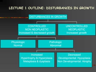
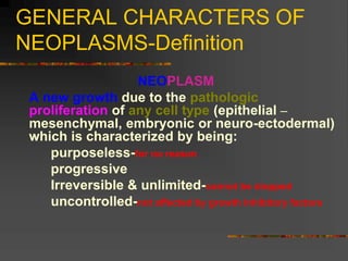
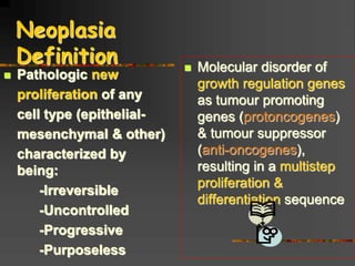
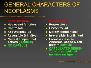
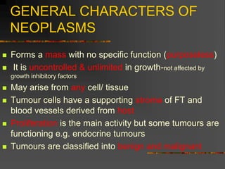
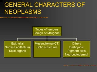
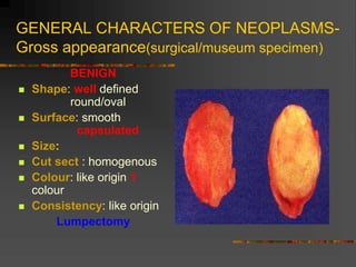
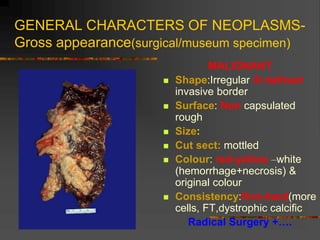
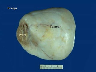
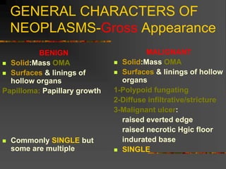
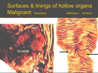
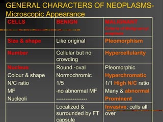
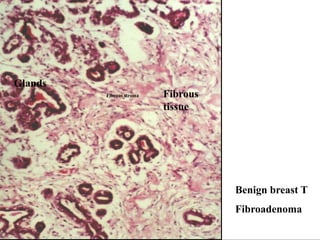
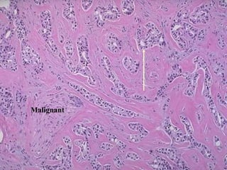
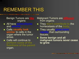
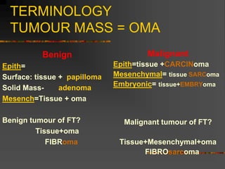
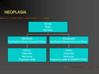
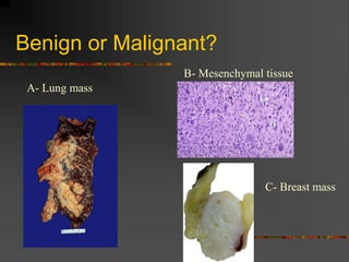
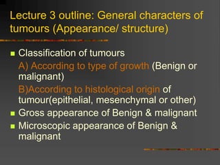
DISTURBANCES IN
GROWTH & TUMOURS:
NEOPLASIA L3
By
Prof. Soheir Mahfouz
NEOPLASMS
 Etiology : Co- carcinogens & carcinogens Lecture 2
 General characters of Benign & malignant
neoplasms(APPEARANCE) & classification of
tumours Lecture 3 pages 157-160
 General characters Behaviour of B & M tumours:–
complications & methods of tumour spread Lecture 4
 Epithelial neoplasms: B& M Lecture 5
 Mesenchymal neoplasms: B& M Lecture 6
 Other neoplasms: B& M Lecture 7
 Types of Malignant tumours their diagnosis,
prognosis & treatment Lecture 8
Physiologic
Normal
Increased
Hypertrophy & Hyperplasia
Metaplasia & dysplasia
Decreased
Developmental: Hypoplasia
Non Developmental: Atrophy
Pathologic
Abnormal
CONTROLLED
NON NEOPLASTIC
Increased & decreased growth
UNCONTROLLED
NEOPLASTIC
Increased growth
DISTURBANCES IN GROWTH
LECTURE 1 OUTLINE: DISTURBANCES IN GROWTH
NON NEOPLASTIC Increased growth
(controlled)
 Hypertrophy:Increase size of cells
 Hyperplasia: Increase number of cells
 Metaplasia: change from one mature cell type to
another mature more resistant cell type of the same
origin
 Dysplasia: Disordered proliferation & maturation ( it is
a preneoplastic change)
All are characterized by:
1-Known stimulus (CELL INJURY)
2-Reversible nature: return to normal after removal of stimulus
3-Intact basement membrane
4-Still under body control
L3
General
characters of
tumours
(Appearance/
structure)
What do they
look like?
GENERAL CHARACTERS OF
NEOPLASMS-Definition
NEOPLASM
A new growth due to the pathologic
proliferation of any cell type (epithelial –
mesenchymal, embryonic or neuro-ectodermal)
which is characterized by being:
purposeless-for no reason
progressive
Irreversible & unlimited-cannot be stopped
uncontrolled-not affected by growth inhibitory factors
Neoplasia
Definition
 Pathologic new
proliferation of any
cell type (epithelial-
mesenchymal & other)
characterized by
being:
-Irreversible
-Uncontrolled
-Progressive
-Purposeless
 Molecular disorder of
growth regulation genes
as tumour promoting
genes (protoncogenes)
& tumour suppressor
(anti-oncogenes),
resulting in a multistep
proliferation &
differentiation sequence
GENERAL CHARACTERS OF
NEOPLASMS
HYPERPLASIA
 Has useful function
 Controlled
 Known stimulus
 Reversible & limited
 Normal shape & cell
pattern (identical)
 NO CAPSULE
NEOPLASIA
 Purposeless
 Uncontrolled
 Mostly spontaneous
 Irreversible & unlimited
 Forms a mass +/-
Abnormal shape & cell
pattern (similar)
 CAPSULATED BENIGN
T. Non capsulated
invasive malignant
GENERAL CHARACTERS OF
NEOPLASMS
 Forms a mass with no specific function (purposeless)
 It is uncontrolled & unlimited in growth-not affected by
growth inhibitory factors
 May arise from any cell/ tissue
 Tumour cells have a supporting stroma of FT and
blood vessels derived from host
 Proliferation is the main activity but some tumours are
functioning e.g. endocrine tumours
 Tumours are classified into benign and malignant
GENERAL CHARACTERS OF
NEOPLASMS The structure & behaviour of any
neoplasm depends on:
Benign
Slow
Expansile
Localized
Malignant
Rapid
Invasive
Spread
GROWTH
1-Rate
2-Mode
3-Spread
GENERAL CHARACTERS OF
NEOPLASMS
Epithelial
Surface epithelium
Solid organs
Mesenchymal(CT)
Solid structures
Others
Embryonic
Pigment cells
Neuroectodermal
Types of tumours
Benign or Malignant
GENERAL CHARACTERS OF NEOPLASMS-
Gross appearance(surgical/museum specimen)
BENIGN
 Shape: well defined
round/oval
 Surface: smooth
capsulated
 Size:
 Cut sect : homogenous
 Colour: like origin 1
colour
 Consistency: like origin
Lumpectomy
GENERAL CHARACTERS OF NEOPLASMS-
Gross appearance(surgical/museum specimen)
MALIGNANT
 Shape:Irregular ill defined
invasive border
 Surface: Non capsulated
rough
 Size:
 Cut sect: mottled
 Colour: red-yellow –white
(hemorrhage+necrosis) &
original colour
 Consistency:firm-hard(more
cells, FT,dystrophic calcific
Radical Surgery +….
GROSS
Solid-OMA
BENIGN MALIGNANT
Benign
ovary
Tumour
Benign
tumour
Small
int.
Capsule
Benign
Malignant
GENERAL CHARACTERS OF
NEOPLASMS-Gross Appearance
BENIGN
 Solid:Mass OMA
 Surfaces & linings of
hollow organs
Papilloma: Papillary growth
 Commonly SINGLE but
some are multiple
MALIGNANT
 Solid:Mass OMA
 Surfaces & linings of hollow
organs
1-Polypoid fungating
2-Diffuse infiltrative/stricture
3-Malignant ulcer:
raised everted edge
raised necrotic Hgic floor
indurated base
 SINGLE
GROSS
Surfaces & linings of hollow organs
Papilloma-Polypoid fungating carcinoma
BENIGN MALIGNANT

Benign
Malignant
Surfaces & linings of hollow organs
Malignant: Ulcerative -infiltrative / stricture
FLOOR
edge
Microscopic appearance
GENERAL CHARACTERS OF NEOPLASMS-
Microscopic Appearance
CELLS BENIGN MALIGNANT
Criteria of Malignancy/
anaplasia
Size & shape Like original Pleomorphism
Number Cellular but no
crowding
Hypercellularity
Nucleus
Colour & shape
N/C ratio
MF
Nucleoli
Round -oval
Normochromic
1/5
-no abnormal MF
-----------------
Pleomorphic
Hyperchromatic
1/1 High N/C ratio
Many & abnormal
Prominent
Localized &
surrounded by FT
capsule
Invasive: cells all
over
Mitotic
figs
Malignant
Fibrous stroma
Glands
Fibrous
tissue
Benign breast T
Fibroadenoma
Malignant
REMEMBER THIS
Benign Tumors are like
organs:
 All have parenchyma
and stroma.
 Cells usually look
similar to cells in the
organ where the tumor
arose.
 Cells will continue to
perform some of the
functions of the parent
organ.
Malignant Tumors are different
from organs:
 They don't contribute to the
homeostasis of the body.
 They usually grow more
rapidly than surrounding
tissues.
 Some benign and all
malignant tumors never cease
to grow.
TERMINOLOGY
TUMOUR MASS = OMA
Benign
Epith=
Surface: tissue + papilloma
Solid Mass- adenoma
Mesench=Tissue + oma
Benign tumour of FT?
Tissue+oma
FIBRoma
Malignant
Epith=tissue +CARCINoma
Mesenchymal= tissue SARComa
Embryonic= tissue+EMBRYoma
Malignant tumour of FT?
Tissue+Mesenchymal+oma
FIBROsarcoma
NEOPLASIA
Other:
Vascular
Nervous
Pigment cells
BENIGN
Epithelial-Mesenchymal-Other
Other:
Vascular
Nervous
Pigment cells & EMBRYONIC
Malignant
Epithelial-Mesenchymal-Other
Mode
Rate
Spread
Benign or Malignant?
A- Lung mass
B- Mesenchymal tissue
C- Breast mass
Quiz Benign or Malignant?
A- Lung mass
Malignant
B- Mesenchymal tissue
Malignant
C- Breast mass
Benign
Now try these questions
 1. What are the main differences between
benign and malignant tumors?
 2. How are benign tumors distinguished
from malignant tumors in practice?
 3. What is anaplasia?
 4. How are neoplasms classified clinically?
 5. What is the basis of histologic or
histogenetic classification of tumors?
Lecture 3 outline: General characters of
tumours (Appearance/ structure)
 Classification of tumours
A) According to type of growth (Benign or
malignant)
B)According to histological origin of
tumour(epithelial, mesenchymal or other)
 Gross appearance of Benign & malignant
 Microscopic appearance of Benign &
malignant
Resources
 Chandrasoma, P. and Taylor, C.R.: Concise Pathology. 3' edition. McGraw Hill
Education 1997
 EI Hindawi, A.: Basic General Pathology. Faculty of Medicine, Cairo University
2009
 Reid,R Callander, R Ramsden I, Roberts,F . MacFarlane,PS. Pathology Illustrated
6th edition, Elsevier Science Health Science div 2005
 Jones, E. & Morris, A. Cell biology & Genetics. Mosby, London Philadelphia SI
Louis, Sydney, Tokyo. 1998
 Kumar,v., Abbas A.K. Cotran, R.S. Robbins,& Cotran Basic Pathology. 7" edition,
W.B. Saunders Co., Philadelphia, London, Toronto, Sydney, New York, Sydney &
Tokyo. 2005
 Stevens,J. & Lowe,J.: Pathology. Mosby. 1998
 Underwood, J.C.E.: General and Systematic Pathology. 3' edition. Churchill
Livingstone. Edinburgh, London, Madrid, Melbourne, New York & Tokyo. 2000
 Ciba collection of medical illustrations 1971
 Sandritter W., and Wartman, WB: Color atlas and textbook of Tissue and
cellular Pathology 5 th edition Year book Medical publications 1975
UNDERGRADUATE LECT 2
36
Resources
WEB
 http://www.medicine.cu.edu.eg/beta/enljcalpro/2428.htmI
 http://www.kasralainy.edu.eg/elearning/
 http://www.pathmax.com/
 http://www-medJib.med.utah.edu/WebPath/LABS/LABMENU.html#2
 http:/Iwww.med.uiuc.edu/PathAtlasf/titlePage.htmI
 http://www.medscape.com/pathologyhome
 http://www.gwumc.edu/deptlpath/2F.HTM
 http://www.path. uiowa.ed u/vi rtualsl idebox/
 http://web.med.unsw.edu.au/pathology/Pathmus/pathmus.htm#1
nteractiveJmages
 http://www.virtualpathology.leeds.ac.ukf
 http://histopathsho.34sp.comfindex.htm
 http://pathology.class.kmu.edu.tw/index.htm
 http://www.gJa.ac.ukffaculties/medicine/teaching/MedCALIist.htm

Undergraduate Pathology (L1)
37

n_l3_general_chracters_app_09pathology.ppt

  • 1.
    DISTURBANCES IN GROWTH &TUMOURS: NEOPLASIA L3 By Prof. Soheir Mahfouz
  • 2.
    NEOPLASMS  Etiology :Co- carcinogens & carcinogens Lecture 2  General characters of Benign & malignant neoplasms(APPEARANCE) & classification of tumours Lecture 3 pages 157-160  General characters Behaviour of B & M tumours:– complications & methods of tumour spread Lecture 4  Epithelial neoplasms: B& M Lecture 5  Mesenchymal neoplasms: B& M Lecture 6  Other neoplasms: B& M Lecture 7  Types of Malignant tumours their diagnosis, prognosis & treatment Lecture 8
  • 3.
    Physiologic Normal Increased Hypertrophy & Hyperplasia Metaplasia& dysplasia Decreased Developmental: Hypoplasia Non Developmental: Atrophy Pathologic Abnormal CONTROLLED NON NEOPLASTIC Increased & decreased growth UNCONTROLLED NEOPLASTIC Increased growth DISTURBANCES IN GROWTH LECTURE 1 OUTLINE: DISTURBANCES IN GROWTH
  • 4.
    NON NEOPLASTIC Increasedgrowth (controlled)  Hypertrophy:Increase size of cells  Hyperplasia: Increase number of cells  Metaplasia: change from one mature cell type to another mature more resistant cell type of the same origin  Dysplasia: Disordered proliferation & maturation ( it is a preneoplastic change) All are characterized by: 1-Known stimulus (CELL INJURY) 2-Reversible nature: return to normal after removal of stimulus 3-Intact basement membrane 4-Still under body control
  • 5.
  • 6.
    GENERAL CHARACTERS OF NEOPLASMS-Definition NEOPLASM Anew growth due to the pathologic proliferation of any cell type (epithelial – mesenchymal, embryonic or neuro-ectodermal) which is characterized by being: purposeless-for no reason progressive Irreversible & unlimited-cannot be stopped uncontrolled-not affected by growth inhibitory factors
  • 7.
    Neoplasia Definition  Pathologic new proliferationof any cell type (epithelial- mesenchymal & other) characterized by being: -Irreversible -Uncontrolled -Progressive -Purposeless  Molecular disorder of growth regulation genes as tumour promoting genes (protoncogenes) & tumour suppressor (anti-oncogenes), resulting in a multistep proliferation & differentiation sequence
  • 8.
    GENERAL CHARACTERS OF NEOPLASMS HYPERPLASIA Has useful function  Controlled  Known stimulus  Reversible & limited  Normal shape & cell pattern (identical)  NO CAPSULE NEOPLASIA  Purposeless  Uncontrolled  Mostly spontaneous  Irreversible & unlimited  Forms a mass +/- Abnormal shape & cell pattern (similar)  CAPSULATED BENIGN T. Non capsulated invasive malignant
  • 9.
    GENERAL CHARACTERS OF NEOPLASMS Forms a mass with no specific function (purposeless)  It is uncontrolled & unlimited in growth-not affected by growth inhibitory factors  May arise from any cell/ tissue  Tumour cells have a supporting stroma of FT and blood vessels derived from host  Proliferation is the main activity but some tumours are functioning e.g. endocrine tumours  Tumours are classified into benign and malignant
  • 10.
    GENERAL CHARACTERS OF NEOPLASMSThe structure & behaviour of any neoplasm depends on: Benign Slow Expansile Localized Malignant Rapid Invasive Spread GROWTH 1-Rate 2-Mode 3-Spread
  • 11.
    GENERAL CHARACTERS OF NEOPLASMS Epithelial Surfaceepithelium Solid organs Mesenchymal(CT) Solid structures Others Embryonic Pigment cells Neuroectodermal Types of tumours Benign or Malignant
  • 12.
    GENERAL CHARACTERS OFNEOPLASMS- Gross appearance(surgical/museum specimen) BENIGN  Shape: well defined round/oval  Surface: smooth capsulated  Size:  Cut sect : homogenous  Colour: like origin 1 colour  Consistency: like origin Lumpectomy
  • 13.
    GENERAL CHARACTERS OFNEOPLASMS- Gross appearance(surgical/museum specimen) MALIGNANT  Shape:Irregular ill defined invasive border  Surface: Non capsulated rough  Size:  Cut sect: mottled  Colour: red-yellow –white (hemorrhage+necrosis) & original colour  Consistency:firm-hard(more cells, FT,dystrophic calcific Radical Surgery +….
  • 14.
  • 15.
  • 16.
  • 17.
  • 18.
    GENERAL CHARACTERS OF NEOPLASMS-GrossAppearance BENIGN  Solid:Mass OMA  Surfaces & linings of hollow organs Papilloma: Papillary growth  Commonly SINGLE but some are multiple MALIGNANT  Solid:Mass OMA  Surfaces & linings of hollow organs 1-Polypoid fungating 2-Diffuse infiltrative/stricture 3-Malignant ulcer: raised everted edge raised necrotic Hgic floor indurated base  SINGLE
  • 19.
  • 20.
    Surfaces & liningsof hollow organs Papilloma-Polypoid fungating carcinoma BENIGN MALIGNANT  Benign Malignant
  • 21.
    Surfaces & liningsof hollow organs Malignant: Ulcerative -infiltrative / stricture FLOOR edge
  • 23.
  • 24.
    GENERAL CHARACTERS OFNEOPLASMS- Microscopic Appearance CELLS BENIGN MALIGNANT Criteria of Malignancy/ anaplasia Size & shape Like original Pleomorphism Number Cellular but no crowding Hypercellularity Nucleus Colour & shape N/C ratio MF Nucleoli Round -oval Normochromic 1/5 -no abnormal MF ----------------- Pleomorphic Hyperchromatic 1/1 High N/C ratio Many & abnormal Prominent Localized & surrounded by FT capsule Invasive: cells all over
  • 25.
  • 26.
  • 27.
  • 28.
    REMEMBER THIS Benign Tumorsare like organs:  All have parenchyma and stroma.  Cells usually look similar to cells in the organ where the tumor arose.  Cells will continue to perform some of the functions of the parent organ. Malignant Tumors are different from organs:  They don't contribute to the homeostasis of the body.  They usually grow more rapidly than surrounding tissues.  Some benign and all malignant tumors never cease to grow.
  • 29.
    TERMINOLOGY TUMOUR MASS =OMA Benign Epith= Surface: tissue + papilloma Solid Mass- adenoma Mesench=Tissue + oma Benign tumour of FT? Tissue+oma FIBRoma Malignant Epith=tissue +CARCINoma Mesenchymal= tissue SARComa Embryonic= tissue+EMBRYoma Malignant tumour of FT? Tissue+Mesenchymal+oma FIBROsarcoma
  • 30.
  • 31.
    Benign or Malignant? A-Lung mass B- Mesenchymal tissue C- Breast mass
  • 32.
    Quiz Benign orMalignant? A- Lung mass Malignant B- Mesenchymal tissue Malignant C- Breast mass Benign
  • 33.
    Now try thesequestions  1. What are the main differences between benign and malignant tumors?  2. How are benign tumors distinguished from malignant tumors in practice?  3. What is anaplasia?  4. How are neoplasms classified clinically?  5. What is the basis of histologic or histogenetic classification of tumors?
  • 34.
    Lecture 3 outline:General characters of tumours (Appearance/ structure)  Classification of tumours A) According to type of growth (Benign or malignant) B)According to histological origin of tumour(epithelial, mesenchymal or other)  Gross appearance of Benign & malignant  Microscopic appearance of Benign & malignant
  • 35.
    Resources  Chandrasoma, P.and Taylor, C.R.: Concise Pathology. 3' edition. McGraw Hill Education 1997  EI Hindawi, A.: Basic General Pathology. Faculty of Medicine, Cairo University 2009  Reid,R Callander, R Ramsden I, Roberts,F . MacFarlane,PS. Pathology Illustrated 6th edition, Elsevier Science Health Science div 2005  Jones, E. & Morris, A. Cell biology & Genetics. Mosby, London Philadelphia SI Louis, Sydney, Tokyo. 1998  Kumar,v., Abbas A.K. Cotran, R.S. Robbins,& Cotran Basic Pathology. 7" edition, W.B. Saunders Co., Philadelphia, London, Toronto, Sydney, New York, Sydney & Tokyo. 2005  Stevens,J. & Lowe,J.: Pathology. Mosby. 1998  Underwood, J.C.E.: General and Systematic Pathology. 3' edition. Churchill Livingstone. Edinburgh, London, Madrid, Melbourne, New York & Tokyo. 2000  Ciba collection of medical illustrations 1971  Sandritter W., and Wartman, WB: Color atlas and textbook of Tissue and cellular Pathology 5 th edition Year book Medical publications 1975 UNDERGRADUATE LECT 2 36
  • 36.
    Resources WEB  http://www.medicine.cu.edu.eg/beta/enljcalpro/2428.htmI  http://www.kasralainy.edu.eg/elearning/ http://www.pathmax.com/  http://www-medJib.med.utah.edu/WebPath/LABS/LABMENU.html#2  http:/Iwww.med.uiuc.edu/PathAtlasf/titlePage.htmI  http://www.medscape.com/pathologyhome  http://www.gwumc.edu/deptlpath/2F.HTM  http://www.path. uiowa.ed u/vi rtualsl idebox/  http://web.med.unsw.edu.au/pathology/Pathmus/pathmus.htm#1 nteractiveJmages  http://www.virtualpathology.leeds.ac.ukf  http://histopathsho.34sp.comfindex.htm  http://pathology.class.kmu.edu.tw/index.htm  http://www.gJa.ac.ukffaculties/medicine/teaching/MedCALIist.htm  Undergraduate Pathology (L1) 37