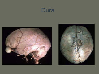
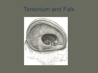
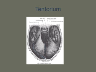
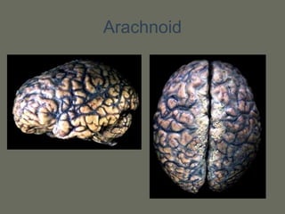
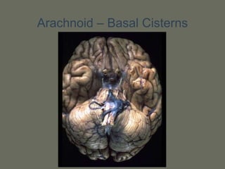
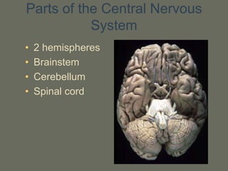
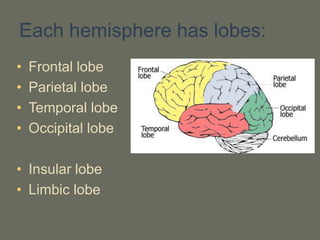
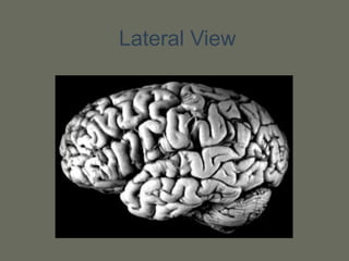
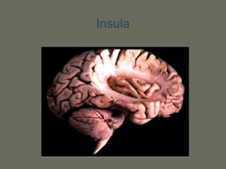
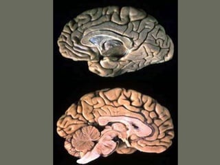
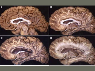
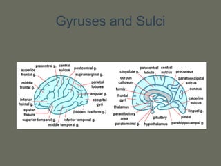
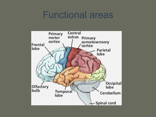
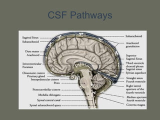
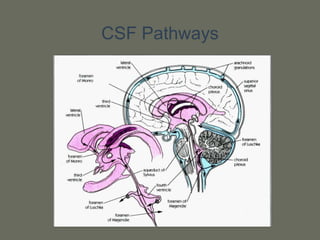
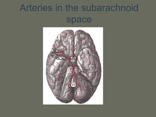
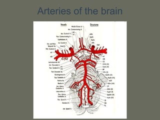
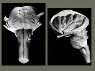
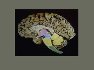
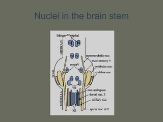
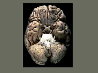
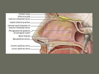
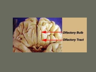
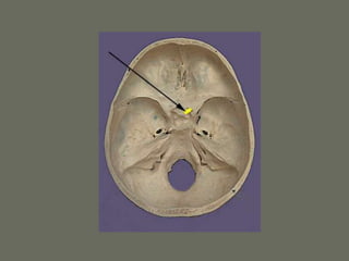
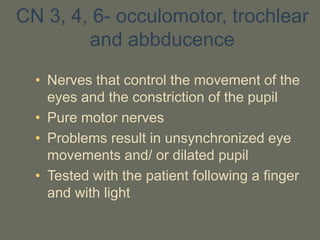
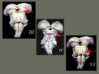
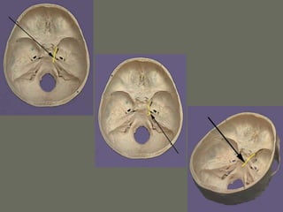
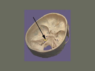
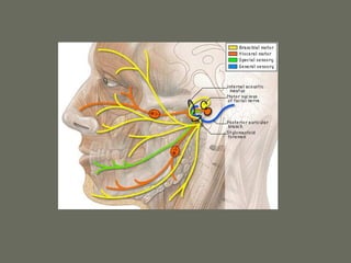
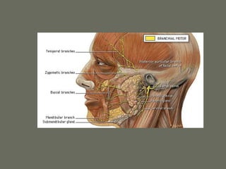
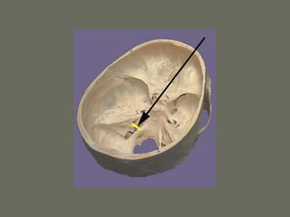
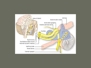
Brain Anatomy and
Physiology
Dr. Nevo Margalit – Neurosurgery
Tel Aviv Sourasky Medical Center
Scalp
• Skin
• Connective tissue (superficial fascia)
• Epicranial aponeurosis (galea
aponeurotica)
• Loose areolar tissue
• Pericranium
• Occipitofrontalis muscle
Scalp
The Skull
The Skull
The skull
The Menings
The 3 layers covering the brain
• Dura: covers the brain and spinal cord. The
venous sinuses run through the dura. 2
additional Parts of the dura are the Tentorium
and the Falx Cerebri.
• Arachnoid: Transparent, thin web structure.
Creates a space around the brain called the
sub arachnoid space where the CSF and the
main blood vessels pass.
• Pia: a very thin layer. Coats the brain and
spinal cord and is inseparable from them.
Dura
Tentorium and Falx
Tentorium
Arachnoid
Arachnoid – Basal Cisterns
Parts of the Central Nervous
System
• 2 hemispheres
• Brainstem
• Cerebellum
• Spinal cord
Each hemisphere has lobes:
• Frontal lobe
• Parietal lobe
• Temporal lobe
• Occipital lobe
• Insular lobe
• Limbic lobe
Lateral View
Insula
Gyruses and sulci
• Each lobe is composed of gyruses
separated by sulci
• The topography can be defined by
histology, anatomical relationships or
function
Map Brodmann
Gyruses and Sulci
Functional areas
Function 2
Function 3
f-MRI
The Motor Strip
Fibers
Ventricles and CSF
Ventricles and CSF
• CSF is produced by the choroid plexus
in the ventricles
• The circulation is from the lateral
ventricles to the third through the
Monroe- aqueduct- IV ventricle- sub
arachnoid space in skull and spine-
absorption in sss and other sinuses
through arachnoid granulations
CSF Pathways
CSF Pathways
Arteries of the brain
• Anterior circulation – internal
carotid artery, from common
carotid in the neck. Bifurcates to
MCA and ACA
• Posterior circulation – vertebral
arteries that join to form the basilar
artery that will then bifurcate to 2
PCA
Circle of Willis
• Communication between 2 sides –
anterior communicating (a-com)
• Communication between anterior
and posterior circulation – posterior
communicating (p-com)
• Many anomalies may exist
Arteries in the neck
Arteries in the subarachnoid
space
Arteries of the brain
Physiology
Blood supply to the brain
• The brain gets 15% of the cardiac output
and 20% of the oxygen consumption
• The brain tissue gets in average 50ml of
blood per 100gr of tissue per minute. The
gray matter receives about 3 to 4 times
more then the white matter
• Total blood supply to the brain is about 500-
600ml per minute
Factors Affecting the blood
supply
• Autoregulation
• Biochemical changes – O2 and CO2
• Blood brain barrier - BBB
Autoregulation
• Maintains a regular blood supply to the
brain in changing blood pressures
• The range is 50-130 mm mercury
• Possible mechanisms are the
myogenic control, neurogenic and
biochemichal control
CO2
• The most important and powerful
mechanism that controls brain blood
flow
• A change in 1mm PCO2 changes the
flow in 4-5%
• PCO2 of 70 gives a maximal
vasodilatation. Above that the flow is
pressure dependent
Hyperventilation
• Hyperventilation lowers the PCO2
• It has a strong effect but it is limited in
time
• Could be dangerous if not regulated-
ischemia
• Can be regulated with a jugular bulb
oximeter
BLOOD BRAIN BARRIER
• The BBB is composed of the tight
junctions in the endothelium cells of
the blood vessels
• Prevents passage of large molecules
and even small ions like Na and Cl
• Specific substances pass the BBB like
glucose and amino acids
BLOOD BRAIN BARRIER
• Because of the BBB, in the brain
hydrostatic and oncotic pressures are
not significant. The important
parameter is the osmotic pressure
• The BBB is damages in trauma, tumor,
infarct, SAH and infection
BLOOD BRAIN BARRIER
Brainstem and Cranial Nerves
Brain stem
• Has 3 parts: midbrain, pons and medulla
• Transports all the information to and from
the brain
• Centers for breathing and blood pressure
• The origin or endpoint for cranial nerves
• Contains the center of consciousness
• Creates connections to the cerebellum
Nuclei in the brain stem
Cranial nerves
• 12 pairs of nerves
• All cranial nerves except I and II
originate from the brainstem
• The nerves are sensory, motor or
mixed
• There are nuclei in the brainstem that
are the origin or the endpoint of the
cranial nerves
CN 1- Olfactory Nerve
• The sensation of smell
• Pure sensory nerve
• From the nose to the forebrain
• Very developed in some animals
• Tested an odorous substance
CN 2 - Optic N
• Visual information from the eye to the
brain
• Pure sensory nerve
• Belongs to the CNS and is not part of
the PNS
• Problems could result in field problem,
acuity problem and more
• Tested with a vision chart and field
exam on confrontation
CN 3, 4, 6- occulomotor, trochlear
and abbducence
• Nerves that control the movement of the
eyes and the constriction of the pupil
• Pure motor nerves
• Problems result in unsynchronized eye
movements and/ or dilated pupil
• Tested with the patient following a finger
and with light
CN 5 – Trigeminal N
• The largest CN
• Sensory and motor
• Sensation from the face eyes, mouth,
and motor for mastication muscles
• Tested with touch and pin on face
CN 7 – Facial N
• Motor nerve of the face and taste from
the tongue
• Motor and sensory
• Tested with movement of the facial
muscles
CN 8 – Vestibulocochlear N
• The nerves for hearing and balance
• Sensory nerves
• The most common origin of a
schwanoma
• Tested by hearing test and balance
function
CN 9 – Glossopharyngeal N
• Sensation from the pharynx
• Mainly sensory and small motor part
• Problems result in aspiration
• Tested with the gag reflex
CN 10 – Vagus N
• Motor to the muscles larynx and
pharynx and the parasympathetic of
the body
• Motor nerve
• Problems result in hoarseness
• Tested by opening mouth wide and
saying “AH“
CN 11 – Accessory N
• Nerve to the trapezius and
sternomastoid muscles
• Motor nerve
• Tested with shoulder raise or turning
the head
CN 12 – Hypoglossal N
• Nerve to the muscle of the tongue
• Motor nerve
• Tested with movement of the tongue
Human brain anatomy and physiologypptx.ppt

Human brain anatomy and physiologypptx.ppt

  • 1.
    Brain Anatomy and Physiology Dr.Nevo Margalit – Neurosurgery Tel Aviv Sourasky Medical Center
  • 2.
    Scalp • Skin • Connectivetissue (superficial fascia) • Epicranial aponeurosis (galea aponeurotica) • Loose areolar tissue • Pericranium • Occipitofrontalis muscle
  • 3.
  • 4.
  • 5.
  • 6.
  • 7.
    The Menings The 3layers covering the brain • Dura: covers the brain and spinal cord. The venous sinuses run through the dura. 2 additional Parts of the dura are the Tentorium and the Falx Cerebri. • Arachnoid: Transparent, thin web structure. Creates a space around the brain called the sub arachnoid space where the CSF and the main blood vessels pass. • Pia: a very thin layer. Coats the brain and spinal cord and is inseparable from them.
  • 8.
  • 10.
  • 11.
  • 12.
  • 13.
  • 14.
    Parts of theCentral Nervous System • 2 hemispheres • Brainstem • Cerebellum • Spinal cord
  • 15.
    Each hemisphere haslobes: • Frontal lobe • Parietal lobe • Temporal lobe • Occipital lobe • Insular lobe • Limbic lobe
  • 16.
  • 17.
  • 20.
    Gyruses and sulci •Each lobe is composed of gyruses separated by sulci • The topography can be defined by histology, anatomical relationships or function
  • 21.
  • 22.
  • 23.
  • 24.
  • 25.
  • 26.
  • 27.
  • 28.
  • 29.
  • 30.
    Ventricles and CSF •CSF is produced by the choroid plexus in the ventricles • The circulation is from the lateral ventricles to the third through the Monroe- aqueduct- IV ventricle- sub arachnoid space in skull and spine- absorption in sss and other sinuses through arachnoid granulations
  • 31.
  • 32.
  • 33.
  • 34.
    Arteries of thebrain • Anterior circulation – internal carotid artery, from common carotid in the neck. Bifurcates to MCA and ACA • Posterior circulation – vertebral arteries that join to form the basilar artery that will then bifurcate to 2 PCA
  • 35.
    Circle of Willis •Communication between 2 sides – anterior communicating (a-com) • Communication between anterior and posterior circulation – posterior communicating (p-com) • Many anomalies may exist
  • 36.
  • 37.
    Arteries in thesubarachnoid space
  • 38.
  • 39.
  • 40.
    Blood supply tothe brain • The brain gets 15% of the cardiac output and 20% of the oxygen consumption • The brain tissue gets in average 50ml of blood per 100gr of tissue per minute. The gray matter receives about 3 to 4 times more then the white matter • Total blood supply to the brain is about 500- 600ml per minute
  • 41.
    Factors Affecting theblood supply • Autoregulation • Biochemical changes – O2 and CO2 • Blood brain barrier - BBB
  • 42.
    Autoregulation • Maintains aregular blood supply to the brain in changing blood pressures • The range is 50-130 mm mercury • Possible mechanisms are the myogenic control, neurogenic and biochemichal control
  • 43.
    CO2 • The mostimportant and powerful mechanism that controls brain blood flow • A change in 1mm PCO2 changes the flow in 4-5% • PCO2 of 70 gives a maximal vasodilatation. Above that the flow is pressure dependent
  • 44.
    Hyperventilation • Hyperventilation lowersthe PCO2 • It has a strong effect but it is limited in time • Could be dangerous if not regulated- ischemia • Can be regulated with a jugular bulb oximeter
  • 45.
    BLOOD BRAIN BARRIER •The BBB is composed of the tight junctions in the endothelium cells of the blood vessels • Prevents passage of large molecules and even small ions like Na and Cl • Specific substances pass the BBB like glucose and amino acids
  • 46.
    BLOOD BRAIN BARRIER •Because of the BBB, in the brain hydrostatic and oncotic pressures are not significant. The important parameter is the osmotic pressure • The BBB is damages in trauma, tumor, infarct, SAH and infection
  • 47.
  • 48.
  • 49.
    Brain stem • Has3 parts: midbrain, pons and medulla • Transports all the information to and from the brain • Centers for breathing and blood pressure • The origin or endpoint for cranial nerves • Contains the center of consciousness • Creates connections to the cerebellum
  • 53.
    Nuclei in thebrain stem
  • 55.
    Cranial nerves • 12pairs of nerves • All cranial nerves except I and II originate from the brainstem • The nerves are sensory, motor or mixed • There are nuclei in the brainstem that are the origin or the endpoint of the cranial nerves
  • 56.
    CN 1- OlfactoryNerve • The sensation of smell • Pure sensory nerve • From the nose to the forebrain • Very developed in some animals • Tested an odorous substance
  • 60.
    CN 2 -Optic N • Visual information from the eye to the brain • Pure sensory nerve • Belongs to the CNS and is not part of the PNS • Problems could result in field problem, acuity problem and more • Tested with a vision chart and field exam on confrontation
  • 64.
    CN 3, 4,6- occulomotor, trochlear and abbducence • Nerves that control the movement of the eyes and the constriction of the pupil • Pure motor nerves • Problems result in unsynchronized eye movements and/ or dilated pupil • Tested with the patient following a finger and with light
  • 68.
    CN 5 –Trigeminal N • The largest CN • Sensory and motor • Sensation from the face eyes, mouth, and motor for mastication muscles • Tested with touch and pin on face
  • 71.
    CN 7 –Facial N • Motor nerve of the face and taste from the tongue • Motor and sensory • Tested with movement of the facial muscles
  • 76.
    CN 8 –Vestibulocochlear N • The nerves for hearing and balance • Sensory nerves • The most common origin of a schwanoma • Tested by hearing test and balance function
  • 79.
    CN 9 –Glossopharyngeal N • Sensation from the pharynx • Mainly sensory and small motor part • Problems result in aspiration • Tested with the gag reflex
  • 81.
    CN 10 –Vagus N • Motor to the muscles larynx and pharynx and the parasympathetic of the body • Motor nerve • Problems result in hoarseness • Tested by opening mouth wide and saying “AH“
  • 83.
    CN 11 –Accessory N • Nerve to the trapezius and sternomastoid muscles • Motor nerve • Tested with shoulder raise or turning the head
  • 85.
    CN 12 –Hypoglossal N • Nerve to the muscle of the tongue • Motor nerve • Tested with movement of the tongue