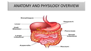
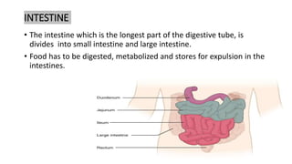
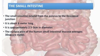
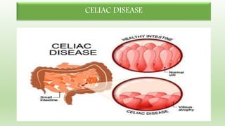
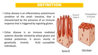
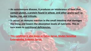
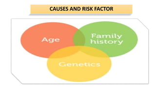
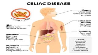
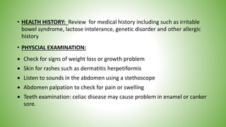
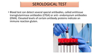
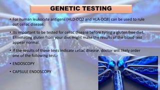
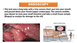
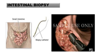
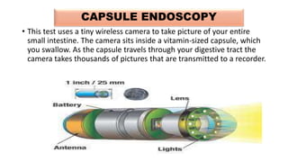
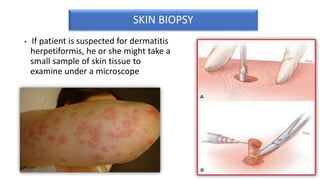
CLASS PRESENTATION
ON
CELIAC DISEASE
PRESENTED BY
BHAVYA SHARMA
M.Sc. Nursing First Year
ANATOMY AND PHYSILOGY OVERVIEW
INTESTINE
• The intestine which is the longest part of the digestive tube, is
divides into small intestine and large intestine.
• Food has to be digested, metabolized and stores for expulsion in the
intestines.
THE SMALL INTESTINE
• The small intestine extend from the pylorus to the ileocaecal
junction
• It is about 6 meter long
• It is approximately 2.5-3cm in diameter.
• The surface area of the human small intestinal mucosa averages
30square meter.
• Small intestine is divided into 3
parts
Duodenum: Widest, shortest and
most flexed part of small intestine
and about 25 cm.
Jejunum: is the middle part and
about 8ft long
Ileum: is longest part 12 ft of small
intestine. It finally open in the
caecum in the lower part of the
abdominal cavity.
HISTOLOGY OF GUT
• The gut wall ( especially of intestine) is
formed of 4 coats, each which has
different types of cells.
• 1. VISCERAL PERITONEUM OF SEROSA: It
is the outermost coat and is formed of a
squamour epithelium. It is protective in
function.
• 2. MUSCULAR COAT: Outer layer- with
smooth longitudinal muscle fibers. And
Inner Layer- with smooth circular muscle
fibers.
• 3. SUBMUCOSA: Formed of aerolar
connective tissue and has blood
capillaries, lymph capillaries ( lacteals),
nerve fibers.
4.MUCOSA: It is innermost coat. In intestine it is highly adapted to
ensure maximum absorption.
• Inner mucosa of small intestine raised into about 4 millions of minute
finger like projections called villi. Villus is the unit of absorption of
food.
• The free surface of these cells have numerous electron microscopic
evaginations, called microvilli which form a brush border.
• Villi and microvilli increases the surface area of digestion and
absorption of food.
CELIAC DISEASE
INTRODUCTION
• The word ‘celiac’ comes from Greek word meaning ‘Suffering in the
bowels’
• Celiac disease is a digestive disease that damages the small
intestine and interferes with absorption of nutrients from food.
People who have celiac disease cannot tolerate Gluten, a protein in
wheat, rye and barley.
• Gluten is found mainly in food but may also be found in everyday
products such as medicines, vitamins and lip balms.
DEFINITION
• Celiac disease is an inflammatory autoimmune
condition of the small- intestine, that is
characterized by the presence of an immune
response that is triggered by ingesting gluten.
• Celiac disease is an immune mediated
systemic disorder elicited by wheat gluten and
related prolamines. It occurs mainly in
genetically (mainly HLA) susceptible
individuals.
• An autoimmune disease, it produces an intolerance of food that
contain gluten, a protein found in wheat, and other grains such as
barley, rye, and triticale.
• It causes an immune reaction in the small intestine that damages
the lining and lowers the absorption levels of nutrients. This in
turn leads to nutritional deficiencies.
• This condition is also know as Celiac Sprue, Gluten Sensitive
Enteropathy, Endemic Sprue.
CAUSES AND RISK FACTOR
GENETICS
• Patient with celiac disease may or may not possess genetic markers
such as HUMAN LEUKOCYTE ANTIGEN ALLELES HLD-DQ2 and HLA-
DQ8 . The reason these genes produce an increase in risk of celiac
disease is that the receptors formed by these genes bind to gliadin
peptides more tightly than other forms of the antigen presenting
receptor.
PROLAMINS
• Prolamins are plant proteins that have a high proline content which
makes them difficult to fully digest.
• Gliadin a component of gluten is a prolamin found in wheat and
upon ingestion causes inflammation due to stimulation of T cell.
TISSUE TRANSGLUTAMINASE
• Tissue transglutaminase is a calcium dependent ubiquitous enzyme
which catalyses posttranslational modification of proteins and is
released from cells during inflammation.
• 2 Crucial role in celiac disease: 1. As a deamidating enzyme that can
enhances the immunostimulatory effects of gluten 2nd As a target
autoantigen in the immune response
VILLOUS ATROPHY AND MALABSORPTION
• The inflammatory process, mediated by T cells, leads to disruption of
the structure and function of the small bowels mucosal lining and
causes malabsorption as it impairs the body’s ability to absorb
nutrients, minerals and fat soluble vitamins A D E K and from food.
Other Factors
• Family history
• Persons of Europeans ancestry.
• Smoking
• Malignant disease: Lymphomas.
• Type 1 Diabetes mellitus.
• Few responds to steroids.
• Age :It is recognized as a common disorder that can be diagnosed at
any age but commonly occurs at the age of 1-5 year old.
PATHOPHYSIOLOGY
• When people with autoimmune condition eat foods containing gluten
|
• Activation of immune system
|
• This causes formation of Antibodies ( Anti-tTG antiendomysial and antigliadin
antibodies) to gluten which attack the intestinal lining.
|
• Inflammation in the intestine
|
• Epithelial cell injury
|
• If inflammation remain long that causes damage to the villia, the hair like
structure on the linings of the small intestine
• (Celiac Sprue)
• (Nutrients from food are normally absorbed by the Villi)
|
• The mucosal damage results in the deficiency of enzymes on the mucosal
surface, such as disaccharidase and peptidase.
|
• If villa are damage the person cannot absorb nutrients properly and ends up
malnourished
The Classic pathology changes of celiac disease in the small bowel are
categorized by ‘MARSH CLASSIFICATION”
• Marsh Stage 0: Normal mucosa.
• Marsh stage 1: Intestinal lining has been infiltrates with IELS ( Intraepithelial Lymphocytes) seen in
patients on a gluten free diet along with Dermatitis Herpetiformis is seen
• Marsh Stage 2: proliferation of the crypts of Lieberkuhn.
• Marsh Stage 3: Patrial or complete villous atrophy.
• Marsh stage 4: Hypoplasia of the small bowel architecture
CLINICAL MENIFESTATIONS
 The most commonly recognized symptoms of
celiac disease related to the improper
absorption of food in the GIT.
 Patient presents with diarrhoea (50%),
steatorrhea, flatulence, distended abdomen,
weight loss and generalized weakness.
 Unrecognized celiac disease may cause
malabsorption, iron deficiency anaemia,
osteoporosis, osteomalacia causing bone
fracture, pain and bony deformities
 People with celiac disease may also
experience lactose intolerance due to lactose
enzyme deficiency
DERMATITIS HERPETIFORMIS
• Dermatitis Herpetiformis Is the skin manifestation of celiac disease.
It is an intensely itchy rash that occurs in the hands fingers,
forearms, buttocks or scalp or anywhere on the body The rash
typically consists of intensely itchy, small red dots that may develop
into blisters or pimples.
CELIAC DISEASE AND LUNGS
• Extrinsic allergic alveolitis was found in combination with celiac
disease and it may be considered that both these disease are based
on one common immunologic disorder.
Digestive symptoms are more common infants and young children
and may include
 Abdominal bloating and pain
 Chronic Diarrhoea.
 Vomiting
 Constipation
 Pale , foul smelling or fatty stool
 Weight loss
 Defects in the tooth enamel and changes in tooth colour
 Delayed puberty
 Irritable and fussy behaviour
 Poor Weight gain
 Slowed growth and shorter than normal height for their age.
Adults are less likely to have digestive symptoms may instead have
one or more of the following:
 Unexplained iron deficiency anaemia
 Fatigue
 Bone or joint pain
 Bone loss or osteoporosis
 Depression or anxiety
 Tingling numbness in the hand and feet.
 Seizures
 Missed menstrual periods
 Canker sores inside the mouth
 An itchy skin rash called dermatitis herpetiformis
DIAGNOSTIC FINDINGS
• HEALTH HISTORY: Review for medical history including such as irritable
bowel syndrome, lactose intolerance, genetic disorder and other allergic
history
• PHYSCIAL EXAMINATION:
 Check for signs of weight loss or growth problem
 Skin for rashes such as dermatitis herpetiformis
 Listen to sounds in the abdomen using a stethoscope
 Abdomen palpation to check for pain or swelling
 Teeth examination: celiac disease may cause problem in enamel or canker
sore.
SEROLOGICAL TEST
• Blood test can detect several special antibodies, called antitissue
transglutaminase antibodies (tTGA) or anti- endomysium antibodies
(EMA). Elevated levels of certain antibody proteins indicate an
immune reaction gluten.
GENETIC TESTING
• For human leukocyte antigens (HLD-DQ2 and HLA-DQ8) can be used to rule
out celiac disease.
• Its important to be tested for celiac disease before tyring a gluten free diet.
Eliminating gluten from your diet might make the results of the blood test
appear normal.
• If the results of these tests indicate celiac disease, doctor will likely order
one of the following test.-
• ENDOSCOPY
• CAPSULE ENDOSCOPY
ENDOSCOPY
• This test uses a long tube with a tiny camera that’s put into your mouth
and passed down your throat (upper endoscopy). The camera enables
your doctor to view your small intestine and take a small tissue sample
(Biopsy) to analyse for damage to the villi
INTESTINAL BIOPSY
CAPSULE ENDOSCOPY
• This test uses a tiny wireless camera to take picture of your entire
small intestine. The camera sits inside a vitamin-sized capsule, which
you swallow. As the capsule travels through your digestive tract the
camera takes thousands of pictures that are transmitted to a recorder.
SKIN BIOPSY
• If patient is suspected for dermatitis
herpetiformis, he or she might take a
small sample of skin tissue to
examine under a microscope
OTHER DIAGNOSITIC EVALUTION
• CT SCAN
• MRI
• ABDOMINAL XRAY
• ABDOMINAL ULTRASOUND
MANAGEMENT
>MEDICAL MANAGEMNT
>NUTRITIONAL MANAGEMENT
> NURSING MANAGEMENT
• Goal:
 To relieve symptoms
 To reduce the underlying cause
 To improve the health status of the individual.
 To achieve the Celiac disease free status of the client.
 To improve the patients functional status and quality of the life
There is no medication that treat celiac disease. To avoid the health
problems that is can cause you will need to go completely gluten free
diet.
• LIFE LONG GLUTEN FREE DIET:
a. Avoid all foods contain wheat, rye and barley and their derivatives
because even a small amount, such as a trace contaminant , may cause
a pathologic response.
b. Oats are naturally gluten fee, however they are often cross
contaminated during the manufacturing process, so their exclusion may
ne recommended.
c. Clinical signs of improvement are often seen days to weeks after proper
diet is initiated, but can take up to 1 year in the case of more insidious
symptoms such as delayed liner growth.
Adequate caloric and nutrient intake.
Supplemental vitamins and minerals may be used
clinically indicated although there is currently no
standard for use.
a. Folic acids if low level is suspected or
detected.
b. Vitamins A and D
c. Iron up to 3 months if anaemic.
d. Vitamin K if evidence of hypo -
prothrmobinemia and bleeding
e. Calcium if milk is restricted.
• Temporary restriction of lactose and sucrose ( disaccharidases) from
diet for 6 to 8 weeks may be indicated in some cases, if these
products are shown to worsen symptoms.
• Corticosteroids: To damp down the
immune system .
• Immunotherapy: Antigen bases
immunotherapy could be a feasible
option. The immunodominant gluten
epitopes are well characterized and are
recognized by pathogenic CD4+ T cell that
could be desensitized with
immunotherapy. An intradermal adjuvant
free, formulation of three specific gluten
peptides ( Nexvax 2) showed a good
safety profile,
If constipation persists, try rice bran or laxative called Fybogel
which is available with or without a prescription.
Antiemetic : Emeset can be given to reduce nausea and
vomiting.
Antidiarrheal : Antibiotic Metoclopramide can be given
NURSING MANAGEMENT
NURSING ASSESSMENT
• HEALTH HISTORY:
1. Obtain family dietary history as it related to other of symptoms.
2. Assess nutritional status.
3. Check for sign of infection
4. Assess growth and development parameters.
PHYSICAL EXAMINATION:
 Missed menstrual periods
 Canker sores inside the mouth
 An itchy skin rash called dermatitis herpetiformis
• Extrinsic allergic alveolitis
• Rapid Weight Loss
 Defects in the tooth enamel and changes in tooth colour
 Delayed puberty
 Irritable and fussy behaviour
NURSING DIAGNOSIS
1. Diarrhoea related to intestinal inflammation secondary to Celiac
disease.
2. Imbalanced nutrition less than body requirement related to reduced
absorption of nutrients secondary to Celiac disease.
3. Impaired parenting related to inability to control behavioural
problems.
4. Ineffective coping related disease condition.
5. Risk for deficient fluid volume related to diarrhoea secondary to
celiac disease.
NURSING INTERVENTION
• Provide gluten free diet being careful to avoid accidental exposure via cross
contamination while food is prepared and or cooked.
• An initial diet high in protein, relatively low in fat, low in lactose and free from
gluten may be necessary if the newly diagnosed child exhibits severe
malabsorption.
• Initiate a referral with a dietitian for precise diet recommendations.
• Soya milk or lactide drop may be indicated if the patient is exhibiting signs of
lactose intolerance.
• Maintain NPO status during the initial treatment of celiac crisis or during
diagnostic testing. Take special precaution to ensure proper restriction if the child
is ambulatory.
• Encourage small frequent appetizing meal, but do not force eating if
the child has anorexia
• A gluten challenged diet is not routinely indicated.
• Provide the family with assistance to obtain a variety of glutem free
foods. Studies
• Create a daily weight chart and a food and fluid chart .
• Administer supplements of vitamins and minerals as prescribed,
• EVALUATION: EXPECTED OUTCOMES:
• Tolerates gluten free diet well, poor growth is not the symptom for
now.
• GI disturbance is reduced to some extent.
NURSING RESEARCH
• A Quasi experimental study to assess the different profiles of wheat
antigens in patients suffering from coeliac disease and IgE-mediated
food allergy
• Claudia Constantin , Wolf Dietrich Huber, Gerhard Granditsch, Margit
Weghofer, Rudolf Valenta November 2005
• Background: Dietary intake of wheat can cause two distinct
immunologically mediated diseases with severe gastrointestinal
manifestations, coeliac disease (CD) and IgE-mediated food allergy.
The pathomechanisms underlying these diseases are different, but
the profile of the target antigens in wheat has not been compared for
the two diseases.
• Methods: We compared IgA- and IgE-reactive antigens in wheat using
sera from patients with coeliac disease (n = 35) and food allergy to
wheat (n = 16) by one- and two-dimensional immunoblotting.
Furthermore, the IgG subclass (IgG1-IgG4) reactivity to wheat
antigens was studied by enzyme-linked immunosorbent assay.
• Results: IgA antibodies from CD patients and IgE antibodies from
allergic patients recognised distinct profiles of wheat antigens.
Furthermore, the IgG subclass responses to wheat antigens were
different in CD and wheat-allergic patients.
• Conclusion: This study thus demonstrates that wheat contains
antigens/epitopes which are preferentially recognised by CD patients,
whereas others elicit IgE-mediated food allergy. This finding suggests
that the nature of a food antigen may influence the quality of the
pathological immune response in the gut and has implications for the
diagnosis and therapy of hypersensitivity to wheat.
Celiac disease   copy

Celiac disease copy

  • 1.
    CLASS PRESENTATION ON CELIAC DISEASE PRESENTEDBY BHAVYA SHARMA M.Sc. Nursing First Year
  • 2.
  • 3.
    INTESTINE • The intestinewhich is the longest part of the digestive tube, is divides into small intestine and large intestine. • Food has to be digested, metabolized and stores for expulsion in the intestines.
  • 4.
    THE SMALL INTESTINE •The small intestine extend from the pylorus to the ileocaecal junction • It is about 6 meter long • It is approximately 2.5-3cm in diameter. • The surface area of the human small intestinal mucosa averages 30square meter.
  • 5.
    • Small intestineis divided into 3 parts Duodenum: Widest, shortest and most flexed part of small intestine and about 25 cm. Jejunum: is the middle part and about 8ft long Ileum: is longest part 12 ft of small intestine. It finally open in the caecum in the lower part of the abdominal cavity.
  • 6.
    HISTOLOGY OF GUT •The gut wall ( especially of intestine) is formed of 4 coats, each which has different types of cells. • 1. VISCERAL PERITONEUM OF SEROSA: It is the outermost coat and is formed of a squamour epithelium. It is protective in function. • 2. MUSCULAR COAT: Outer layer- with smooth longitudinal muscle fibers. And Inner Layer- with smooth circular muscle fibers. • 3. SUBMUCOSA: Formed of aerolar connective tissue and has blood capillaries, lymph capillaries ( lacteals), nerve fibers.
  • 7.
    4.MUCOSA: It isinnermost coat. In intestine it is highly adapted to ensure maximum absorption. • Inner mucosa of small intestine raised into about 4 millions of minute finger like projections called villi. Villus is the unit of absorption of food. • The free surface of these cells have numerous electron microscopic evaginations, called microvilli which form a brush border. • Villi and microvilli increases the surface area of digestion and absorption of food.
  • 8.
  • 9.
    INTRODUCTION • The word‘celiac’ comes from Greek word meaning ‘Suffering in the bowels’ • Celiac disease is a digestive disease that damages the small intestine and interferes with absorption of nutrients from food. People who have celiac disease cannot tolerate Gluten, a protein in wheat, rye and barley. • Gluten is found mainly in food but may also be found in everyday products such as medicines, vitamins and lip balms.
  • 10.
    DEFINITION • Celiac diseaseis an inflammatory autoimmune condition of the small- intestine, that is characterized by the presence of an immune response that is triggered by ingesting gluten. • Celiac disease is an immune mediated systemic disorder elicited by wheat gluten and related prolamines. It occurs mainly in genetically (mainly HLA) susceptible individuals.
  • 11.
    • An autoimmunedisease, it produces an intolerance of food that contain gluten, a protein found in wheat, and other grains such as barley, rye, and triticale. • It causes an immune reaction in the small intestine that damages the lining and lowers the absorption levels of nutrients. This in turn leads to nutritional deficiencies. • This condition is also know as Celiac Sprue, Gluten Sensitive Enteropathy, Endemic Sprue.
  • 12.
  • 13.
    GENETICS • Patient withceliac disease may or may not possess genetic markers such as HUMAN LEUKOCYTE ANTIGEN ALLELES HLD-DQ2 and HLA- DQ8 . The reason these genes produce an increase in risk of celiac disease is that the receptors formed by these genes bind to gliadin peptides more tightly than other forms of the antigen presenting receptor.
  • 14.
    PROLAMINS • Prolamins areplant proteins that have a high proline content which makes them difficult to fully digest. • Gliadin a component of gluten is a prolamin found in wheat and upon ingestion causes inflammation due to stimulation of T cell.
  • 15.
    TISSUE TRANSGLUTAMINASE • Tissuetransglutaminase is a calcium dependent ubiquitous enzyme which catalyses posttranslational modification of proteins and is released from cells during inflammation. • 2 Crucial role in celiac disease: 1. As a deamidating enzyme that can enhances the immunostimulatory effects of gluten 2nd As a target autoantigen in the immune response
  • 16.
    VILLOUS ATROPHY ANDMALABSORPTION • The inflammatory process, mediated by T cells, leads to disruption of the structure and function of the small bowels mucosal lining and causes malabsorption as it impairs the body’s ability to absorb nutrients, minerals and fat soluble vitamins A D E K and from food.
  • 17.
    Other Factors • Familyhistory • Persons of Europeans ancestry. • Smoking • Malignant disease: Lymphomas. • Type 1 Diabetes mellitus. • Few responds to steroids. • Age :It is recognized as a common disorder that can be diagnosed at any age but commonly occurs at the age of 1-5 year old.
  • 18.
    PATHOPHYSIOLOGY • When peoplewith autoimmune condition eat foods containing gluten | • Activation of immune system | • This causes formation of Antibodies ( Anti-tTG antiendomysial and antigliadin antibodies) to gluten which attack the intestinal lining. | • Inflammation in the intestine | • Epithelial cell injury |
  • 19.
    • If inflammationremain long that causes damage to the villia, the hair like structure on the linings of the small intestine • (Celiac Sprue) • (Nutrients from food are normally absorbed by the Villi) | • The mucosal damage results in the deficiency of enzymes on the mucosal surface, such as disaccharidase and peptidase. | • If villa are damage the person cannot absorb nutrients properly and ends up malnourished
  • 20.
    The Classic pathologychanges of celiac disease in the small bowel are categorized by ‘MARSH CLASSIFICATION” • Marsh Stage 0: Normal mucosa. • Marsh stage 1: Intestinal lining has been infiltrates with IELS ( Intraepithelial Lymphocytes) seen in patients on a gluten free diet along with Dermatitis Herpetiformis is seen • Marsh Stage 2: proliferation of the crypts of Lieberkuhn. • Marsh Stage 3: Patrial or complete villous atrophy. • Marsh stage 4: Hypoplasia of the small bowel architecture
  • 21.
  • 22.
     The mostcommonly recognized symptoms of celiac disease related to the improper absorption of food in the GIT.  Patient presents with diarrhoea (50%), steatorrhea, flatulence, distended abdomen, weight loss and generalized weakness.  Unrecognized celiac disease may cause malabsorption, iron deficiency anaemia, osteoporosis, osteomalacia causing bone fracture, pain and bony deformities  People with celiac disease may also experience lactose intolerance due to lactose enzyme deficiency
  • 23.
    DERMATITIS HERPETIFORMIS • DermatitisHerpetiformis Is the skin manifestation of celiac disease. It is an intensely itchy rash that occurs in the hands fingers, forearms, buttocks or scalp or anywhere on the body The rash typically consists of intensely itchy, small red dots that may develop into blisters or pimples.
  • 24.
    CELIAC DISEASE ANDLUNGS • Extrinsic allergic alveolitis was found in combination with celiac disease and it may be considered that both these disease are based on one common immunologic disorder.
  • 25.
    Digestive symptoms aremore common infants and young children and may include  Abdominal bloating and pain  Chronic Diarrhoea.  Vomiting  Constipation  Pale , foul smelling or fatty stool  Weight loss
  • 26.
     Defects inthe tooth enamel and changes in tooth colour  Delayed puberty  Irritable and fussy behaviour  Poor Weight gain  Slowed growth and shorter than normal height for their age.
  • 27.
    Adults are lesslikely to have digestive symptoms may instead have one or more of the following:  Unexplained iron deficiency anaemia  Fatigue  Bone or joint pain  Bone loss or osteoporosis  Depression or anxiety  Tingling numbness in the hand and feet.  Seizures
  • 28.
     Missed menstrualperiods  Canker sores inside the mouth  An itchy skin rash called dermatitis herpetiformis
  • 30.
  • 31.
    • HEALTH HISTORY:Review for medical history including such as irritable bowel syndrome, lactose intolerance, genetic disorder and other allergic history • PHYSCIAL EXAMINATION:  Check for signs of weight loss or growth problem  Skin for rashes such as dermatitis herpetiformis  Listen to sounds in the abdomen using a stethoscope  Abdomen palpation to check for pain or swelling  Teeth examination: celiac disease may cause problem in enamel or canker sore.
  • 32.
    SEROLOGICAL TEST • Bloodtest can detect several special antibodies, called antitissue transglutaminase antibodies (tTGA) or anti- endomysium antibodies (EMA). Elevated levels of certain antibody proteins indicate an immune reaction gluten.
  • 33.
    GENETIC TESTING • Forhuman leukocyte antigens (HLD-DQ2 and HLA-DQ8) can be used to rule out celiac disease. • Its important to be tested for celiac disease before tyring a gluten free diet. Eliminating gluten from your diet might make the results of the blood test appear normal. • If the results of these tests indicate celiac disease, doctor will likely order one of the following test.- • ENDOSCOPY • CAPSULE ENDOSCOPY
  • 34.
    ENDOSCOPY • This testuses a long tube with a tiny camera that’s put into your mouth and passed down your throat (upper endoscopy). The camera enables your doctor to view your small intestine and take a small tissue sample (Biopsy) to analyse for damage to the villi
  • 35.
  • 36.
    CAPSULE ENDOSCOPY • Thistest uses a tiny wireless camera to take picture of your entire small intestine. The camera sits inside a vitamin-sized capsule, which you swallow. As the capsule travels through your digestive tract the camera takes thousands of pictures that are transmitted to a recorder.
  • 37.
    SKIN BIOPSY • Ifpatient is suspected for dermatitis herpetiformis, he or she might take a small sample of skin tissue to examine under a microscope
  • 38.
    OTHER DIAGNOSITIC EVALUTION •CT SCAN • MRI • ABDOMINAL XRAY • ABDOMINAL ULTRASOUND
  • 39.
  • 40.
    • Goal:  Torelieve symptoms  To reduce the underlying cause  To improve the health status of the individual.  To achieve the Celiac disease free status of the client.  To improve the patients functional status and quality of the life
  • 41.
    There is nomedication that treat celiac disease. To avoid the health problems that is can cause you will need to go completely gluten free diet. • LIFE LONG GLUTEN FREE DIET:
  • 42.
    a. Avoid allfoods contain wheat, rye and barley and their derivatives because even a small amount, such as a trace contaminant , may cause a pathologic response. b. Oats are naturally gluten fee, however they are often cross contaminated during the manufacturing process, so their exclusion may ne recommended. c. Clinical signs of improvement are often seen days to weeks after proper diet is initiated, but can take up to 1 year in the case of more insidious symptoms such as delayed liner growth.
  • 43.
    Adequate caloric andnutrient intake.
  • 44.
    Supplemental vitamins andminerals may be used clinically indicated although there is currently no standard for use. a. Folic acids if low level is suspected or detected. b. Vitamins A and D c. Iron up to 3 months if anaemic. d. Vitamin K if evidence of hypo - prothrmobinemia and bleeding e. Calcium if milk is restricted.
  • 45.
    • Temporary restrictionof lactose and sucrose ( disaccharidases) from diet for 6 to 8 weeks may be indicated in some cases, if these products are shown to worsen symptoms.
  • 46.
    • Corticosteroids: Todamp down the immune system . • Immunotherapy: Antigen bases immunotherapy could be a feasible option. The immunodominant gluten epitopes are well characterized and are recognized by pathogenic CD4+ T cell that could be desensitized with immunotherapy. An intradermal adjuvant free, formulation of three specific gluten peptides ( Nexvax 2) showed a good safety profile,
  • 47.
    If constipation persists,try rice bran or laxative called Fybogel which is available with or without a prescription. Antiemetic : Emeset can be given to reduce nausea and vomiting. Antidiarrheal : Antibiotic Metoclopramide can be given
  • 48.
  • 49.
    NURSING ASSESSMENT • HEALTHHISTORY: 1. Obtain family dietary history as it related to other of symptoms. 2. Assess nutritional status. 3. Check for sign of infection 4. Assess growth and development parameters. PHYSICAL EXAMINATION:  Missed menstrual periods  Canker sores inside the mouth  An itchy skin rash called dermatitis herpetiformis
  • 50.
    • Extrinsic allergicalveolitis • Rapid Weight Loss  Defects in the tooth enamel and changes in tooth colour  Delayed puberty  Irritable and fussy behaviour
  • 51.
    NURSING DIAGNOSIS 1. Diarrhoearelated to intestinal inflammation secondary to Celiac disease. 2. Imbalanced nutrition less than body requirement related to reduced absorption of nutrients secondary to Celiac disease. 3. Impaired parenting related to inability to control behavioural problems. 4. Ineffective coping related disease condition. 5. Risk for deficient fluid volume related to diarrhoea secondary to celiac disease.
  • 52.
    NURSING INTERVENTION • Providegluten free diet being careful to avoid accidental exposure via cross contamination while food is prepared and or cooked. • An initial diet high in protein, relatively low in fat, low in lactose and free from gluten may be necessary if the newly diagnosed child exhibits severe malabsorption. • Initiate a referral with a dietitian for precise diet recommendations. • Soya milk or lactide drop may be indicated if the patient is exhibiting signs of lactose intolerance. • Maintain NPO status during the initial treatment of celiac crisis or during diagnostic testing. Take special precaution to ensure proper restriction if the child is ambulatory.
  • 53.
    • Encourage smallfrequent appetizing meal, but do not force eating if the child has anorexia • A gluten challenged diet is not routinely indicated. • Provide the family with assistance to obtain a variety of glutem free foods. Studies • Create a daily weight chart and a food and fluid chart . • Administer supplements of vitamins and minerals as prescribed,
  • 54.
    • EVALUATION: EXPECTEDOUTCOMES: • Tolerates gluten free diet well, poor growth is not the symptom for now. • GI disturbance is reduced to some extent.
  • 55.
    NURSING RESEARCH • AQuasi experimental study to assess the different profiles of wheat antigens in patients suffering from coeliac disease and IgE-mediated food allergy • Claudia Constantin , Wolf Dietrich Huber, Gerhard Granditsch, Margit Weghofer, Rudolf Valenta November 2005
  • 56.
    • Background: Dietaryintake of wheat can cause two distinct immunologically mediated diseases with severe gastrointestinal manifestations, coeliac disease (CD) and IgE-mediated food allergy. The pathomechanisms underlying these diseases are different, but the profile of the target antigens in wheat has not been compared for the two diseases.
  • 57.
    • Methods: Wecompared IgA- and IgE-reactive antigens in wheat using sera from patients with coeliac disease (n = 35) and food allergy to wheat (n = 16) by one- and two-dimensional immunoblotting. Furthermore, the IgG subclass (IgG1-IgG4) reactivity to wheat antigens was studied by enzyme-linked immunosorbent assay. • Results: IgA antibodies from CD patients and IgE antibodies from allergic patients recognised distinct profiles of wheat antigens. Furthermore, the IgG subclass responses to wheat antigens were different in CD and wheat-allergic patients.
  • 58.
    • Conclusion: Thisstudy thus demonstrates that wheat contains antigens/epitopes which are preferentially recognised by CD patients, whereas others elicit IgE-mediated food allergy. This finding suggests that the nature of a food antigen may influence the quality of the pathological immune response in the gut and has implications for the diagnosis and therapy of hypersensitivity to wheat.