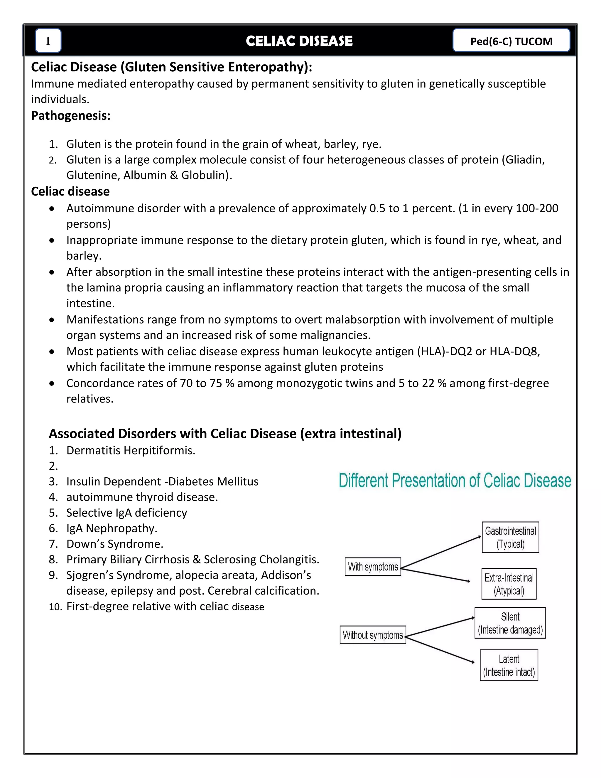
CELIAC DISEASE1 Ped(6-C) TUCOM
Celiac Disease (Gluten Sensitive Enteropathy):
Immune mediated enteropathy caused by permanent sensitivity to gluten in genetically susceptible
individuals.
Pathogenesis:
1. Gluten is the protein found in the grain of wheat, barley, rye.
2. Gluten is a large complex molecule consist of four heterogeneous classes of protein (Gliadin,
Glutenine, Albumin & Globulin).
Celiac disease
• Autoimmune disorder with a prevalence of approximately 0.5 to 1 percent. (1 in every 100-200
persons)
• Inappropriate immune response to the dietary protein gluten, which is found in rye, wheat, and
barley.
• After absorption in the small intestine these proteins interact with the antigen-presenting cells in
the lamina propria causing an inflammatory reaction that targets the mucosa of the small
intestine.
• Manifestations range from no symptoms to overt malabsorption with involvement of multiple
organ systems and an increased risk of some malignancies.
• Most patients with celiac disease express human leukocyte antigen (HLA)-DQ2 or HLA-DQ8,
which facilitate the immune response against gluten proteins
• Concordance rates of 70 to 75 % among monozygotic twins and 5 to 22 % among first-degree
relatives.
Associated Disorders with Celiac Disease (extra intestinal)
1. Dermatitis Herpitiformis.
2.
3. Insulin Dependent -Diabetes Mellitus
4. autoimmune thyroid disease.
5. Selective IgA deficiency
6. IgA Nephropathy.
7. Down’s Syndrome.
8. Primary Biliary Cirrhosis & Sclerosing Cholangitis.
9. Sjogren’s Syndrome, alopecia areata, Addison’s
disease, epilepsy and post. Cerebral calcification.
10. First-degree relative with celiac disease
CELIAC DISEASE2 Ped(6-C) TUCOM
Clinical Features:
1. Age of onset: variable, most children present between one & five years of age but they may
present for the first time at any time from infancy to old age. Classic GI pediatric cases usually
appear in children aged 9-18 months .
2. “Latent interval”: the time period between the introduction of gluten into the diet and the
development of clinical manifestation, varies from months to years.
3. Silent Celiac Disease: Abnormal small bowel mucosa characteristic of celiac disease but the child
is Asymptomatic.
4.
5. Latent Celiac Disease: The small intestinal mucosa shows no flat villi but abnormal in the form of
increasing intraepithelial lymphocyte in addition to positive circulating antigliadin or
antiendomysial antibodies.
Signs and Symptoms:
A- Common:
1. Diarrhea
2. Fatigue and muscle wasting
3. Borborygmus
4. Abdominal pain
5. Weight loss, FTT and short stature
6. Abdominal distention
7. Flatulence
B- Uncommon:
1. Osteopenia/ osteoporosis
2. Abnormal liver function
3. Nausea and Vomiting
4. Dental enamel hypoplasia
5. Iron-deficiency anemia resistant to treatment
6. Neurologic dysfunction: ataxia, epilepsy, PNP
7. Constipation
8. Delayed puberty
9. Psychiatric disorders
10.Up to 38 % Asymptomatic
Diarrhea:
• The most common presentation
• Acute, chronic or recurrent
• Stool is characteristically pale, loose and very offensive, often one large bulky stool, but could
more frequent, some children might have recurrent attacks of more severe diarrhea. Few
children with CD have constipation.
CELIAC DISEASE3 Ped(6-C) TUCOM
Diagnosis of Celiac Disease:
Clinical picture:
1- Small intestinal biopsy: the gold standard.
2- Serological markers: IgA Antiendomysial, IgA and IgG antigliadin, and IgA Anti-tissue
transglutaminase antibodies.
• + Tissue transglutaminase (tTG) antibodies had sensitivity and specificity > 95%.
• +Testing for gliadin antibodies is no longer recommended because of the low sensitivity and
specificity for celiac disease.
• +The tTG antibody test is less costly because it uses an enzyme-linked immunosorbent assay;
it is the recommended single serologic test for celiac disease screening in the primary care
setting.
3- SMALL BOWEL BIOPSY
• Required to confirm the diagnosis of celiac disease for most patients.
• Should also be considered in patients with negative serologic test results who are at high risk or
in whom the physician strongly suspects celiac disease.
Findings:
• -Short, flat villi
• -increased number of lymphocytes in the epithelial layer
• -crypt hyperplasia
Variations in the severity of pathologic changes on biopsy may obscure the typical changes found in
celiac disease, and patients with latent celiac disease may have normal results on small bowel biopsy.
Reported causes of flat small intestinal mucosa in childhood:
1- Celiac Disease
2- Cow’s milk and Soy protein allergy.
3- Gastroenteritis and post enteritis syndromes.
4- Giardiasis.
5- Autoimmune enteropathy.
6- Microvillous atrophy.
7- Acquired hypogammaglobulinemia.
8- Protein-energy malnutrition.
Figure 3. Endoscopic and biopsy findings in patients with and without celiac disease. (A) High-definition endoscopic photo
of normal small intestine. The villi are clearly visible with no evidence of atrophy or scalloping of the folds. (B) Biopsy
specimen of normal small intestine (hematoxylin-eosin; original magnification, × 100). (C) PillCam image of small intestine
in a patient with celiac disease, showing scalloping of the mucosal folds (arrows) characteristic of a malabsorption pattern.
There is also evidence of villous atrophy compared with normal. (D) Biopsy specimen of small intestine in a patient with
celiac disease (hematoxylin-eosin; original magnification, × 100). Note the loss of villous architecture.
CELIAC DISEASE4 Ped(6-C) TUCOM
Differential Diagnosis of Celiac Disease:
1- Anorexia nervosa
2- Autoimmune enteropathy
3- Bacterial overgrowth
4- Collagenous sprue
5- Crohn's disease
6- Giardiasis
7- Human immunodeficiency (virus enteropathy)
8- Hypogammaglobulinemia
9- Infective gastroenteritis
10- Intestinal lymphoma
11- Irritable bowel syndrome
12- Ischemic enteritis
13- Lactose intolerance
14- Pancreatic insufficiency
15- Soy protein intolerance
16- Tropical sprue
17- Tuberculosis
18- Whipple's disease
19- Zollinger-Ellison syndrome
CELIAC DISEASE5 Ped(6-C) TUCOM
Treatment:
1- Avoidance of food products that contain gluten proteins for life.
2- Improve the appetite,
3- decrease the diarrhea,
4- reverse osteopenia,
5- prevent lymphoma,
6- enhance appropriate growth and puberty
• Key elements to successful treatment include the motivation of the patient, the attentiveness of the
physician to comorbidities that need to be addressed.
• Formal consultation with a trained dietitian is necessary.
• National celiac disease support organizations can provide patients invaluable resources for information
and support.
• Replacement of nutritional deficiencies as vitamins, iron, calories
COMORBIDITIES:
 Osteoporosis (common finding)
 Thyroid dysfunction
 Deficiencies in folic acid, vitamin B12, fat-soluble vitamins, and iron
 Increased mortality due to increased risk of malignancy
Intestinal lymphoma (3-6x more likely)
Most common is intestinal non- Hodgkin's lymphoma.
Screening:
1- Screening an asymptomatic patient for celiac disease must be weighed against the psychological,
emotional, and economic impact of a false positive result.
2- Also, it would necessitate further evaluation with small bowel biopsy.
3- The need to follow a strict diet indefinitely can adversely affect the patient's perceived quality of
life.
4- Routine screening of the general population is not recommended.
5- Persons at high risk for celiac disease who exhibit any level of symptoms, appropriate testing is
indicated.
AHMED E ALBAYATY, 2017-07-05
Name:

Celiac disease

  • 1.
    CELIAC DISEASE1 Ped(6-C)TUCOM Celiac Disease (Gluten Sensitive Enteropathy): Immune mediated enteropathy caused by permanent sensitivity to gluten in genetically susceptible individuals. Pathogenesis: 1. Gluten is the protein found in the grain of wheat, barley, rye. 2. Gluten is a large complex molecule consist of four heterogeneous classes of protein (Gliadin, Glutenine, Albumin & Globulin). Celiac disease • Autoimmune disorder with a prevalence of approximately 0.5 to 1 percent. (1 in every 100-200 persons) • Inappropriate immune response to the dietary protein gluten, which is found in rye, wheat, and barley. • After absorption in the small intestine these proteins interact with the antigen-presenting cells in the lamina propria causing an inflammatory reaction that targets the mucosa of the small intestine. • Manifestations range from no symptoms to overt malabsorption with involvement of multiple organ systems and an increased risk of some malignancies. • Most patients with celiac disease express human leukocyte antigen (HLA)-DQ2 or HLA-DQ8, which facilitate the immune response against gluten proteins • Concordance rates of 70 to 75 % among monozygotic twins and 5 to 22 % among first-degree relatives. Associated Disorders with Celiac Disease (extra intestinal) 1. Dermatitis Herpitiformis. 2. 3. Insulin Dependent -Diabetes Mellitus 4. autoimmune thyroid disease. 5. Selective IgA deficiency 6. IgA Nephropathy. 7. Down’s Syndrome. 8. Primary Biliary Cirrhosis & Sclerosing Cholangitis. 9. Sjogren’s Syndrome, alopecia areata, Addison’s disease, epilepsy and post. Cerebral calcification. 10. First-degree relative with celiac disease
  • 2.
    CELIAC DISEASE2 Ped(6-C)TUCOM Clinical Features: 1. Age of onset: variable, most children present between one & five years of age but they may present for the first time at any time from infancy to old age. Classic GI pediatric cases usually appear in children aged 9-18 months . 2. “Latent interval”: the time period between the introduction of gluten into the diet and the development of clinical manifestation, varies from months to years. 3. Silent Celiac Disease: Abnormal small bowel mucosa characteristic of celiac disease but the child is Asymptomatic. 4. 5. Latent Celiac Disease: The small intestinal mucosa shows no flat villi but abnormal in the form of increasing intraepithelial lymphocyte in addition to positive circulating antigliadin or antiendomysial antibodies. Signs and Symptoms: A- Common: 1. Diarrhea 2. Fatigue and muscle wasting 3. Borborygmus 4. Abdominal pain 5. Weight loss, FTT and short stature 6. Abdominal distention 7. Flatulence B- Uncommon: 1. Osteopenia/ osteoporosis 2. Abnormal liver function 3. Nausea and Vomiting 4. Dental enamel hypoplasia 5. Iron-deficiency anemia resistant to treatment 6. Neurologic dysfunction: ataxia, epilepsy, PNP 7. Constipation 8. Delayed puberty 9. Psychiatric disorders 10.Up to 38 % Asymptomatic Diarrhea: • The most common presentation • Acute, chronic or recurrent • Stool is characteristically pale, loose and very offensive, often one large bulky stool, but could more frequent, some children might have recurrent attacks of more severe diarrhea. Few children with CD have constipation.
  • 3.
    CELIAC DISEASE3 Ped(6-C)TUCOM Diagnosis of Celiac Disease: Clinical picture: 1- Small intestinal biopsy: the gold standard. 2- Serological markers: IgA Antiendomysial, IgA and IgG antigliadin, and IgA Anti-tissue transglutaminase antibodies. • + Tissue transglutaminase (tTG) antibodies had sensitivity and specificity > 95%. • +Testing for gliadin antibodies is no longer recommended because of the low sensitivity and specificity for celiac disease. • +The tTG antibody test is less costly because it uses an enzyme-linked immunosorbent assay; it is the recommended single serologic test for celiac disease screening in the primary care setting. 3- SMALL BOWEL BIOPSY • Required to confirm the diagnosis of celiac disease for most patients. • Should also be considered in patients with negative serologic test results who are at high risk or in whom the physician strongly suspects celiac disease. Findings: • -Short, flat villi • -increased number of lymphocytes in the epithelial layer • -crypt hyperplasia Variations in the severity of pathologic changes on biopsy may obscure the typical changes found in celiac disease, and patients with latent celiac disease may have normal results on small bowel biopsy. Reported causes of flat small intestinal mucosa in childhood: 1- Celiac Disease 2- Cow’s milk and Soy protein allergy. 3- Gastroenteritis and post enteritis syndromes. 4- Giardiasis. 5- Autoimmune enteropathy. 6- Microvillous atrophy. 7- Acquired hypogammaglobulinemia. 8- Protein-energy malnutrition. Figure 3. Endoscopic and biopsy findings in patients with and without celiac disease. (A) High-definition endoscopic photo of normal small intestine. The villi are clearly visible with no evidence of atrophy or scalloping of the folds. (B) Biopsy specimen of normal small intestine (hematoxylin-eosin; original magnification, × 100). (C) PillCam image of small intestine in a patient with celiac disease, showing scalloping of the mucosal folds (arrows) characteristic of a malabsorption pattern. There is also evidence of villous atrophy compared with normal. (D) Biopsy specimen of small intestine in a patient with celiac disease (hematoxylin-eosin; original magnification, × 100). Note the loss of villous architecture.
  • 4.
    CELIAC DISEASE4 Ped(6-C)TUCOM Differential Diagnosis of Celiac Disease: 1- Anorexia nervosa 2- Autoimmune enteropathy 3- Bacterial overgrowth 4- Collagenous sprue 5- Crohn's disease 6- Giardiasis 7- Human immunodeficiency (virus enteropathy) 8- Hypogammaglobulinemia 9- Infective gastroenteritis 10- Intestinal lymphoma 11- Irritable bowel syndrome 12- Ischemic enteritis 13- Lactose intolerance 14- Pancreatic insufficiency 15- Soy protein intolerance 16- Tropical sprue 17- Tuberculosis 18- Whipple's disease 19- Zollinger-Ellison syndrome
  • 5.
    CELIAC DISEASE5 Ped(6-C)TUCOM Treatment: 1- Avoidance of food products that contain gluten proteins for life. 2- Improve the appetite, 3- decrease the diarrhea, 4- reverse osteopenia, 5- prevent lymphoma, 6- enhance appropriate growth and puberty • Key elements to successful treatment include the motivation of the patient, the attentiveness of the physician to comorbidities that need to be addressed. • Formal consultation with a trained dietitian is necessary. • National celiac disease support organizations can provide patients invaluable resources for information and support. • Replacement of nutritional deficiencies as vitamins, iron, calories COMORBIDITIES:  Osteoporosis (common finding)  Thyroid dysfunction  Deficiencies in folic acid, vitamin B12, fat-soluble vitamins, and iron  Increased mortality due to increased risk of malignancy Intestinal lymphoma (3-6x more likely) Most common is intestinal non- Hodgkin's lymphoma. Screening: 1- Screening an asymptomatic patient for celiac disease must be weighed against the psychological, emotional, and economic impact of a false positive result. 2- Also, it would necessitate further evaluation with small bowel biopsy. 3- The need to follow a strict diet indefinitely can adversely affect the patient's perceived quality of life. 4- Routine screening of the general population is not recommended. 5- Persons at high risk for celiac disease who exhibit any level of symptoms, appropriate testing is indicated. AHMED E ALBAYATY, 2017-07-05 Name: