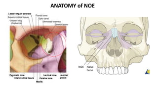
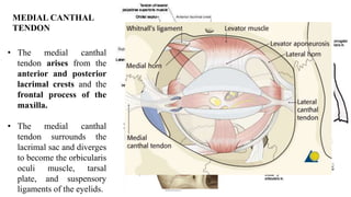
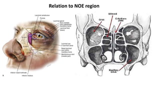
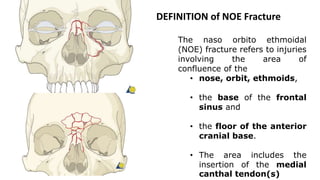
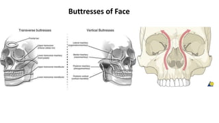
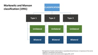
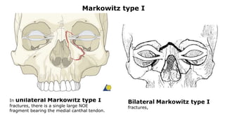
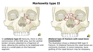
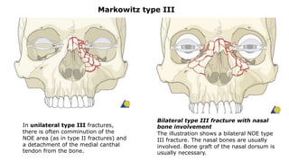
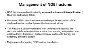
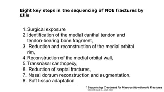
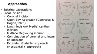
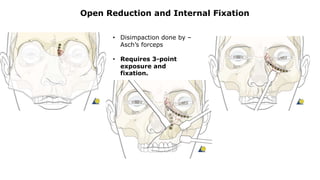
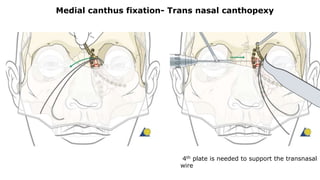
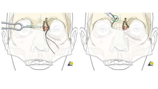
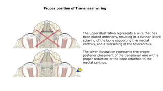
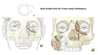
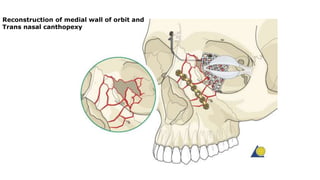
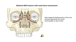
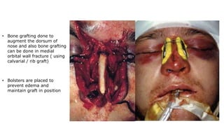
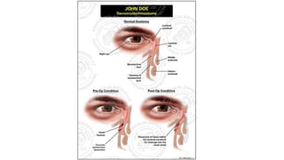
Naso Orbito Ethmoid (NOE)
Complex Fracture
Presented by : Sailesh Kumar. R
PG Trainee
•Dawson and Fordyce coined the term " fracture of the
ethmoids" to denote an injury more severe than a
simple nasal fracture.
• Converse and Smith used the term “naso-orbital”.
•Stranc preferred the term “naso-ethmoid”.
• In 1973 Epker coined the term "naso-orbito-ethmoid“,
(NOE) the most popular term in use today."
Evolution of the term NOE
•Manson et al have used two terms for these injuries;
"naso-orbital-ethmoid" and "nasoethmoid orbital”.
•Gruss has also used the term "nasoethmoid- orbital”.
•Ian Jackson used the term "orbitoethmoid," believing
that the orbital component needed to be stressed in
these injuries because it accounted for the most
difficult complications."
ANATOMY of NOE
• The medial canthal
tendon arises from the
anterior and posterior
lacrimal crests and the
frontal process of the
maxilla.
• The medial canthal
tendon surrounds the
lacrimal sac and diverges
to become the orbicularis
oculi muscle, tarsal
plate, and suspensory
ligaments of the eyelids.
MEDIAL CANTHAL
TENDON
Relation to NOE region
DEFINITION of NOE Fracture
The naso orbito ethmoidal
(NOE) fracture refers to injuries
involving the area of
confluence of the
• nose, orbit, ethmoids,
• the base of the frontal
sinus and
• the floor of the anterior
cranial base.
• The area includes the
insertion of the medial
canthal tendon(s)
PATHOPHYSIOLOGY
Buttresses of Face
CLASSIFICATION
Type 1
Unilateral
Bilateral
Type 2
Unilateral
Bilateral
Type 3
Unilateral
Bilateral
Markowitz and Manson
classification (1991)
*Management of medial canthal tendon in nasoorbital ethmoid fractures: In importance of the central
fragment in classification and treatment.
Markowitz et al, plastic and reconstructive surgery,1991, vol 87
In unilateral Markowitz type I
fractures, there is a single large NOE
fragment bearing the medial canthal tendon.
Markowitz type I
Bilateral Markowitz type I
fractures,
In unilateral type II fractures, there is often
comminution of the NOE area, but the canthal
tendon remains attached to a fragment of
bone, allowing the canthus to be stabilized with
wires or a small plate on the fractured
segment.
Bilateral type II fracture with nasal bone
involvement
The illustration shows a bilateral NOE type II
fracture. In bilateral fractures the nasal bones are
commonly involved. In some instances, bone
grafting of the nasal dorsum may be necessary.
Markowitz type II
In unilateral type III fractures,
there is often comminution of the
NOE area (as in type II fractures) and
a detachment of the medial canthal
tendon from the bone.
Bilateral type III fracture with nasal
bone involvement
The illustration shows a bilateral NOE type
III fracture. The nasal bones are usually
involved. Bone graft of the nasal dorsum is
usually necessary.
Markowitz type III
Signs and Symptoms
• Decreased dorsal nasal projection
• Upturned nasal tip (pig snout)
• Periorbital ecchymosis and oedema
• Epistaxis
• CSF rhinorrhoea
• Epiphora
• Anosmia
• Nasal airway obstruction
• Traumatic telecanthus
• Traumatic hypertelorism
• Orbital dystopia
Type 1 &
Type 2
Type 3
Evaluation of intercanthal distance
Clinical
examination
Ideal nasofrontal angle – 115
to 130 degrees
Ideal nasal projection is 1:1 • Average intercanthal
distance – 28 to 35mm
(differ with Race)
• Found to be half the value
of interpupillary distance
• Evaluate pre injury photos
Clinical
examination
• Bimanual palpation using
Kelly clamp
• Determines whether the
canthal-bearing bone
fragment is displaced and
mobile
Furnas traction test/ Bow string test/
Eyelash traction test
Evaluating Crepitus
CSF Leakage
Examination of the Eye should also be done along with NOE examination
 A study reported by Holt et al, 67% of 727
patients with facial fractures sustained some
degree of ocular injury
Radiological examination
Management of NOE fractures
• NOE fractures are best treated by open reduction and internal fixation (
Dingman and Natvig 1964)
• Mustarde(1966) described an open technique for restoration of the
displaced medial canthal ligament by transnasal wiring
• This fracture is better overtreated than undertreated because the
secondary deformities (soft tissue retraction, scarring, malposition and
displaced bony fragments) that accompany inadequate therapy are
extremely difficult to correct
• Major reason for treating NOE fracture is esthetics
1.Surgical exposure
2.Identification of the medial canthal tendon and
tendon-bearing bone fragment,
3. Reduction and reconstruction of the medial orbital
rim,
4.Reconstruction of the medial orbital wall,
5.Transnasal canthopexy,
6. Reduction of septal fractures,
7. Nasal dorsum reconstruction and augmentation,
8. Soft tissue adaptation
Eight key steps in the sequencing of NOE fractures by
Ellis
* Sequencing Treatment for Naso-orbito-ethmoid Fractures
EDWARD ELLIS III , JOMS 1993
Approaches
• Existing Lacerations
• Local incision
• Coronal incision
• Open Sky Approach (Converse &
Hogan,1970)
• Lynch incision/ Medial canthal
incision
• Midface Degloving incision
• Combination of coronal and lower
lid incisions
• Extended Glabellar approach
(Horizontal Y approach)
Surgical landmarks along the dissection of
the medial orbital wall are the:
•Anterior lacrimal crest
•Anterior ethmoidal artery (15 mm from the
crest)
•Posterior ethmoidal artery (25 mm from the
crest)
Pitfall:To avoid injury to the optic nerve
Dissection should stop at the posterior
ethmoidal artery in order not to endanger the
optic nerve at the optic foramen (located at
about 35-40 mm from the crest)
Extended Glabellar approach
(Horizontal Y approach)
• The incision should be planned in the glabellar furrows or, if appropriate, in the
region camouflaged by the bridge of eyeglasses.
• The incision is then extended from the lateral nasal bridge to about 3 mm
medial to the skin edge of the caruncle.
• From there it bifurcates into an upper and lower eyelid incision.
• These two extensions can be taken up to the midline of the globe.
Wide visualization of
• medial canthal area,
• lacrimal sac, and
• medial orbital wall
Midfacial degloving approach
The horseshoe type bilateral maxillary vestibular
incision is combined with a circumferential incision
(intercartilaginous, transfixion, and nasal floor)
inside both nostrils.
This enables lifting the soft-tissue envelope all the
way up to the nasal dorsum, radix and ethmoid
region.
Access area is extended cranially into the nasal
dorsum and ethmoid area as well as the entire
zygomatic body and the lower portion of lateral
orbital rim
The circular endonasal incision and soft-tissue
dissection is achieved using a combination of three
techniques:
•Intercartilaginous incision (A)
•Transfixion incision (B)
•Incision of the nasal floor along the piriform
aperture (C)
After intranasal freeing, the soft-tissue
envelope over the nose and the midface can be
lifted in a subperiosteal and subperichondrial
plane all the way up into the ethmoid region.
Coronal approach
Access areas
Entire calvarial vault
•Anterior and lateral skull base
•Frontal sinus/Ethmoid
•Zygoma
•Zygomatic arch
•Orbit (lateral/cranial/medial)
•Nasal dorsum
•Temporomandibular joint (TMJ)
•Condyle and subcondylar region
Lower lid incision - Transcutaneous
There are three basic approaches through the
external skin of the lower eyelid to give access to
the inferior, lower medial, and lateral aspects of
the orbital cavity:
•Subciliary (A, synonym: lower blepharoplasty)
•Subtarsal (B, synonym: lower or mideyelid)
•Infraorbital (C, synonym: inferior orbital rim)
The subciliary approach can be extended laterally
to gain access to the lateral orbital rim (D).
Hypertrophic scarring and keloid formation is very
uncommon following lower lid skin incisions. In
general, the scars become inconspicuous with
time.
Open Reduction and Internal Fixation
(Dingman and Natvig 1964)
Open Reduction and Internal Fixation
• Disimpaction done by –
Asch’s forceps
• Requires 3-point
exposure and
fixation.
Medial canthus fixation- Trans nasal canthopexy
4th plate is needed to support the transnasal
wire
The upper illustration represents a wire that has
been placed anteriorly, resulting in a further lateral
splaying of the bone supporting the medial
canthus, and a worsening of the telecanthus.
The lower illustration represents the proper
posterior placement of the transnasal wire with a
proper reduction of the bone attached to the
medial canthus.
Proper position of Transnasal wiring
Barb ended wire for Trans nasal canthopexy
Reconstruction of medial wall of orbit and
Trans nasal canthopexy
Bilateral NOE fracture with nasal bone involvement
With bilateral displacement of the two
canthal ligaments are wired
transnasally to each other.
• Bone grafting done to
augment the dorsum of
nose and also bone grafting
can be done in medial
orbital wall fracture ( using
calvarial / rib graft)
• Bolsters are placed to
prevent edema and
maintain graft in position
Post operative management
• Nose-blowing should be avoided for at least 10 days following NOE fracture
repair.
• Medications - Ophthalmic ointment, Steroids, Antibiotics and analgesics
• Ophthalmological Examination
• Vision
• Extraocular motion (motility)
• Diplopia
• Globe position
• Visual field test
• Lid position
• If the patient complains of epiphora (tear overflow), the lacrimal duct must be
checked
• If the patient complains of eye pain, evaluate for corneal abrasion
• Periodic monitoring of globe position, vision and nasal airway obstruction
Evaluating the patency of lacrimal apparatus
post operatively
• Dye test – flouriscin dye used
• Jones test I & II
• Direct
• Indirect
• Dacrocystography – lacrimal system is injected with contrast and the
midface is scanned by CT (Ashenhurst et al)
Differential diagnosis of Epiphora:
• Aging, with resultant pulling away from the puncta,
• Paralysis of CN VII,
• Disruption of the medial canthal ligament, and
• Obstruction of hasner’s valve
Dacrocystorhinotomy (DCR)
• DCR is done to bypass the nasolacrimal duct by
anastomosing the lacrimal sac with the nasal
mucosa.
The sac is dissected free from its bony
attachments and the bony ostium is
made medial to the lower part of the
sac with a 10-mm trephine bur.
The lacrimal bone and part of the
anterior lacrimal crest are removed.
The posterior nasal and sac flaps are
sutured, as are the corresponding
anterior flaps.
Hollwich has modified – that the
anterior mucosal flap of the sac is
sutured to the overlying subcuticular
skin.
• The nasal bone opening must be large enough, its borders must be
smooth so that granulomas do not form, and daily lavage with
Ringer’s solution should be started on the postoperative day 2 and
continued for approximately 4 weeks.
• DCR usually can be performed safely 3 to 4 months after the initial
reconstruction if the lacrimal obstruction was initially left unnoticed
References
• AO Foundation. “Nasal/NOE”. https://www2.aofoundation.org
• Rowe and Williams’ Maxillofacial Injuries
• Fonseca walker, 4th ed
• Management of medial canthal tendon in nasoorbital ethmoid fractures: In importance
of the central fragment in classification and treatment. Markowitz et al, plastic and
reconstructive surgery,1991, vol 87
• Sequencing Treatment for Naso-orbito-ethmoid Fractures , EDWARD ELLIS III , JOMS
1993
• Grays Anatomy, 40th ed
*
THANK YOU

Naso orbito ethmoid (noe) complex fracture

  • 1.
    Naso Orbito Ethmoid(NOE) Complex Fracture Presented by : Sailesh Kumar. R PG Trainee
  • 2.
    •Dawson and Fordycecoined the term " fracture of the ethmoids" to denote an injury more severe than a simple nasal fracture. • Converse and Smith used the term “naso-orbital”. •Stranc preferred the term “naso-ethmoid”. • In 1973 Epker coined the term "naso-orbito-ethmoid“, (NOE) the most popular term in use today." Evolution of the term NOE
  • 3.
    •Manson et alhave used two terms for these injuries; "naso-orbital-ethmoid" and "nasoethmoid orbital”. •Gruss has also used the term "nasoethmoid- orbital”. •Ian Jackson used the term "orbitoethmoid," believing that the orbital component needed to be stressed in these injuries because it accounted for the most difficult complications."
  • 4.
  • 5.
    • The medialcanthal tendon arises from the anterior and posterior lacrimal crests and the frontal process of the maxilla. • The medial canthal tendon surrounds the lacrimal sac and diverges to become the orbicularis oculi muscle, tarsal plate, and suspensory ligaments of the eyelids. MEDIAL CANTHAL TENDON
  • 6.
  • 7.
    DEFINITION of NOEFracture The naso orbito ethmoidal (NOE) fracture refers to injuries involving the area of confluence of the • nose, orbit, ethmoids, • the base of the frontal sinus and • the floor of the anterior cranial base. • The area includes the insertion of the medial canthal tendon(s)
  • 8.
  • 9.
  • 10.
    CLASSIFICATION Type 1 Unilateral Bilateral Type 2 Unilateral Bilateral Type3 Unilateral Bilateral Markowitz and Manson classification (1991) *Management of medial canthal tendon in nasoorbital ethmoid fractures: In importance of the central fragment in classification and treatment. Markowitz et al, plastic and reconstructive surgery,1991, vol 87
  • 11.
    In unilateral Markowitztype I fractures, there is a single large NOE fragment bearing the medial canthal tendon. Markowitz type I Bilateral Markowitz type I fractures,
  • 12.
    In unilateral typeII fractures, there is often comminution of the NOE area, but the canthal tendon remains attached to a fragment of bone, allowing the canthus to be stabilized with wires or a small plate on the fractured segment. Bilateral type II fracture with nasal bone involvement The illustration shows a bilateral NOE type II fracture. In bilateral fractures the nasal bones are commonly involved. In some instances, bone grafting of the nasal dorsum may be necessary. Markowitz type II
  • 13.
    In unilateral typeIII fractures, there is often comminution of the NOE area (as in type II fractures) and a detachment of the medial canthal tendon from the bone. Bilateral type III fracture with nasal bone involvement The illustration shows a bilateral NOE type III fracture. The nasal bones are usually involved. Bone graft of the nasal dorsum is usually necessary. Markowitz type III
  • 14.
    Signs and Symptoms •Decreased dorsal nasal projection • Upturned nasal tip (pig snout) • Periorbital ecchymosis and oedema • Epistaxis • CSF rhinorrhoea • Epiphora • Anosmia • Nasal airway obstruction • Traumatic telecanthus • Traumatic hypertelorism • Orbital dystopia Type 1 & Type 2 Type 3
  • 15.
    Evaluation of intercanthaldistance Clinical examination Ideal nasofrontal angle – 115 to 130 degrees Ideal nasal projection is 1:1 • Average intercanthal distance – 28 to 35mm (differ with Race) • Found to be half the value of interpupillary distance • Evaluate pre injury photos
  • 16.
    Clinical examination • Bimanual palpationusing Kelly clamp • Determines whether the canthal-bearing bone fragment is displaced and mobile Furnas traction test/ Bow string test/ Eyelash traction test Evaluating Crepitus CSF Leakage
  • 17.
    Examination of theEye should also be done along with NOE examination  A study reported by Holt et al, 67% of 727 patients with facial fractures sustained some degree of ocular injury
  • 18.
  • 19.
    Management of NOEfractures • NOE fractures are best treated by open reduction and internal fixation ( Dingman and Natvig 1964) • Mustarde(1966) described an open technique for restoration of the displaced medial canthal ligament by transnasal wiring • This fracture is better overtreated than undertreated because the secondary deformities (soft tissue retraction, scarring, malposition and displaced bony fragments) that accompany inadequate therapy are extremely difficult to correct • Major reason for treating NOE fracture is esthetics
  • 20.
    1.Surgical exposure 2.Identification ofthe medial canthal tendon and tendon-bearing bone fragment, 3. Reduction and reconstruction of the medial orbital rim, 4.Reconstruction of the medial orbital wall, 5.Transnasal canthopexy, 6. Reduction of septal fractures, 7. Nasal dorsum reconstruction and augmentation, 8. Soft tissue adaptation Eight key steps in the sequencing of NOE fractures by Ellis * Sequencing Treatment for Naso-orbito-ethmoid Fractures EDWARD ELLIS III , JOMS 1993
  • 21.
    Approaches • Existing Lacerations •Local incision • Coronal incision • Open Sky Approach (Converse & Hogan,1970) • Lynch incision/ Medial canthal incision • Midface Degloving incision • Combination of coronal and lower lid incisions • Extended Glabellar approach (Horizontal Y approach)
  • 22.
    Surgical landmarks alongthe dissection of the medial orbital wall are the: •Anterior lacrimal crest •Anterior ethmoidal artery (15 mm from the crest) •Posterior ethmoidal artery (25 mm from the crest) Pitfall:To avoid injury to the optic nerve Dissection should stop at the posterior ethmoidal artery in order not to endanger the optic nerve at the optic foramen (located at about 35-40 mm from the crest)
  • 23.
    Extended Glabellar approach (HorizontalY approach) • The incision should be planned in the glabellar furrows or, if appropriate, in the region camouflaged by the bridge of eyeglasses. • The incision is then extended from the lateral nasal bridge to about 3 mm medial to the skin edge of the caruncle. • From there it bifurcates into an upper and lower eyelid incision. • These two extensions can be taken up to the midline of the globe. Wide visualization of • medial canthal area, • lacrimal sac, and • medial orbital wall
  • 24.
    Midfacial degloving approach Thehorseshoe type bilateral maxillary vestibular incision is combined with a circumferential incision (intercartilaginous, transfixion, and nasal floor) inside both nostrils. This enables lifting the soft-tissue envelope all the way up to the nasal dorsum, radix and ethmoid region. Access area is extended cranially into the nasal dorsum and ethmoid area as well as the entire zygomatic body and the lower portion of lateral orbital rim
  • 25.
    The circular endonasalincision and soft-tissue dissection is achieved using a combination of three techniques: •Intercartilaginous incision (A) •Transfixion incision (B) •Incision of the nasal floor along the piriform aperture (C) After intranasal freeing, the soft-tissue envelope over the nose and the midface can be lifted in a subperiosteal and subperichondrial plane all the way up into the ethmoid region.
  • 26.
    Coronal approach Access areas Entirecalvarial vault •Anterior and lateral skull base •Frontal sinus/Ethmoid •Zygoma •Zygomatic arch •Orbit (lateral/cranial/medial) •Nasal dorsum •Temporomandibular joint (TMJ) •Condyle and subcondylar region
  • 27.
    Lower lid incision- Transcutaneous There are three basic approaches through the external skin of the lower eyelid to give access to the inferior, lower medial, and lateral aspects of the orbital cavity: •Subciliary (A, synonym: lower blepharoplasty) •Subtarsal (B, synonym: lower or mideyelid) •Infraorbital (C, synonym: inferior orbital rim) The subciliary approach can be extended laterally to gain access to the lateral orbital rim (D). Hypertrophic scarring and keloid formation is very uncommon following lower lid skin incisions. In general, the scars become inconspicuous with time.
  • 28.
    Open Reduction andInternal Fixation (Dingman and Natvig 1964)
  • 30.
    Open Reduction andInternal Fixation • Disimpaction done by – Asch’s forceps • Requires 3-point exposure and fixation.
  • 31.
    Medial canthus fixation-Trans nasal canthopexy 4th plate is needed to support the transnasal wire
  • 33.
    The upper illustrationrepresents a wire that has been placed anteriorly, resulting in a further lateral splaying of the bone supporting the medial canthus, and a worsening of the telecanthus. The lower illustration represents the proper posterior placement of the transnasal wire with a proper reduction of the bone attached to the medial canthus. Proper position of Transnasal wiring
  • 34.
    Barb ended wirefor Trans nasal canthopexy
  • 35.
    Reconstruction of medialwall of orbit and Trans nasal canthopexy
  • 36.
    Bilateral NOE fracturewith nasal bone involvement With bilateral displacement of the two canthal ligaments are wired transnasally to each other.
  • 37.
    • Bone graftingdone to augment the dorsum of nose and also bone grafting can be done in medial orbital wall fracture ( using calvarial / rib graft) • Bolsters are placed to prevent edema and maintain graft in position
  • 38.
    Post operative management •Nose-blowing should be avoided for at least 10 days following NOE fracture repair. • Medications - Ophthalmic ointment, Steroids, Antibiotics and analgesics • Ophthalmological Examination • Vision • Extraocular motion (motility) • Diplopia • Globe position • Visual field test • Lid position • If the patient complains of epiphora (tear overflow), the lacrimal duct must be checked • If the patient complains of eye pain, evaluate for corneal abrasion • Periodic monitoring of globe position, vision and nasal airway obstruction
  • 39.
    Evaluating the patencyof lacrimal apparatus post operatively • Dye test – flouriscin dye used • Jones test I & II • Direct • Indirect • Dacrocystography – lacrimal system is injected with contrast and the midface is scanned by CT (Ashenhurst et al) Differential diagnosis of Epiphora: • Aging, with resultant pulling away from the puncta, • Paralysis of CN VII, • Disruption of the medial canthal ligament, and • Obstruction of hasner’s valve
  • 40.
    Dacrocystorhinotomy (DCR) • DCRis done to bypass the nasolacrimal duct by anastomosing the lacrimal sac with the nasal mucosa.
  • 41.
    The sac isdissected free from its bony attachments and the bony ostium is made medial to the lower part of the sac with a 10-mm trephine bur. The lacrimal bone and part of the anterior lacrimal crest are removed. The posterior nasal and sac flaps are sutured, as are the corresponding anterior flaps. Hollwich has modified – that the anterior mucosal flap of the sac is sutured to the overlying subcuticular skin.
  • 43.
    • The nasalbone opening must be large enough, its borders must be smooth so that granulomas do not form, and daily lavage with Ringer’s solution should be started on the postoperative day 2 and continued for approximately 4 weeks. • DCR usually can be performed safely 3 to 4 months after the initial reconstruction if the lacrimal obstruction was initially left unnoticed
  • 44.
    References • AO Foundation.“Nasal/NOE”. https://www2.aofoundation.org • Rowe and Williams’ Maxillofacial Injuries • Fonseca walker, 4th ed • Management of medial canthal tendon in nasoorbital ethmoid fractures: In importance of the central fragment in classification and treatment. Markowitz et al, plastic and reconstructive surgery,1991, vol 87 • Sequencing Treatment for Naso-orbito-ethmoid Fractures , EDWARD ELLIS III , JOMS 1993 • Grays Anatomy, 40th ed *
  • 45.