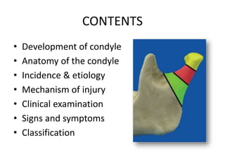
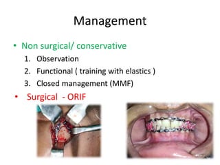
orthognathic surgery is very intresting and well knowing branch in oral surgery ....this presentation is dealing with jaw correction surgery in upper jaw.
This seminar explains about the development, relations, ligaments, various attachments, vascular and nervous supply and various surgical approaches and its modifications to TMJ
Dr. Ahmed M. Adawy, Professor Emeritus, Dep. Oral & Maxillofacial Surgery. Former Dean, Faculty of Dental Medicine, Al-Azhar University. Condylar fractures represent one of the most controversial issues in maxillofacial traumatology regarding classification, diagnoses and therapeutic management. Classification systems of condylar fracture is discussed. Diagnosis is usually based on history clinical examination and radiographic finding. Treatment ranges from observation, jaw exercises to closed or opened interventions. For years closed reduction was thought to be essentially complication-free. Several serious complications however have been reported including temporomandibular joint ankyloses, malocclusion, mandibular deviation and the generative joint pathology. The absolute and relative indications for open reduction is given. The debate between supporters of open or closed reduction is still continuing and the issue has not been resolved. However, the final choice treatment modality should takes into account the location of the fracture, age of the patient, presence or absence of other associated injuries, cosmetic impact of the surgery and presence of other systemic medical conditions.
The Layout of this Presentation is as follows,
Hemifacial Microsomia, Definition, Etiology, classification, Investigations, Treatment Options, Step wise Treatment and Case Presentation, Some references for Further study and the end take Home message.
For Contact: sulaimankhankcd@gmail.com
Condylar fractures /certified fixed orthodontic courses by Indian dental aca...Indian dental academy
The Indian Dental Academy is the Leader in continuing dental education , training dentists in all aspects of dentistry and offering a wide range of dental certified courses in different formats.
Indian dental academy provides dental crown & Bridge,rotary endodontics,fixed orthodontics,
Dental implants courses.for details pls visit www.indiandentalacademy.com ,or call
0091-9248678078
This PowerPoint presentation provides a concise and technical exploration of NOE fractures, encompassing fracture classifications, diagnostic modalities, and treatment approaches. Delve into the intricacies of fracture pathology, radiological assessments, and surgical interventions
New Directions in Targeted Therapeutic Approaches for Older Adults With Mantl...i3 Health
i3 Health is pleased to make the speaker slides from this activity available for use as a non-accredited self-study or teaching resource.
This slide deck presented by Dr. Kami Maddocks, Professor-Clinical in the Division of Hematology and
Associate Division Director for Ambulatory Operations
The Ohio State University Comprehensive Cancer Center, will provide insight into new directions in targeted therapeutic approaches for older adults with mantle cell lymphoma.
STATEMENT OF NEED
Mantle cell lymphoma (MCL) is a rare, aggressive B-cell non-Hodgkin lymphoma (NHL) accounting for 5% to 7% of all lymphomas. Its prognosis ranges from indolent disease that does not require treatment for years to very aggressive disease, which is associated with poor survival (Silkenstedt et al, 2021). Typically, MCL is diagnosed at advanced stage and in older patients who cannot tolerate intensive therapy (NCCN, 2022). Although recent advances have slightly increased remission rates, recurrence and relapse remain very common, leading to a median overall survival between 3 and 6 years (LLS, 2021). Though there are several effective options, progress is still needed towards establishing an accepted frontline approach for MCL (Castellino et al, 2022). Treatment selection and management of MCL are complicated by the heterogeneity of prognosis, advanced age and comorbidities of patients, and lack of an established standard approach for treatment, making it vital that clinicians be familiar with the latest research and advances in this area. In this activity chaired by Michael Wang, MD, Professor in the Department of Lymphoma & Myeloma at MD Anderson Cancer Center, expert faculty will discuss prognostic factors informing treatment, the promising results of recent trials in new therapeutic approaches, and the implications of treatment resistance in therapeutic selection for MCL.
Target Audience
Hematology/oncology fellows, attending faculty, and other health care professionals involved in the treatment of patients with mantle cell lymphoma (MCL).
Learning Objectives
1.) Identify clinical and biological prognostic factors that can guide treatment decision making for older adults with MCL
2.) Evaluate emerging data on targeted therapeutic approaches for treatment-naive and relapsed/refractory MCL and their applicability to older adults
3.) Assess mechanisms of resistance to targeted therapies for MCL and their implications for treatment selection
Prix Galien International 2024 Forum ProgramLevi Shapiro
June 20, 2024, Prix Galien International and Jerusalem Ethics Forum in ROME. Detailed agenda including panels:
- ADVANCES IN CARDIOLOGY: A NEW PARADIGM IS COMING
- WOMEN’S HEALTH: FERTILITY PRESERVATION
- WHAT’S NEW IN THE TREATMENT OF INFECTIOUS,
ONCOLOGICAL AND INFLAMMATORY SKIN DISEASES?
- ARTIFICIAL INTELLIGENCE AND ETHICS
- GENE THERAPY
- BEYOND BORDERS: GLOBAL INITIATIVES FOR DEMOCRATIZING LIFE SCIENCE TECHNOLOGIES AND PROMOTING ACCESS TO HEALTHCARE
- ETHICAL CHALLENGES IN LIFE SCIENCES
- Prix Galien International Awards Ceremony
263778731218 Abortion Clinic /Pills In Harare ,sisternakatoto
263778731218 Abortion Clinic /Pills In Harare ,ABORTION WOMEN’S CLINIC +27730423979 IN women clinic we believe that every woman should be able to make choices in her pregnancy. Our job is to provide compassionate care, safety,affordable and confidential services. That’s why we have won the trust from all generations of women all over the world. we use non surgical method(Abortion pills) to terminate…Dr.LISA +27730423979women Clinic is committed to providing the highest quality of obstetrical and gynecological care to women of all ages. Our dedicated staff aim to treat each patient and her health concerns with compassion and respect.Our dedicated group ABORTION WOMEN’S CLINIC +27730423979 IN women clinic we believe that every woman should be able to make choices in her pregnancy. Our job is to provide compassionate care, safety,affordable and confidential services. That’s why we have won the trust from all generations of women all over the world. we use non surgical method(Abortion pills) to terminate…Dr.LISA +27730423979women Clinic is committed to providing the highest quality of obstetrical and gynecological care to women of all ages. Our dedicated staff aim to treat each patient and her health concerns with compassion and respect.Our dedicated group of receptionists, nurses, and physicians have worked together as a teamof receptionists, nurses, and physicians have worked together as a team wwww.lisywomensclinic.co.za/
Title: Sense of Smell
Presenter: Dr. Faiza, Assistant Professor of Physiology
Qualifications:
MBBS (Best Graduate, AIMC Lahore)
FCPS Physiology
ICMT, CHPE, DHPE (STMU)
MPH (GC University, Faisalabad)
MBA (Virtual University of Pakistan)
Learning Objectives:
Describe the primary categories of smells and the concept of odor blindness.
Explain the structure and location of the olfactory membrane and mucosa, including the types and roles of cells involved in olfaction.
Describe the pathway and mechanisms of olfactory signal transmission from the olfactory receptors to the brain.
Illustrate the biochemical cascade triggered by odorant binding to olfactory receptors, including the role of G-proteins and second messengers in generating an action potential.
Identify different types of olfactory disorders such as anosmia, hyposmia, hyperosmia, and dysosmia, including their potential causes.
Key Topics:
Olfactory Genes:
3% of the human genome accounts for olfactory genes.
400 genes for odorant receptors.
Olfactory Membrane:
Located in the superior part of the nasal cavity.
Medially: Folds downward along the superior septum.
Laterally: Folds over the superior turbinate and upper surface of the middle turbinate.
Total surface area: 5-10 square centimeters.
Olfactory Mucosa:
Olfactory Cells: Bipolar nerve cells derived from the CNS (100 million), with 4-25 olfactory cilia per cell.
Sustentacular Cells: Produce mucus and maintain ionic and molecular environment.
Basal Cells: Replace worn-out olfactory cells with an average lifespan of 1-2 months.
Bowman’s Gland: Secretes mucus.
Stimulation of Olfactory Cells:
Odorant dissolves in mucus and attaches to receptors on olfactory cilia.
Involves a cascade effect through G-proteins and second messengers, leading to depolarization and action potential generation in the olfactory nerve.
Quality of a Good Odorant:
Small (3-20 Carbon atoms), volatile, water-soluble, and lipid-soluble.
Facilitated by odorant-binding proteins in mucus.
Membrane Potential and Action Potential:
Resting membrane potential: -55mV.
Action potential frequency in the olfactory nerve increases with odorant strength.
Adaptation Towards the Sense of Smell:
Rapid adaptation within the first second, with further slow adaptation.
Psychological adaptation greater than receptor adaptation, involving feedback inhibition from the central nervous system.
Primary Sensations of Smell:
Camphoraceous, Musky, Floral, Pepperminty, Ethereal, Pungent, Putrid.
Odor Detection Threshold:
Examples: Hydrogen sulfide (0.0005 ppm), Methyl-mercaptan (0.002 ppm).
Some toxic substances are odorless at lethal concentrations.
Characteristics of Smell:
Odor blindness for single substances due to lack of appropriate receptor protein.
Behavioral and emotional influences of smell.
Transmission of Olfactory Signals:
From olfactory cells to glomeruli in the olfactory bulb, involving lateral inhibition.
Primitive, less old, and new olfactory systems with different path
Recomendações da OMS sobre cuidados maternos e neonatais para uma experiência pós-natal positiva.
Em consonância com os ODS – Objetivos do Desenvolvimento Sustentável e a Estratégia Global para a Saúde das Mulheres, Crianças e Adolescentes, e aplicando uma abordagem baseada nos direitos humanos, os esforços de cuidados pós-natais devem expandir-se para além da cobertura e da simples sobrevivência, de modo a incluir cuidados de qualidade.
Estas diretrizes visam melhorar a qualidade dos cuidados pós-natais essenciais e de rotina prestados às mulheres e aos recém-nascidos, com o objetivo final de melhorar a saúde e o bem-estar materno e neonatal.
Uma “experiência pós-natal positiva” é um resultado importante para todas as mulheres que dão à luz e para os seus recém-nascidos, estabelecendo as bases para a melhoria da saúde e do bem-estar a curto e longo prazo. Uma experiência pós-natal positiva é definida como aquela em que as mulheres, pessoas que gestam, os recém-nascidos, os casais, os pais, os cuidadores e as famílias recebem informação consistente, garantia e apoio de profissionais de saúde motivados; e onde um sistema de saúde flexível e com recursos reconheça as necessidades das mulheres e dos bebês e respeite o seu contexto cultural.
Estas diretrizes consolidadas apresentam algumas recomendações novas e já bem fundamentadas sobre cuidados pós-natais de rotina para mulheres e neonatos que recebem cuidados no pós-parto em unidades de saúde ou na comunidade, independentemente dos recursos disponíveis.
É fornecido um conjunto abrangente de recomendações para cuidados durante o período puerperal, com ênfase nos cuidados essenciais que todas as mulheres e recém-nascidos devem receber, e com a devida atenção à qualidade dos cuidados; isto é, a entrega e a experiência do cuidado recebido. Estas diretrizes atualizam e ampliam as recomendações da OMS de 2014 sobre cuidados pós-natais da mãe e do recém-nascido e complementam as atuais diretrizes da OMS sobre a gestão de complicações pós-natais.
O estabelecimento da amamentação e o manejo das principais intercorrências é contemplada.
Recomendamos muito.
Vamos discutir essas recomendações no nosso curso de pós-graduação em Aleitamento no Instituto Ciclos.
Esta publicação só está disponível em inglês até o momento.
Prof. Marcus Renato de Carvalho
www.agostodourado.com
These simplified slides by Dr. Sidra Arshad present an overview of the non-respiratory functions of the respiratory tract.
Learning objectives:
1. Enlist the non-respiratory functions of the respiratory tract
2. Briefly explain how these functions are carried out
3. Discuss the significance of dead space
4. Differentiate between minute ventilation and alveolar ventilation
5. Describe the cough and sneeze reflexes
Study Resources:
1. Chapter 39, Guyton and Hall Textbook of Medical Physiology, 14th edition
2. Chapter 34, Ganong’s Review of Medical Physiology, 26th edition
3. Chapter 17, Human Physiology by Lauralee Sherwood, 9th edition
4. Non-respiratory functions of the lungs https://academic.oup.com/bjaed/article/13/3/98/278874
TEST BANK for Operations Management, 14th Edition by William J. Stevenson, Ve...kevinkariuki227
TEST BANK for Operations Management, 14th Edition by William J. Stevenson, Verified Chapters 1 - 19, Complete Newest Version.pdf
Flu Vaccine Alert in Bangalore Karnatakaaddon Scans
As flu season approaches, health officials in Bangalore, Karnataka, are urging residents to get their flu vaccinations. The seasonal flu, while common, can lead to severe health complications, particularly for vulnerable populations such as young children, the elderly, and those with underlying health conditions.
Dr. Vidisha Kumari, a leading epidemiologist in Bangalore, emphasizes the importance of getting vaccinated. "The flu vaccine is our best defense against the influenza virus. It not only protects individuals but also helps prevent the spread of the virus in our communities," he says.
This year, the flu season is expected to coincide with a potential increase in other respiratory illnesses. The Karnataka Health Department has launched an awareness campaign highlighting the significance of flu vaccinations. They have set up multiple vaccination centers across Bangalore, making it convenient for residents to receive their shots.
To encourage widespread vaccination, the government is also collaborating with local schools, workplaces, and community centers to facilitate vaccination drives. Special attention is being given to ensuring that the vaccine is accessible to all, including marginalized communities who may have limited access to healthcare.
Residents are reminded that the flu vaccine is safe and effective. Common side effects are mild and may include soreness at the injection site, mild fever, or muscle aches. These side effects are generally short-lived and far less severe than the flu itself.
Healthcare providers are also stressing the importance of continuing COVID-19 precautions. Wearing masks, practicing good hand hygiene, and maintaining social distancing are still crucial, especially in crowded places.
Protect yourself and your loved ones by getting vaccinated. Together, we can help keep Bangalore healthy and safe this flu season. For more information on vaccination centers and schedules, residents can visit the Karnataka Health Department’s official website or follow their social media pages.
Stay informed, stay safe, and get your flu shot today!
Ethanol (CH3CH2OH), or beverage alcohol, is a two-carbon alcohol
that is rapidly distributed in the body and brain. Ethanol alters many
neurochemical systems and has rewarding and addictive properties. It
is the oldest recreational drug and likely contributes to more morbidity,
mortality, and public health costs than all illicit drugs combined. The
5th edition of the Diagnostic and Statistical Manual of Mental Disorders
(DSM-5) integrates alcohol abuse and alcohol dependence into a single
disorder called alcohol use disorder (AUD), with mild, moderate,
and severe subclassifications (American Psychiatric Association, 2013).
In the DSM-5, all types of substance abuse and dependence have been
combined into a single substance use disorder (SUD) on a continuum
from mild to severe. A diagnosis of AUD requires that at least two of
the 11 DSM-5 behaviors be present within a 12-month period (mild
AUD: 2–3 criteria; moderate AUD: 4–5 criteria; severe AUD: 6–11 criteria).
The four main behavioral effects of AUD are impaired control over
drinking, negative social consequences, risky use, and altered physiological
effects (tolerance, withdrawal). This chapter presents an overview
of the prevalence and harmful consequences of AUD in the U.S.,
the systemic nature of the disease, neurocircuitry and stages of AUD,
comorbidities, fetal alcohol spectrum disorders, genetic risk factors, and
pharmacotherapies for AUD.
NVBDCP.pptx Nation vector borne disease control programSapna Thakur
NVBDCP was launched in 2003-2004 . Vector-Borne Disease: Disease that results from an infection transmitted to humans and other animals by blood-feeding arthropods, such as mosquitoes, ticks, and fleas. Examples of vector-borne diseases include Dengue fever, West Nile Virus, Lyme disease, and malaria.
These lecture slides, by Dr Sidra Arshad, offer a quick overview of physiological basis of a normal electrocardiogram.
Learning objectives:
1. Define an electrocardiogram (ECG) and electrocardiography
2. Describe how dipoles generated by the heart produce the waveforms of the ECG
3. Describe the components of a normal electrocardiogram of a typical bipolar leads (limb II)
4. Differentiate between intervals and segments
5. Enlist some common indications for obtaining an ECG
Study Resources:
1. Chapter 11, Guyton and Hall Textbook of Medical Physiology, 14th edition
2. Chapter 9, Human Physiology - From Cells to Systems, Lauralee Sherwood, 9th edition
3. Chapter 29, Ganong’s Review of Medical Physiology, 26th edition
4. Electrocardiogram, StatPearls - https://www.ncbi.nlm.nih.gov/books/NBK549803/
5. ECG in Medical Practice by ABM Abdullah, 4th edition
6. ECG Basics, http://www.nataliescasebook.com/tag/e-c-g-basics
Report Back from SGO 2024: What’s the Latest in Cervical Cancer?bkling
Are you curious about what’s new in cervical cancer research or unsure what the findings mean? Join Dr. Emily Ko, a gynecologic oncologist at Penn Medicine, to learn about the latest updates from the Society of Gynecologic Oncology (SGO) 2024 Annual Meeting on Women’s Cancer. Dr. Ko will discuss what the research presented at the conference means for you and answer your questions about the new developments.
ARTIFICIAL INTELLIGENCE IN HEALTHCARE.pdfAnujkumaranit
Artificial intelligence (AI) refers to the simulation of human intelligence processes by machines, especially computer systems. It encompasses tasks such as learning, reasoning, problem-solving, perception, and language understanding. AI technologies are revolutionizing various fields, from healthcare to finance, by enabling machines to perform tasks that typically require human intelligence.
2. CONTENTS
• Development of condyle
• Anatomy of the condyle
• Incidence & etiology
• Mechanism of injury
• Clinical examination
• Signs and symptoms
• Classification
3. • Management options
• Criteria for Open reduction
• Surgical Considerations
a. Approaches
b. Reduction maneuvers
c. Types of Fixation
• Condyle with associated fractures
• Head fractures
• Pediatric fractures
• Delayed fractures
• Complications
• Conclusion
4. DEVELOPMENT
• Meckel’s cartilage
• Intramembranous
ossification
• Condensation of
mesenchyme just lateral
to Meckel’s cartilage
• Cartilage disappears as
bony mandible develops
5. Anatomy of condyle
• Represents inferior articular surface of
TMJ
• Its axis is perpendicular to ramus, to
which connected by thin neck (collum)
• Oblique directed medially and slightly
backwards, forming angle of 25◦ with
frontal plane
• Surface is convex & displays 3 parts from front to back
1. Front- fossa of lateral pterygoid muscle
2. Anterior slope of condyle, inlined at angle of 45◦ from
horizontal & meeting the temporal articular tubercle
3. Back – posterior side of condyle, which descends 5mm
beneath the apex or rounded crest separating the two
slopes
6. • Has 2 poles – lateral & medial to
which disc is attached with strong
ligaments
• Average dimensions of condyle:
AP 8.5 mm & ML 21mm
• Collum that it supports is 10mm
AP & 7mm ML diameter
• Base is 22mm in sagittal AP & only
5mm in transverse diameter-
extremely fragile & vulnerable to
# in adults
7. Parameter Child Adult
Cortical bone Thin Thick
Condylar neck Broad Thin
Articular surface Thin Thick
Capsule Highly vascular Less vascular
Periosteum Highly active
osteogenic phase
Less active in latent
stage
Intracapsular fracture
& hemarthrosis
Very common Less common
Remodelling capacity
following trauma
Present Absent
Disturbance in
growth
Likely Rare
8. Blood Supply
• Branches from Superficial
temporal artery
• Deep auricular artery-
branch of maxillary artery
Venous drainage
Superficial temporal vein
10. Incidence
Haug, R. H., Prather, J., & Thomas Indresano, A.
(1990). An epidemiologic survey of facial fractures
and concomitant injuries. Journal of Oral and
Maxillofacial
11. Etiology
• Trauma
• Motor vehicle accident
• Interpersonal violence
• Fall from heights
• Ballistic injuries
• Pathological process
12. Rowe & Williams Vol 1
MECHANISM OF INJURY
As Proposed by Lindahl in 1977
13. Clinical Examination
• Preauricular pain & swelling
• Restricted mandibular excursion
• Deviation on ipsilateral side on
opening
• Posterior gagging of occlusion
• Anterior open bite
• Laceration of EAC – ear bleed
• CSF rhinorrhea & otorrhea with
associated skull base #
16. Radiographic Evaluation
• Conventional – reverse townes & Panoramic
• CT – gold standard
diagnostic accuracy CT 90 % , OPG 73 %
Chacon GE, A comparative study of 2 imaging
techniques for the diagnosis of condylar fractures in
children. J Oral Maxillofac Surg. 2003
17. • Other Adjunct investigation
• MRI- assess non osseous TMJ & disc
• CT Angiography - close relationship of condyle
with ICA
• When foreign body is close to maxillary artery
mean distance between the artery and the
medial border of the subcondylar portion of
the mandible was 6.8 mm
Orbay 2007 ,Maxillary Artery: Anatomical Landmarks and
Relationship with the Mandibular Subcondyle. Plastic and
Reconstructive Surgery
18. Classification
• Displacement – shifting between fracture bony
fragments
• Dislocation –shifting between components of
TMJ
• Proposed by Loukota et al 2005 & as adopted by
Strasbourg Osteosynthesis Research
Group(SORG)
19. Wassmund -1934
• Type I – 10◦ to 45◦ angulation of condylar head with
bony contact between the fragments. Reduce
spontaneously
• Type II – 45◦ to 90 ◦ angulation of condylar head with
slight bony contact between the fragments. Tearing of
medial portion of joint capsule
• Type III – Severe medial displacement with no contact
between the bony fragments. Open reduction
advocated.
• Type IV – Fractured head articulates on/ forward to
eminence
• Type V – Vertical / oblique # through head of condyle -
rare
20. MacLennan 1952
• Class I: no deviation (bending)
• Class II: deviation (bending) at the fracture
level
• Class III: displacement (condylar head remains
within fossa)
• Class IV: dislocation (condylar head outside of
fossa)
21. Rowe and Killey, 1955
• Intracapsular fractures
• Extracapsular fractures
• Fractures associated with the TMJ capsule,
TMJ ligaments, articulating disk, and bony
structures surrounding the TMJ
22. Dingman and Natvig , 1964
• High condylar neck fracture: fracture line is at or
above the level of the lateral pterygoid
attachment on the fovea of the condylar
apparatus
• Intermediate condylar neck fracture: fracture
line is below the level of insertion of the lateral
pterygoid
• Low condylar neck fracture: fracture begins at or
below the sigmoid notch and extends to the
posterior border of the mandibular ramus
23. Spiessl and Schroll, 1972
• Type I: condylar neck fracture
without deviation / displacement
• Type II: low condylar neck fracture
with deviation/displacement
• Type III: high condylar neck
fracture with deviation /
displacement
IIIa: ventral
IIIb: medial
IIIc: lateral
IIId: dorsal
24. • Type IV: low condylar neck
fracture with dislocation
• Type V: high condylar neck
fracture with dislocation
• Type VI: intracapsular
fracture of the condylar
head
26. • 2: Relationship of condylar fragment to mandible
2a: angulation with medial override
2b: angualtion with lateral override
2c: angulation without override
2d: nondisplaced / fissure #
• 3: Relationship of condylar head to fossa
no displacement (0)
slight displacement (1)
moderate displacement(2)
Dislocation (3)
27. Ellis et al, 1999
• Condylar head fracture:
intracapsular fracture
• Condylar neck fracture:
fracture below the
condylar head, but on or
above the lowest point
of the sigmoid notch
• Condylar base fracture:
fracture in which the
fracture line is located
below the lowest point
of the sigmoid notch
28. Loukota et al -2005, SORG
• Diacapitular fracture: the fracture line starts in
the articular surface and may extend outside the
TMJ capsule
• Condylar neck: the fracture line starts
somewhere above Line A and runs above Line A
for more than half of its length
• Condylar base: the fracture line extends behind
the mandibular foramen and runs below Line A
for more than half of its length
• Minimal displacement: displacement of less than
10 or overlap of the bone edges by less than 2
mm, or both
29. Dicapitular FractureLine A- perpendicular line through the
sigmoid notch to tangent of the ramus
Condylar neck fracture Condylar base (subcondylar) fracture
30. AO modification of Ellis ,2010
Line 1- parallels the posterior border
of mandible
Line 2- runs perpendicular to line 1
at deepest portion of the sigmoid
notch
Line 3- below lateral pole of
condylar head, also perpendicular to
line 1
A line is drawn half way between
the lateral pole line and the sigmoid
notch line
“high-neck” # is above this line,
“low-neck” # is below the line
31. MRI based classification 2018
A- without loss of ramal height – no disk displacement
B- without loss of ramal height – with disk displacement
C – with loss of ramal height – regardless of disk
Ying et al. BJOMS(2018)
32. Management
• Non surgical/ conservative
1. Observation
2. Functional ( training with elastics )
3. Closed management (MMF)
• Surgical - ORIF
34. Goals of Therapy
1. Obtain stable occlusion.
2. Restore interincisal opening and mandibular excursive
movements.
3. Establish a full range of mandibular excursive
movements.
4. Minimize deviation of the mandible.
5. Produce a pain-free articular apparatus at rest and
during function.
6. Avoid internal derangement of the temporomandibular
joint on the injured or the contralateral side.
7. Avoid the long-term complication of growth
disturbance.
Peterson 2nd edition
35. Indications for ORIF
Zide and Kent’s indications for open reduction (1983)
For Both Adults & Children
Mainly for Adults
36. Mathes Treatment protocol 1983
OPEN REDUCTION
• Malocclusion with CR
• Fragment angulation:
> 30°
• Bone gap: ≥ 4-5mm
Lateral override
• Lack of contact of #
fragment
PREFERRED FOR OPEN REDUCTION
• Any low, dislocated subcondylar #
• Low condylar # with multiply mandible
/ Le Fort #
• Low condylar # with head out of fossa
• Condylar fragment 14°- medial tilt
• Ramus shortening - 5%
• Bilateral fracture with open bite
• Gross fracture end mal-alignment
• Fracture – dislocation
• Abnormal function, malocclusion
37. Management of subcondylar # base on
SORG (2007)
• Two proven indication
1. Ramus Height shortening
2. Displacement of condyle
•Class 1 – Closed reduction
ramus shortening <2mm,
displacement <10o
•Class 2 – ORIF
ramus shortening 2-15mm,
displacement 10o to 45o
•Class 3 – ORIF
ramus shotening > 15mm,
displacement >45o
38. Nonsurgical Management
• Functional Training with elastics
• Neuromuscular adjustment for
proper occlusion
• For period of 4-6 weeks
Unilateral #
1-2 class II elastics on fractured site
to draw mandible anteriorly.
Less elastics to promote active use
of mandible
Bilateral #
B/L class II elastics with vertical
elastics anteriorly
Complete elastics at night
Condylar Process Fractures of the Mandible
Edward Ellis III, Facial plastic surgery clinics 2000
39. • Goals
MO > 40mm, without deviation
Lateral excursions >10mm
Protrusive excursions >5mm
• Weaning from elastics
2-3 weeks: Obtain pre traumatic occlusion
Next 2-3 weeks : Used only while sleeping
Arch bars left in place
U/L – 2 months
B/L – 3 months
Functional Training with elastics
40. MMF Period for Condylar #
• Recommended period: 7 days – 6 weeks
• Children: 7-10 days ( to prevent ankylosis )
Killey’s Fractures of Mandible 3rd Edition
41. Closed management Maneuvers
• Hypomochlion : Hypo- small, Mochlion - lever
Thelekkat, Yeshaswini & Aravindakshan, shyam mohan. (2014). Hypomochlion
aided reduction for sub-condylar fractures. Kerala dental journal
Class III Lever
42. AAOMS Parameters of Care 2017
Closed reduction
• Nondisplaced/displaced #
where from/function can be
restored
• # dislocations / comminution
in growing child where
form/function can be restored
• Medical contraindications to
ORIF
Open reduction ( including
Endoscopic )
• # dislocation of condyle
• Mechanical interference with
function
• # with loss of AP & vertical
dimension that cannot be
managed by closed reduction (eg,
edentulous patient, multiple facial
fractures)
• Compound #
• Displacement into middle cranial
fossa
• Patient/surgeon preference
• Prevention of Complication of
MMF
Ochs, M., Chung, W., & Powers, D. (2017). Trauma
Surgery. Journal of Oral and Maxillofacial Surgery
43. Surgical Considerations
1. Ramus must be distracted.
2. Proximal condyle must be controlled &
manipulated.
3. # must be anatomically reduced & plated with
more than one screw on the proximal
segment
45. 1. Preauricular approach
• Indications
– Wire fixation of high, anteromedially
displaced proximal fragment
• Advantages.
– access to superiormost portion of joint.
• Disadvantages.
– not indicated for placement of plate and
screw fixation.
– no access to distract the ramus inferiorly;
– osteosynthesis plate placement extremely
difficult
– increased risk of necrosis of condyle
46. • Incision: skin fold along the entire
length of ear
• Dissect along anterior portion of EAC
to avoid damage to the superficial
temporal vessels and
auriculotemporal nerve
• Incise the superficial layer of the
temporalis fascia just anterior to the
tragus at the zygoma, continuing in
antero-superior direction
• Oblique incision made through
capsule near root of zygoma to enter
joint capsule & Condylar #
48. 3. Submandibular Approach
• Indications
– axial anchor screw fixation.
• Advantages
– ability to distract mandibular ramus
and direct access of the gonial angle.
• Disadvantages
– limited surgical site exposure (the
incision is distant from the fracture),
– difficult to reduce medially displaced
condyles
– plate and screw fixation restricted
without a transfacial trocar.
49.
50. Retromandibular Approach
• Indications
– any fracture that is large enough to be
reduced and stabilized by ORIF using
plates and screws.
• Advantages.
– best access to the fracture site
– no need for a transfacial trocar
– facial scar is less noticeable than
submandibular incision
– effective in patients with edema
– access for an osteotomy if required
• Disadvantages
– scar is more noticeable than with a
preauricular incision.
51. Rhytidectomy Approach
• same access as that of the
retromandibular approach,
with better cosmesis.
• This approach must be
drained with closed suction
drainage postoperatively
52. TransMasseteric anteroparotid
Approach ( Peri-angular )
• Indications
– provide access to high & low
subcondylar and ramus fractures
• Advantages
– quick and direct access to fracture sites
for direct plating and screw fixation,
– with excellent exposure and the ability
to distract mandibular ramus because of
access to the gonial angle,
– better access
• Disadvantages
– a visible scar that is more noticeable
than with the other approaches and
there is potential damage to the facial
nerve.
53. • Make the incision at the line
connecting the intersections of
the last third on the posterior
ramus line and first third on the
inferior border line, the
intersection closest to the gonial
angle in each direction
• The initial incision is to the depth
of the parotidomasseteric fascia
(SMAS), followed by extensive
subcutaneous undermining in all
directions to allow for maximal
exposure.
54. Intraoral Approach
• Indications.
– low subcondylar fractures.
– Axial anchor screws /miniplate fixation may
be used.
• Advantages.
– visible scar avoided & damage to facial nerve
is minimized.
• Disadvantages
• Intraoral Approach without Endoscope.
– poorest access of all the approaches,
– difficult to ascertain the adequacy of
reduction
– high rate of complications.
• Endoscope-Assisted Intraoral Approach.
– More time-consuming, steep learning curve,
– poor visibility of the posterior ramus,
– difficulty in reducing certain fracture types
56. Matching fracture type to surgical
access
Emam ,Matching Surgical Approach to Condylar Fracture Type.
Atlas of the Oral and Maxillofacial Surgery Clinics (2017)
57. • Most important
factor in determining
approach used: Level
of Fracture
• Modifying Factors:
Degree of
displacemennt or
dislocation
Time elapsed after
trauma
58. Approaches
A. Displaced, not dislocated
1. Base # : High submandibular incision/
endoscopy assisted
2. Neck : Retromandibular
3. Head : Preauricular
B. Dislocated
Approach : One level Higher
C. Mal-united at any level
Preauricular ± Combination
59. Condyle Fixation Techniques
Single Miniplate Double MIniplate Indirect Lag screw- Kernel
Direct Lag screw -
Brown
K wire- Stephenson
& Graham
Trans osseous
wiring - Masser
Lag screw &
Pin- Rasse
60. What is Ideal Fixation ?
Meyer’s Line of Osteosynthesis
Christopher Meyer
J Cranio-Maxillofac Surg 2002
Single Miniplate
Center/ Long axis of condyle
Close to Posterior Border
Two Miniplates
In Triangular Fashion
1st plate below Sigmoid Notch
2nd plate Along Posterior Border
Fixation with specific
implants for the
subcondylar region.
61. What to do first ?
Dentate First
More force needed to
distract mandible
Condyle First
Less force needed to distract
mandible
Condyle reduction is easier
62. Inferior Distraction of Ramus
• Manual Method – with surgeon’s hand intraorally
• Instruments – Allies, Towel clip
• Specialised retractors
• Screw with wire in 16 G Cannula
63. Bilateral Condyle Fractures
Problems:
●Mandible in 4 pieces ● ↑ intergonial width
● Lingual splay ● Flaring of Rami
Chen, Functional outcomes following surgical treatment
of bilateral mandibular condylar fractures. IJOMS (2011)
64. 1. Ellis Maneuver
Facial width problems associated with rigid fixation
of mandibular fractures: Case reports. JOMS, Ellis, E.,
& Tharanon, W. (1992).
65. 2.Muselet Technique
Temiz (2015) A useful method for the reduction of laterally displaced
mandibular condyles: The muselet technique. Journal of Plastic,
Reconstructive & Aesthetic Surgery
66. What if Head # is not treated ?
• TMJ internal derangement
• Bifid condyle
• Ankylosis
• Asymmetry
• Restricted / painful mouth
opening
• Functional remodelling of
condyle
67. Classification of Head #
• Neff, 2002
A : Medial
B : Lateral
M: Comminuted
• Dongmei He, 2009
A :Lateral, with ramal height reduction
B :Middle, without
C : Medial, without
M : Comminuted
He, D (2009). Intracapsular Condylar Fracture of the
Mandible: Our Classification and Open Treatment
Experience. JOMS
68. Treatment of Condylar Head #
• Type A – Open
• Type B – Closed in most cases ( fragment small
& deep)
• Type C – Closed / Removal of fragment
69. Injury to Articular apparatus
• Effusion : Hemorrhagic or serous
• Soft tissue injury : Disk
Capsule
Ligaments
• Dislocation of the condyle from the fossa
Without fracture
With fracture other than condyle
With associated condylar fracture
• Fracture
Non-displaced Dislocated
Deviated Comminuted
Displaced Involving adjacent bony structures
• Combinations of the above
Peterson 2nd Edition
70. MRI based articular soft tissue injury
• Grade I - hemarthrosis only – best outcome
• Grade II - hemarthrosis and disc displacement
• Grade III - hemarthrosis, disc displacement &
capsular tear
• Grade IV - disc perforation in association with
Grade I,II or III
All fractures managed non-surgically, 100 % had
restricted joint movement & joint noise
Tripathi (2015). Severity of Soft Tissue Injury Within the Temporomandibular
Joint Following Condylar Fracture as Seen on Magnetic Resonance Imaging
and Its Impact on Outcome of Functional Management. JOMS
71. Pediatric Condylar Fractures
Concerns :
• Imaging problems
• Early management &
long term follow up
• Protocol of IMF
• 2nd surgery for
hardware removal
• Growth disturbance
• Ankylosis
• RESTITUTIONAL
REMODELING – Role
of Condylar Cartilage
72. Grunwaldt et al,Pediatric facial fractures: demographics, injury
patterns, and associated injuries in 772 consecutive patients. Plast
Reconstr Surg 2011;
73. Costello (2012). Growth and Development Considerations for Craniomaxillofacial
Surgery. Oral and Maxillofacial Surgery Clinics of North America
75. Current Concepts in the Mandibular Condyle Fracture Management Part II: Open
ReductionVs Closed Reduction, Choi et al , Archieves of Plastic Surgery 2012
76. Mal-united Condylar fractures
• Malocclusion
• Reduced bite force
• Deviation in mouth
opening
• Asymmetry due to loss
of ramus height
• Temporal & masseteric
pain- hypertonic facial
muscles
• TMJ dysfunction
• Ankylosis
Common causes :
• Closed management
• Mismanaged primary
ORIF
77. Time elapsed
after trauma
Recommended treatment Author
< 3 months Conservative treatment Ellis E, Walker
RV Craniomax
Trauma Recon
2009
> 3 to 6
months
Sub-condylar osteotomy Chenn, Ann
Plast Surg
2013
> 9 months Normal Ramus Orthognathic
Surgery
Ellis E, Walker
RV 2009
Comminuted
ramal # or
Shortening of
Ramus height
TMJ
Reconstruction
79. Objectives of Sialocele management
1. Reduce secretion
To facilitate rapid healing
• Nil per oral
• Avoid tangy food
• Local injections
• Hypertonic saline
• Botox
• Systemic drugs
• Scopolamine patch
2. Reduce dead space &
approximate tissues
To eliminate space for saliva
collection
• Repeated aspirations
• Pressure dressing
3. Induce Fibrosis
• To strengthen tissues around
salivary pool
• To block outflow through
subcutaneous tissues -
Sclerosants
4. Divert secretions into oral
cavity
• Surgical procedures
80. Conclusion
• Paradigm shift in century old tradition of non surgical
treatment of condylar #
• Improvement in
CT Diagnostics
Indication specific Osteosynthesis – 3D plates & resorbable
systems
Newer approaches
• Recent consensus on improved evidence
IBRA 2012
AAOMS Parameters of Care 2017
• ORIF – Gold standard in base & neck # in adults
• Endoscopic approach for base with lateral lateral displacement
• Growing tendency to open Head #
• ORIF displaced/ dislocated # in children with mixed dentition