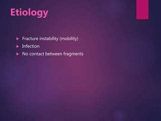
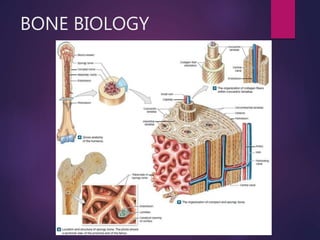
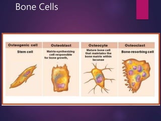
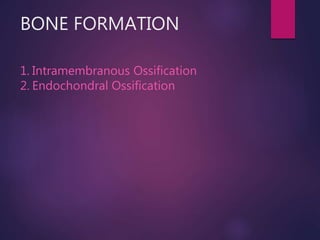
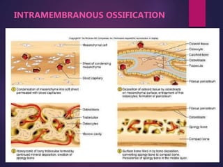
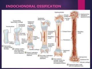
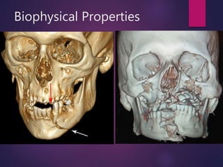
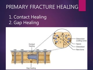
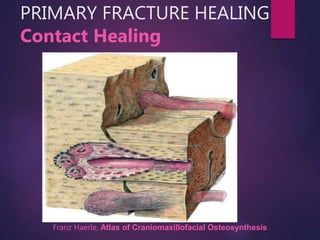
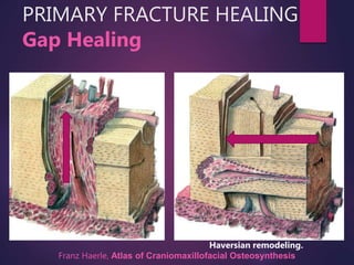
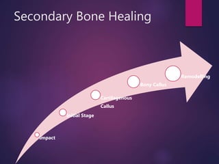
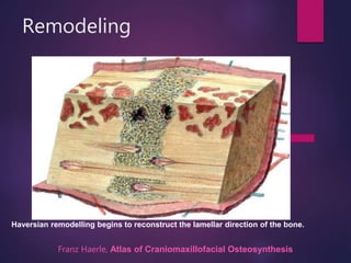
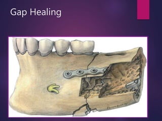
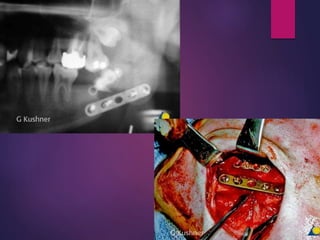
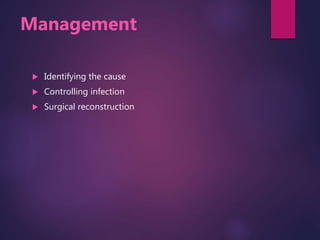
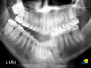
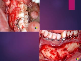
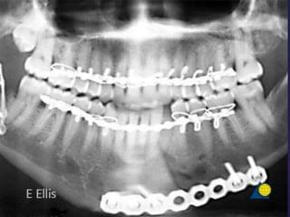
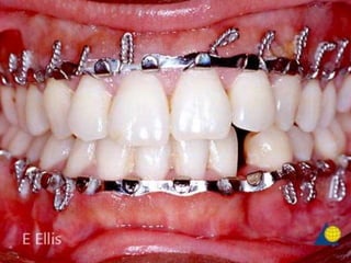
Fracture is a break in the structural continuity of bone, And starts immediately after the fracture occurs.
fracture results in a well-defined progression of tissue responses that are designed to remove tissue debris, to reestablish vascular supply and to produce a new skeletal matrix.
Fracture Healing,Introduction,Pathology&Stages,Factors influencing osteogenesis,differences in healing of fractured bone by conservative&operative management.
Bone morphogenetic proteins (BMPs) is the generic name for a family of proteins that can form bone de novo, for which they are considered osteoinductive
Stages of Bone healing and madalities to enhance bone healing Surya Vijay Singh
Bone healing, direct bone healing, indirect bone healing, primary and secondary bone healing, stages of bone healing, substitute of bone healing, autografting and allograft, fracture healing
Orthobiologics - PRP, BMC the real story so far!Vaibhav Bagaria
A basic presentation on the role of orthobiologics, PRP, Bone marrow aspirate concentrate in orthopaedics. Insights, and future research directions in a rapidly evolving field.
Distraction osteogenesis is a method of producing unlimited quantities of living bone directly from a special osteotomy by controlled mechanical distraction. The new bone spontaneously bridges the gap and rapidly remodels to a normal macrostructure for the local bone.
Clinical & surgical management of the mandibular condylar process fractures has generated a great deal of controversy in maxillofacial trauma and there are many various approaches to treat this injury. Before, many surgeons seem to favor closed treatment with maxillomandibular fixation (MMF), but recently open treatment of condylar fractures with rigid internal fixation (RIF) has become more common & acceptable. The objective of this presentation was to evaluate the factors that determine the choice of method for treatment of condylar fractures: open or closed, pointing out their indications, contra-indications, advantages and disadvantages.
The Indian Dental Academy is the Leader in continuing dental education , training dentists in all aspects of dentistry and offering a wide range of dental certified courses in different formats.
Indian dental academy provides dental crown & Bridge,rotary endodontics,fixed orthodontics,
Dental implants courses.for details pls visit www.indiandentalacademy.com ,or call
00919248678078
“The study of orthodontia is indissolubly connected with that of art as related to the human face.” -Dr. Edward H. Angle.
Each person shares with the rest of the population a great many characteristics, but there are enough differences to make each human being a unique individual. Such limitless variation in the size, shape and relationship of the dental, skeletal and soft tissue facial structures are important in providing each individual with his or her own identity.
Face – Difficult object to measure accurately because of
– complex morphology
– sensitivity to eyes
– its soft nature.
One of goals of orthodontic treatment is creating a balanced & harmonious facial appearance. Craniofacial symmetry is one of the aspect of this harmony. Subject of symmetry or lack of symmetry of human face has been of considerable interest, particularly in the field of Orthodontics. Minor variation is a desirable variation of craniofacial structure which is perceived as esthetically pleasing and has no esthetic or functional significance. Asymmetry becomes important when it affects the function or esthetics of the person.
Supplemental corticosteroids for dental patients with adrenal insufficiencyR...DrKamini Dadsena
Primary Adrenal Insufficiency:
It is caused by a progressive destruction of the adrenal cortex, usually of an idiopathic nature (most commonly autoimmune), but also results from hemorrhage, sepsis, infectious diseases (such as tuberculosis, human immunodeficiency virus, cytomegalovirus and fungal infection), malignancy, adrenalectomy, amyloidosis or drugs.
Clinical assessment scoring system for tracheostomy (CASST) criterion: Objec...DrKamini Dadsena
Tracheotomy has been used for many centuries as a means to bypass upper airway obstruction.
Head and neck cancers are often associated with anatomic changes which can create a potentially difficult airway.
CARNOY’S SOLUTION AS A SURGICAL MEDICAMENT IN THETREATMENT OF KERATOCYSTIC O...DrKamini Dadsena
The term keratocyst was coined by Philipsen in 1956.
Unlike the other cystic lesion KOT, has got strong tendency for recurrence.
Treatment of these lesions remains controversial and has a number of dilemmas about the choice of treatment whether to use carnoys solution as an adjunct therapy after removal of the lesion.
Temporomandibular disorders (TMDs) are considered the major cause of orofacial pain. Internal derangement (ID) of the temporomandibular joint (TMJ), which is classified as disc displacement with or without reduction, is one of the disorders of the TMJ that is frequently seen.
Displacement of the articular disc can result in decreased joint space, joint noise (clicking, popping, or crepitation), arthritis, condylar resorption, inflammation, and compression of the bilaminar tissue, all of which can cause various degrees of pain and dysfunction.
Changing Guidelines of CPR & BLS For General Dental Practitioners & O...DrKamini Dadsena
The tolerance of the heart to anoxia is relatively high, but the central nervous system will show irreversible lesions if anoxia lasts for more than 3–4 min.
Though unusual, there are reports of deaths due to CPA in dental offices during dental treatment.
Cardiopulmonary resuscitation (CPR) is a vital skill which must be mastered by all health care professionals.
Therefore the thorough knowledge of CPR and Basic Life Support is of utmost importance to the dentist.
Neck Dissection: Nomenclature, Classification, and TechniqueDrKamini Dadsena
Removal of the at-risk lymphatic basins serves two important purposes.
First, it allows the removal and identification of occult metastasis in patients in whom cervical metastasis are a risk, - Elective neck dissection.
Secondly, it allows the removal of disease in patients in whom metastasis are highly suspected based on imaging, clinical examination or fine needle aspiration, - Therapeutic neck dissection.
Clinical use of botulinum toxins in oral and maxillofacial surgeryDrKamini Dadsena
Purified botulinum toxin (BTX) was the first bacterial toxin used as a medicine. Since its introduction into clinical use, over 30 years ago, it has become a versatile drug in various fields of medicine.
Its mechanism of inhibiting acetylcholine release at neuromuscular junctions following local injection is unique for the treatment of facial wrinkles.
Other dose-dependent anti-neuroinflammatory effects and vascular modulating properties have extended its spectrum of applications.
Cavernous sinus thrombosis represents a rare but devastating disease process that may be associated with significant long-term patient morbidity or mortality. The prompt recognition and management of this problem is critical.
In 1989, Shetty and Freymiller [7] reviewed indications for removal of teeth in the line of fracture. They recommended the following indications:
1. Significant periodontal disease with gross mobility and periapical pathology
2. Partially erupted third molars with pericoronitis or cystic areas
3. Teeth preventing the reduction of fractures
4. Teeth with fractured roots
5. Teeth with exposed root apices or teeth in which the entire root surface from the apex to the gingival margin is exposed
6. Excessive delay from the time of fracture to the time of definitive treatment
In addition to these indications, another indication that requires extraction of teeth in the line of fracture is an acute, recurring abscess at the site of the fracture despite antibiotic therapy(8)
Assessment of lingual nerve injury using different surgical variables for man...DrKamini Dadsena
Assessment of lingual nerve injury using different surgical variables for mandibular third molar surgery
The objective of this study was to investigate the incidence of sensory impairment of the lingual nerves following lower third molar surgery and to compare the outcome with various operative variables.
Factors that predicted lingual nerve injury were lingual flap retraction, tooth sectioning, and buccal guttering.
Instruments in major oral and maxillofacial surgeryDrKamini Dadsena
A surgical instrument is a specially designed tool or device for performing specific actions and carrying out desired effects during surgery or operations.
Traditional classification were given 100 years back when RTA , assaults, sports injuries, industrial accidents were minimal.
Over the past 100 years RTA (high speed & Low speed) assaults, sports injuries (high contact/ low contact), industrial accidents have increased.
Fracture patterns which are not matching the traditional injuries pattern.
Can speed up diagnosis and treatment planning
Cohorting / clubbing of complication to Specific Fractures.
It facilitate communication between peers and assist documentation and research.
It also have prognostic value for patients and assist Surgeons in planning their management.
It serves as a basis for treatment and for evaluation of the results.
Different fractures/ Areas of fracture has different treatment plan / approaches.
Undisplaced fracture : conservative/ surgical
Displaced Fractures: Surgical/ conservative with traction
Trauma is a global problem and continues to be a leading cause of disability and death.
Approximately 25% to 30% of deaths caused by trauma can be prevented when a systematic and organized approach is used.
The main goal of the initial assessment
Recognize the patient who does have life-threatening injuries
Establish treatment priorities, and
Manage them aggressively
A flap is a unit of tissue that is transferred from one site (donor site) to another (recipient site) while maintaining its own blood supply or from a anastomised vessel.
Flaps come in many different shapes and forms. They range from simple advancements of skin to composites of many different types of tissue
The International Association for the Study of Pain (IASP)1 defines trigeminal neuralgia (TN) as a sudden, usually unilateral, severe brief stabbing recurrent pain in one or more branches of the fifth cranial nerve
synonyms
Idiopathic trigeminal neuralgia / Tic Doulourex.
Trifacial Neuralgia.
Fothergell’s disease.
In 1677 John Locke, a American physician and philosopher, accurately identified the major clinical features of TN
In 1756 the French physician Nicolaus Andre coined the term “Tic douloureux” to the condition.
The English physician John Fothergill in 1773 published detailed description of TN, since then, it has been referred to as ‘Fothergill’s disease’.
Peripheral injections
Long acting LA
Alcohol
Glycerol
Peripheral neurectomy/ nerve avulsion
Cryotherapy
Gasserian ganglion procedures
Percutaneous stereotactic radiofrequency thermal lesioning of the trigeminal ganglion and/or root (rfl)
percutaneous glycerol gangliolysis of the trigeminal ganglion
percutaneous balloon microcompression of the trigeminal ganglion
Intracranial procedures
MVD
Partial sensory rhizotomy
Gamma knife radiation to the trigeminal root entry zone GKR
An oroantral communication is an unnatural perforation between oral cavity and maxillary sinus.
Oroantral fistula is an epithelized, pathological, communication between these two cavities. A fistulous tract present more than 14 days should be considered as chronic fistula.
The practice of surgery rests on certain fundamental principles which remain unchanged, though to apply them the surgeon may have to modify techniques to suit the anatomical field, the type of operation and the conditions obtaining at the time.
Obstructive sleep apnea (OSA) is a prevalent chronic disease characterized by pharyngeal collapse during sleep.
Sleep disorder that involves cessation or significant decrease in airflow through the upper airway in the presence of breathing effort.
Obstructive sleep apnea is the second most common sleep disorder, insomnia being the most common.
Associated with recurrent oxyhemoglobin desaturations and arousals from sleep
Apnea index- no. of apneas /hr of total sleep time.
AHI (APNEA-HYPOPNEA INDEX)- No of apneas and hypoapneas/hr of total sleep time.
RDI (Respiratory Disturbance Index) – no. of apneas, hypoapneas and respiratory effort related arousals(RERA)/hr of total sleep time.
Pain pathway gate control theory
Pain management
An unpleasant emotional experience usually initiated by noxious stimulus and transmitted over a specialized neural network to CNS where it is interpreted as such.
1. Exteroceptors: arising from receptors from skin & mucosa. sensed at conscious level
E.g. Merkel corpuscles : Tactile receptors.
Free Nerve ending :Perceive superficial pain.
2. Proprioceptors : From musculoskeletal structures.
The presence , positions & movement of body. below conscious levels.
E.g. 1) Muscle spindles : Skeletal muscle fibers. Mechanoreceptors.
2) Free nerve ending : Perceive deep somatic pain & other sensations.
3. Interoceptors : From viscera of body below conscious level.
E.g. Pacinian corpuscles : perception of touch-pressure.
Free nerve ending : Perceive visceral pain & other sensations.
These lecture slides, by Dr Sidra Arshad, offer a quick overview of physiological basis of a normal electrocardiogram.
Learning objectives:
1. Define an electrocardiogram (ECG) and electrocardiography
2. Describe how dipoles generated by the heart produce the waveforms of the ECG
3. Describe the components of a normal electrocardiogram of a typical bipolar leads (limb II)
4. Differentiate between intervals and segments
5. Enlist some common indications for obtaining an ECG
Study Resources:
1. Chapter 11, Guyton and Hall Textbook of Medical Physiology, 14th edition
2. Chapter 9, Human Physiology - From Cells to Systems, Lauralee Sherwood, 9th edition
3. Chapter 29, Ganong’s Review of Medical Physiology, 26th edition
4. Electrocardiogram, StatPearls - https://www.ncbi.nlm.nih.gov/books/NBK549803/
5. ECG in Medical Practice by ABM Abdullah, 4th edition
6. ECG Basics, http://www.nataliescasebook.com/tag/e-c-g-basics
Lung Cancer: Artificial Intelligence, Synergetics, Complex System Analysis, S...Oleg Kshivets
RESULTS: Overall life span (LS) was 2252.1±1742.5 days and cumulative 5-year survival (5YS) reached 73.2%, 10 years – 64.8%, 20 years – 42.5%. 513 LCP lived more than 5 years (LS=3124.6±1525.6 days), 148 LCP – more than 10 years (LS=5054.4±1504.1 days).199 LCP died because of LC (LS=562.7±374.5 days). 5YS of LCP after bi/lobectomies was significantly superior in comparison with LCP after pneumonectomies (78.1% vs.63.7%, P=0.00001 by log-rank test). AT significantly improved 5YS (66.3% vs. 34.8%) (P=0.00000 by log-rank test) only for LCP with N1-2. Cox modeling displayed that 5YS of LCP significantly depended on: phase transition (PT) early-invasive LC in terms of synergetics, PT N0—N12, cell ratio factors (ratio between cancer cells- CC and blood cells subpopulations), G1-3, histology, glucose, AT, blood cell circuit, prothrombin index, heparin tolerance, recalcification time (P=0.000-0.038). Neural networks, genetic algorithm selection and bootstrap simulation revealed relationships between 5YS and PT early-invasive LC (rank=1), PT N0—N12 (rank=2), thrombocytes/CC (3), erythrocytes/CC (4), eosinophils/CC (5), healthy cells/CC (6), lymphocytes/CC (7), segmented neutrophils/CC (8), stick neutrophils/CC (9), monocytes/CC (10); leucocytes/CC (11). Correct prediction of 5YS was 100% by neural networks computing (area under ROC curve=1.0; error=0.0).
CONCLUSIONS: 5YS of LCP after radical procedures significantly depended on: 1) PT early-invasive cancer; 2) PT N0--N12; 3) cell ratio factors; 4) blood cell circuit; 5) biochemical factors; 6) hemostasis system; 7) AT; 8) LC characteristics; 9) LC cell dynamics; 10) surgery type: lobectomy/pneumonectomy; 11) anthropometric data. Optimal diagnosis and treatment strategies for LC are: 1) screening and early detection of LC; 2) availability of experienced thoracic surgeons because of complexity of radical procedures; 3) aggressive en block surgery and adequate lymph node dissection for completeness; 4) precise prediction; 5) adjuvant chemoimmunoradiotherapy for LCP with unfavorable prognosis.
Flu Vaccine Alert in Bangalore Karnatakaaddon Scans
As flu season approaches, health officials in Bangalore, Karnataka, are urging residents to get their flu vaccinations. The seasonal flu, while common, can lead to severe health complications, particularly for vulnerable populations such as young children, the elderly, and those with underlying health conditions.
Dr. Vidisha Kumari, a leading epidemiologist in Bangalore, emphasizes the importance of getting vaccinated. "The flu vaccine is our best defense against the influenza virus. It not only protects individuals but also helps prevent the spread of the virus in our communities," he says.
This year, the flu season is expected to coincide with a potential increase in other respiratory illnesses. The Karnataka Health Department has launched an awareness campaign highlighting the significance of flu vaccinations. They have set up multiple vaccination centers across Bangalore, making it convenient for residents to receive their shots.
To encourage widespread vaccination, the government is also collaborating with local schools, workplaces, and community centers to facilitate vaccination drives. Special attention is being given to ensuring that the vaccine is accessible to all, including marginalized communities who may have limited access to healthcare.
Residents are reminded that the flu vaccine is safe and effective. Common side effects are mild and may include soreness at the injection site, mild fever, or muscle aches. These side effects are generally short-lived and far less severe than the flu itself.
Healthcare providers are also stressing the importance of continuing COVID-19 precautions. Wearing masks, practicing good hand hygiene, and maintaining social distancing are still crucial, especially in crowded places.
Protect yourself and your loved ones by getting vaccinated. Together, we can help keep Bangalore healthy and safe this flu season. For more information on vaccination centers and schedules, residents can visit the Karnataka Health Department’s official website or follow their social media pages.
Stay informed, stay safe, and get your flu shot today!
HOT NEW PRODUCT! BIG SALES FAST SHIPPING NOW FROM CHINA!! EU KU DB BK substit...GL Anaacs
Contact us if you are interested:
Email / Skype : kefaya1771@gmail.com
Threema: PXHY5PDH
New BATCH Ku !!! MUCH IN DEMAND FAST SALE EVERY BATCH HAPPY GOOD EFFECT BIG BATCH !
Contact me on Threema or skype to start big business!!
Hot-sale products:
NEW HOT EUTYLONE WHITE CRYSTAL!!
5cl-adba precursor (semi finished )
5cl-adba raw materials
ADBB precursor (semi finished )
ADBB raw materials
APVP powder
5fadb/4f-adb
Jwh018 / Jwh210
Eutylone crystal
Protonitazene (hydrochloride) CAS: 119276-01-6
Flubrotizolam CAS: 57801-95-3
Metonitazene CAS: 14680-51-4
Payment terms: Western Union,MoneyGram,Bitcoin or USDT.
Deliver Time: Usually 7-15days
Shipping method: FedEx, TNT, DHL,UPS etc.Our deliveries are 100% safe, fast, reliable and discreet.
Samples will be sent for your evaluation!If you are interested in, please contact me, let's talk details.
We specializes in exporting high quality Research chemical, medical intermediate, Pharmaceutical chemicals and so on. Products are exported to USA, Canada, France, Korea, Japan,Russia, Southeast Asia and other countries.
Explore natural remedies for syphilis treatment in Singapore. Discover alternative therapies, herbal remedies, and lifestyle changes that may complement conventional treatments. Learn about holistic approaches to managing syphilis symptoms and supporting overall health.
Knee anatomy and clinical tests 2024.pdfvimalpl1234
This includes all relevant anatomy and clinical tests compiled from standard textbooks, Campbell,netter etc..It is comprehensive and best suited for orthopaedicians and orthopaedic residents.
Report Back from SGO 2024: What’s the Latest in Cervical Cancer?bkling
Are you curious about what’s new in cervical cancer research or unsure what the findings mean? Join Dr. Emily Ko, a gynecologic oncologist at Penn Medicine, to learn about the latest updates from the Society of Gynecologic Oncology (SGO) 2024 Annual Meeting on Women’s Cancer. Dr. Ko will discuss what the research presented at the conference means for you and answer your questions about the new developments.
New Drug Discovery and Development .....NEHA GUPTA
The "New Drug Discovery and Development" process involves the identification, design, testing, and manufacturing of novel pharmaceutical compounds with the aim of introducing new and improved treatments for various medical conditions. This comprehensive endeavor encompasses various stages, including target identification, preclinical studies, clinical trials, regulatory approval, and post-market surveillance. It involves multidisciplinary collaboration among scientists, researchers, clinicians, regulatory experts, and pharmaceutical companies to bring innovative therapies to market and address unmet medical needs.
3. ETIOLOGY
Singaram M et al, Prevalence, pattern, etiology, and management of
maxillofacial trauma in a developing country: a retrospective study. J
Korean Assoc Oral Maxillofac Surg 2016;42:174-181)
11. Factors affecting type of
1. Degree of force
2. Resistance to force offered by facial bones
3. Point of application of the force
4. Cross sectional aea of the object
21. Remodeling
Franz Haerle, Atlas of Craniomaxillofacial Osteosynthesis
Haversian remodelling begins to reconstruct the lamellar direction of the bone.
Fracture is a break in the structural continuity of bone, And starts immediately after the fracture occurs.
fracture results in a well-defined progression of tissue responses that are designed to remove tissue debris, to reestablish vascular supply and to produce a new skeletal matrix.
Fracture may be a sequel of RTA, fall, interpersonal violence, sports related injury, industrial injury , iatrogenic injury or rbcz of pathology
267
Bone is a mineralized connective tissue consisting of type I collagen and noncollagenous, structural matrix proteins.
Characteristic of all bones are a dense outer sheet of compact bone and a central, medullary cavity.The cavity is filled with red or yellow bone marrow that is interrupted, by a network of bone trabeculae, cancellous bone.
Surrounded by a connective tissue layer called periosteum.
Cortical bone consists of:
Multiple columnar bone units (osteons), composed of a central longitudinal canal (Haversian canal) that contains a central blood vessel and nervous tissue.
Transverse nutrient canals (Volkmann canals) connecting adjacent osteons.
Bone is laid down in concentric layers around each Haversian canal.
Osteocytes are scattered throughout osteons, each within its own space (lacuna).
There are 4 types of bone cells
Osteogenic stem cells produce osteoblast
Osteoblast are Matrix synthesizing responsible for bone growth
Osteocytes are Mature bone cells that maintains bone matrix within lacunae
Osteoclast Are Multinucleated bone resorbing giant cell with ruffled border
All bone are mesodermal origin. The process of bone formation is called ossification.
intramembranous ossification bones of the vault of the skull frontal parietal, the mandible and the clavicle. Temporal squamous and tympanic, Sphenoid greater wing + pterigoid laminnae, maxilla, palatine zygoma, nasal lacrimal vomar
Occipital: The interparietal part (lying
above the superior nuchal lines) is formed in membrane; the rest of the bone is formed
by endochondral ossification.
-In some situations, bone is laid down directly in a fibrous membrane. This is called intramembranous ossification
- Mesenchymal cells differentiate into osteoblasts lay down osteoid.- intramembranous ossification bones of the vault of the skull frontal parietal, the mandible maxilla, palatine zygoma, nasal lacrimal vomar. Temporal squamous and tympanic, Sphenoid greater wing + pterigoid laminnae, Occipital: The interparietal part (lying
above the superior nuchal lines) is formed in membrane
When bone formation is preceded by the formation of a cartilaginous model that closely resembles the bone to be formed. And cartilage is subsequently replaced by (not converted into) bone. This kind of bone formation is called endochondral ossification.
Vascular ingrowth
When bone fully mature
Endochondral ethmoid + inferior nasal concae,condyle coronoid of mandi petrous + mastoid
Sphenoid rest rest of occipital
- This phenomena also occurs in healing of fractures.
Energy storage capacity of bone depends upon speed of loading i.e. force of traumatic blow.
Higher the speed more the energy stored.
When force of traumatic blow overcomes the internal strength of bone fracture occurs.
When fracture occurs stored energy is released.
Speed of loading affects the pattern of fracture:
When loading speed is Low energy dissipated through single fracture line less displacement of soft tissue and remaining intact bone.
When loading speed is Higher energy not dissipated thrugh single break Comminuted Fracture and extensive soft tissue damage results
1 directly related to momentum of object responsible for trauma or body
Biology of fracture healing is a complex process that lead to regeneration of normal bone architecture.
There are two types of fracture healing:
Primary fracture healing
Secondary fracture healing
Primary bone healing, requires rigid fixation and immobility of fracture segments with a minimal gap between them (less than 100 μm). Osteoclasts migrate to the fracture site and widen adjacent haversian systems, allowing osteoblasts to deposit bone matrix, or osteoid, eventually to calcify into organized mature lamellar bone.3
Secondary, bone healing, is more complex and occurs when a significant gap or interfragmentary motion is present. Secondary bone healing involves the formation of a fibrocartilaginous intermediary bone callous (Fig. 33-1)
There are four distinct stages of indirect bone healing but the end product is the same as mature bone formed in primary healing. The initial insult propagates the inflammatory stage. A hematoma between and around the fracture develops and stabilizes, drawing inflammatory cells to the site. Necrotic and nonviable bone near the fracture is cellularly débrided and repair is initiated by angiogenesis and the activation of osteoprogenitor cells and fibroblasts. The second, or soft callus, stage is characterized by conversion of the hematoma to a fibrocartilaginous mass to bridge the fracture. Fibroblasts and mesenchymal elements are highly active in laying down new collagen to create the substrate into which the third phase, or hard callus stage, develops. During this period, osteoid is calcified and periosteal and endosteal bone ingrowth starts to replace the soft callus as a result of endochondral bone formation. Finally, in the remodeling stage, the woven bone of the hard callus matures and organizes to a trabecular structure to re-create the native preinjury structure.4
Although distinct, both types of bone healing may occur simultaneously in the same fracture. As three-dimensional structures, bones may have varying levels of contact and fracture reduction in the same general site, resulting in endochondral and lamellar elements in different areas at the same point in time (Fig. 33-2).
Close apposition of bone fragments allow transverse bridging of haversian system.
Requires:
Excellent anatomical reduction
Minimal or no mobility
Excellent blood supply
Precludes the necessity for callus formation.
First reported by Danis (1949) described as sodure autogene
Contact healing of the bone means healing of the fracture line after stable anatomical repositioning, with perfect interfragmentary contact and without the possibility for any cellular or vascular ingrowth
Cutting cones are able to cross this interface from one fragment to the other by remodeling the haversian canal. Haversian canal
remodeling is the main mechanism for restoration of the internal architecture of compact bone
Contact healing takes place over the whole fracture line after perfect anatomical reduction, osteosynthesis, and mechanical rest.
Contact healing is only seen directly beneath the miniplate
Resultant core provides pathway for vessel ingrowth and osteoblastic proliferation to form new bone.
Gap healing takes place in stable or “quiet” gaps with a width greater than the 200-μm osteonal diameter. Ingrowth of vessels and mesenchymal cells starts after surgery.
Osteoblasts deposit osteoid on the fragment ends without osteoclastic resorption. The gaps are filled exclusively with primarily formed, transversely oriented lamellar bone. Replacement is usually completed within 4 to 6 weeks
In the second stage, the transversely oriented bone lamellae are replaced by axially orientated osteons, a process which is referred to as haversian remodeling
After 10 weeks the fracture is replaced by newly reconstructed cortical bone
Osteoblasts deposit osteoid,a nd the gaps are filled with primary formation of transversely orientated lamellar bone.
Haversian remodeling. Transversely oriented bone lamellae are replaced by axially orientated osteons.
Small gaps left
Blood vessels invade, bring mesenchymal osteoblastic precursors.
Bone deposited directly without resorption and immediate callus formation.
0.3 mm gap - lamellar bone directly
0.5- 1 mm gap- woven bone followed by lamellar bone.
Firstly bundles are arranged perpendicular to long axis.
Over the time they arrange themselves along the long axis of repaired bone.
Secondary bone healing occurs via the pluripotential cells located within the cortical and cancellous bone periosteum and associated
soft tissue.
It results from the mechanical instability of the fracture, caused by resorption of fracture ends and callus formation.
seen in cases with or without direct fixation of fracture site
Begins with hematoma formation
Bone fracture leads to the rupture of blood vessels with hematoma formation in the surrounding soft tissue and localized avascularity of the fragment ends.
Further complications are thrombosis of the vessels within the haversian and Volkmann canals a fewmillimeters from the ends
of fragments
Polymorphs, histiocytes and mast cells make their appearance.
Initiation of cellular proliferation..
These are plueripotent.
Capillary ingrowth and fibroblasts migrate into wound and lay down collagen.
Initially, the formation of periosteal callus leads to a decrease in interfragmentary strain, which is followed by interfragmentary and endosteal callus formation. Invading granulation tissue replaces the initial hematoma and is transformed into interfragmentary connective tissue
(Fig. 4.4). The ends of the fragments are resorbed by osteoclasts (Figs. 4.4, 4.5). The more interfragmentary con-nective tissue is remodeled into fibrocartilage (Fig. 4.6).
Since fibrocartilage is more rigid than fibrous tissue, the interfragmentary tissue becomes stiffer and increases the resistance to motion of the fragments.
During this phase, the callus can be comprised of fibrous connective tissue, blood vessels, cartilage.
Low oxygen tension and at the site of fracture.
Continuous compression and tensionat the site of fracture.
Periosteal and endosteal mesenchymal cells fibro-cartilagenous callus
CGF-1 and later CGF-2 Type II collagen and hyaluronic acid.
Macrophages, fibroblasts and osteoblasts release enzyme collagenase.
Cuff or callus formed around the fracture site stabilise the involved area
Depending on location
Anchoring callus developes on the outside surface of the bone near the periosteum. It extends some distance away from the fracture.
Sealing callus developes on the inside surface of the bone across the fractured end. It fills the marrow space and goes out into the fractured site. It forms from endosteal proliferation.
Bridging callus developes on the outside surface between the two fractured ends. This is the only cartilaginous.
Uniting callus forms between the ends of bones and between the area of primary calluses that have formed on the two fractured parts.. It forms by direct ossification, extensive resorption of bone ends occurs by this time. It does not form until the other forms of calluses are well developed
Fibrocartiage undergo calcification into woven bone.
Spaces in the Cartilagenous callus allows vascular ingrowth which changes the environment conductive to formation of osteoblasts.
Deposit osteoid on the spicules of calcified cartilage Undergo calcification, forms bone beginning at peripheryProgresses as homogenous calcificationInitially woven bone is formed
Subsequently the fibrocartilage undergoes mineralization. Vascular invasion of fibrocartilage is combined with resorption of mineralized matrix. Calcified fibrocartilage must undergo resorption before osteoblasts can start to produce osteoid as a base for new bone deposition (Fig. 4.7). Initially the calcified fibrocartilage is replaced by woven bone.
Remodelling
Woven bone undergo organization to form lamellar bone.
In accordance with the wolff’s law.
Wolf law every change in the form and the function of a bone leads to changes in its internal architecture and in its external form
Three windows in a mandibular
fracture with miniplate osteosynthesis
on the tension side.
This shows indirect bone healing in the window on the inferior border of the mandible with the mobile fragment; direct bone healing in the window of the lateral cortex of the mandible by contact healing after perfect and anatomical reduction; direct bone healing
in the window of the inner cortex of the mandible by gap healing.
A nonunion occurs when the fracture does not heal in an appropriate time frame. The result is mobility of the fracture segments present after an adequate healing phase. Patients may also demonstrate malocclusion and infection at the site of fracture.
Etiology
Nonunions are usually the result of one or more of the following factors:
Fracture instability (mobility)
Infection
Inaccurate reduction
No contact between fragments
Treatment
Treatment will consist of:
Identifying the cause
Controlling infection
Surgical reconstruction: removing the existing hardware, debridement of devital bone and/or soft tissues, decortication of bone fragments at the fracture ends, reestablishing occlusion, stabilizing segments using a locking reconstruction plate 2.4, and autogenous bone graft to this area.
: removing the existing hardware, debridement of devital bone and/or soft tissues, decortication of bone fragments at the fracture ends, reestablishing occlusion, stabilizing segments using a locking reconstruction plate 2.4, and autogenous bone graft to this area.
Panoramic x-ray 6 weeks after treatment of left angle fracture with single miniplate. The fracture is grossly mobile, infected, and the plate has become loose
The fracture was debrided, the plate removed, the infection drained, and the patient placed on antibiotics to control infection.
Once infection has subsided, the patient was taken to surgery and the fracture exposed through a submandibular approach. The fibrous tissue between the fragments was debrided and the fragments decorticated.
The occlusion was reestablished with MMF. A reconstruction plate was then adapted and secured to provide load-bearing fixation across the fracture gap..
Particulate autogenous bone was placed into the fracture gap and the incision closed in layers.
Panoramic x-ray taken 10 months postoperatively showing bone filling fracture gap
A malunion results when the bony fracture segments heal in an incorrect or nonanatomic position, which can lead to a deformity.16 For fractures of the jaws, malunion will create a malocclusion.
Intraoperative photograph showing malunion of left parasymphyseal fracture. Rt angle region