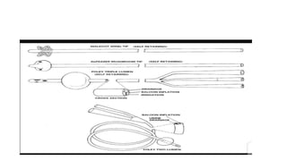
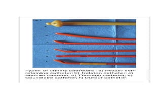
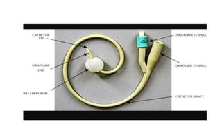
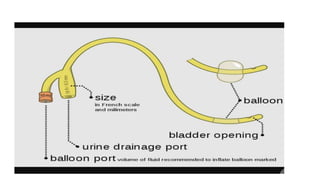
The surgical treatment of an injury or defect within the urethra's walls is known as urethroplasty. The three most frequent factors leading to urethral damage that needs to be repaired are trauma, iatrogenic injury, and infections. The gold standard treatment for urethral strictures is urethroplasty, which has a lower recurrence rate than dilatations and urethrotomies. Although recurrence rates are higher for this challenging treatment group, it is likely the only effective treatment option for chronic and severe strictures.
Urethroplasty is not regarded as a small procedure, taking three to eight hours on average in the operating room. Between 20% and 30% of urethroplasty patients may benefit from the ease of going under the knife for a shorter period of time and going home the same day. On average, hospital stays last two to three days. Seven to ten days may be needed for hospitalization for more complicated surgeries.
Fewer than ten percent of patients experience significant complications after urethroplasty, while complications, particularly recurrences, are more frequent in long and complex strictures.
Bladder injuries are rare. But when present in cases of polytrauma they pose both a diagnostic as well as surgical challenge to the attending surgeon. Understanding the mechanisms underlying bladder injuries is pivotal in developing a diagnostic algorithm in order to avoid missing of any urologic injury. Once the extent and site of damage is diagnosed then prompt surgical intervention is the mainstay of treatment. The pathophysiology and management of bladder injuries is discussed in this paper.
Objectives :
-List the indications and contraindications for urinary catheterization.
- Indicate the appropriate catheter type/size.
- Discuss the risks associated with catheterizations.
-Describe the equipment for female/male/pediatric urinary catheterization.
- Discuss a safe method of performing urinary catheterizations .
Bladder catheters are used for urinary drainage, or as a means to collect urine for measurement.
Alternatives to indwelling urethral catheterization should be considered and include external sheath (ie, condom) catheters, suprapubic catheters, intermittent catheterization, and, in some cases, supportive management with protective garments.
Urinary catheterization
Definition
Types of Urinary catheterization
Effects or risks of Urinary catheterization
Suprapubic catheterization
Intermittent catheterization
Caring for catheters
Signs and symptoms of Urinary tract infections
Pulmonary Thromboembolism - etilogy, types, medical- Surgical and nursing man...VarunMahajani
Disruption of blood supply to lung alveoli due to blockage of one or more pulmonary blood vessels is called as Pulmonary thromboembolism. In this presentation we will discuss its causes, types and its management in depth.
Title: Sense of Smell
Presenter: Dr. Faiza, Assistant Professor of Physiology
Qualifications:
MBBS (Best Graduate, AIMC Lahore)
FCPS Physiology
ICMT, CHPE, DHPE (STMU)
MPH (GC University, Faisalabad)
MBA (Virtual University of Pakistan)
Learning Objectives:
Describe the primary categories of smells and the concept of odor blindness.
Explain the structure and location of the olfactory membrane and mucosa, including the types and roles of cells involved in olfaction.
Describe the pathway and mechanisms of olfactory signal transmission from the olfactory receptors to the brain.
Illustrate the biochemical cascade triggered by odorant binding to olfactory receptors, including the role of G-proteins and second messengers in generating an action potential.
Identify different types of olfactory disorders such as anosmia, hyposmia, hyperosmia, and dysosmia, including their potential causes.
Key Topics:
Olfactory Genes:
3% of the human genome accounts for olfactory genes.
400 genes for odorant receptors.
Olfactory Membrane:
Located in the superior part of the nasal cavity.
Medially: Folds downward along the superior septum.
Laterally: Folds over the superior turbinate and upper surface of the middle turbinate.
Total surface area: 5-10 square centimeters.
Olfactory Mucosa:
Olfactory Cells: Bipolar nerve cells derived from the CNS (100 million), with 4-25 olfactory cilia per cell.
Sustentacular Cells: Produce mucus and maintain ionic and molecular environment.
Basal Cells: Replace worn-out olfactory cells with an average lifespan of 1-2 months.
Bowman’s Gland: Secretes mucus.
Stimulation of Olfactory Cells:
Odorant dissolves in mucus and attaches to receptors on olfactory cilia.
Involves a cascade effect through G-proteins and second messengers, leading to depolarization and action potential generation in the olfactory nerve.
Quality of a Good Odorant:
Small (3-20 Carbon atoms), volatile, water-soluble, and lipid-soluble.
Facilitated by odorant-binding proteins in mucus.
Membrane Potential and Action Potential:
Resting membrane potential: -55mV.
Action potential frequency in the olfactory nerve increases with odorant strength.
Adaptation Towards the Sense of Smell:
Rapid adaptation within the first second, with further slow adaptation.
Psychological adaptation greater than receptor adaptation, involving feedback inhibition from the central nervous system.
Primary Sensations of Smell:
Camphoraceous, Musky, Floral, Pepperminty, Ethereal, Pungent, Putrid.
Odor Detection Threshold:
Examples: Hydrogen sulfide (0.0005 ppm), Methyl-mercaptan (0.002 ppm).
Some toxic substances are odorless at lethal concentrations.
Characteristics of Smell:
Odor blindness for single substances due to lack of appropriate receptor protein.
Behavioral and emotional influences of smell.
Transmission of Olfactory Signals:
From olfactory cells to glomeruli in the olfactory bulb, involving lateral inhibition.
Primitive, less old, and new olfactory systems with different path
- Video recording of this lecture in English language: https://youtu.be/lK81BzxMqdo
- Video recording of this lecture in Arabic language: https://youtu.be/Ve4P0COk9OI
- Link to download the book free: https://nephrotube.blogspot.com/p/nephrotube-nephrology-books.html
- Link to NephroTube website: www.NephroTube.com
- Link to NephroTube social media accounts: https://nephrotube.blogspot.com/p/join-nephrotube-on-social-media.html
These simplified slides by Dr. Sidra Arshad present an overview of the non-respiratory functions of the respiratory tract.
Learning objectives:
1. Enlist the non-respiratory functions of the respiratory tract
2. Briefly explain how these functions are carried out
3. Discuss the significance of dead space
4. Differentiate between minute ventilation and alveolar ventilation
5. Describe the cough and sneeze reflexes
Study Resources:
1. Chapter 39, Guyton and Hall Textbook of Medical Physiology, 14th edition
2. Chapter 34, Ganong’s Review of Medical Physiology, 26th edition
3. Chapter 17, Human Physiology by Lauralee Sherwood, 9th edition
4. Non-respiratory functions of the lungs https://academic.oup.com/bjaed/article/13/3/98/278874
Tom Selleck Health: A Comprehensive Look at the Iconic Actor’s Wellness Journeygreendigital
Tom Selleck, an enduring figure in Hollywood. has captivated audiences for decades with his rugged charm, iconic moustache. and memorable roles in television and film. From his breakout role as Thomas Magnum in Magnum P.I. to his current portrayal of Frank Reagan in Blue Bloods. Selleck's career has spanned over 50 years. But beyond his professional achievements. fans have often been curious about Tom Selleck Health. especially as he has aged in the public eye.
Follow us on: Pinterest
Introduction
Many have been interested in Tom Selleck health. not only because of his enduring presence on screen but also because of the challenges. and lifestyle choices he has faced and made over the years. This article delves into the various aspects of Tom Selleck health. exploring his fitness regimen, diet, mental health. and the challenges he has encountered as he ages. We'll look at how he maintains his well-being. the health issues he has faced, and his approach to ageing .
Early Life and Career
Childhood and Athletic Beginnings
Tom Selleck was born on January 29, 1945, in Detroit, Michigan, and grew up in Sherman Oaks, California. From an early age, he was involved in sports, particularly basketball. which played a significant role in his physical development. His athletic pursuits continued into college. where he attended the University of Southern California (USC) on a basketball scholarship. This early involvement in sports laid a strong foundation for his physical health and disciplined lifestyle.
Transition to Acting
Selleck's transition from an athlete to an actor came with its physical demands. His first significant role in "Magnum P.I." required him to perform various stunts and maintain a fit appearance. This role, which he played from 1980 to 1988. necessitated a rigorous fitness routine to meet the show's demands. setting the stage for his long-term commitment to health and wellness.
Fitness Regimen
Workout Routine
Tom Selleck health and fitness regimen has evolved. adapting to his changing roles and age. During his "Magnum, P.I." days. Selleck's workouts were intense and focused on building and maintaining muscle mass. His routine included weightlifting, cardiovascular exercises. and specific training for the stunts he performed on the show.
Selleck adjusted his fitness routine as he aged to suit his body's needs. Today, his workouts focus on maintaining flexibility, strength, and cardiovascular health. He incorporates low-impact exercises such as swimming, walking, and light weightlifting. This balanced approach helps him stay fit without putting undue strain on his joints and muscles.
Importance of Flexibility and Mobility
In recent years, Selleck has emphasized the importance of flexibility and mobility in his fitness regimen. Understanding the natural decline in muscle mass and joint flexibility with age. he includes stretching and yoga in his routine. These practices help prevent injuries, improve posture, and maintain mobilit
Lung Cancer: Artificial Intelligence, Synergetics, Complex System Analysis, S...Oleg Kshivets
RESULTS: Overall life span (LS) was 2252.1±1742.5 days and cumulative 5-year survival (5YS) reached 73.2%, 10 years – 64.8%, 20 years – 42.5%. 513 LCP lived more than 5 years (LS=3124.6±1525.6 days), 148 LCP – more than 10 years (LS=5054.4±1504.1 days).199 LCP died because of LC (LS=562.7±374.5 days). 5YS of LCP after bi/lobectomies was significantly superior in comparison with LCP after pneumonectomies (78.1% vs.63.7%, P=0.00001 by log-rank test). AT significantly improved 5YS (66.3% vs. 34.8%) (P=0.00000 by log-rank test) only for LCP with N1-2. Cox modeling displayed that 5YS of LCP significantly depended on: phase transition (PT) early-invasive LC in terms of synergetics, PT N0—N12, cell ratio factors (ratio between cancer cells- CC and blood cells subpopulations), G1-3, histology, glucose, AT, blood cell circuit, prothrombin index, heparin tolerance, recalcification time (P=0.000-0.038). Neural networks, genetic algorithm selection and bootstrap simulation revealed relationships between 5YS and PT early-invasive LC (rank=1), PT N0—N12 (rank=2), thrombocytes/CC (3), erythrocytes/CC (4), eosinophils/CC (5), healthy cells/CC (6), lymphocytes/CC (7), segmented neutrophils/CC (8), stick neutrophils/CC (9), monocytes/CC (10); leucocytes/CC (11). Correct prediction of 5YS was 100% by neural networks computing (area under ROC curve=1.0; error=0.0).
CONCLUSIONS: 5YS of LCP after radical procedures significantly depended on: 1) PT early-invasive cancer; 2) PT N0--N12; 3) cell ratio factors; 4) blood cell circuit; 5) biochemical factors; 6) hemostasis system; 7) AT; 8) LC characteristics; 9) LC cell dynamics; 10) surgery type: lobectomy/pneumonectomy; 11) anthropometric data. Optimal diagnosis and treatment strategies for LC are: 1) screening and early detection of LC; 2) availability of experienced thoracic surgeons because of complexity of radical procedures; 3) aggressive en block surgery and adequate lymph node dissection for completeness; 4) precise prediction; 5) adjuvant chemoimmunoradiotherapy for LCP with unfavorable prognosis.
NVBDCP.pptx Nation vector borne disease control programSapna Thakur
NVBDCP was launched in 2003-2004 . Vector-Borne Disease: Disease that results from an infection transmitted to humans and other animals by blood-feeding arthropods, such as mosquitoes, ticks, and fleas. Examples of vector-borne diseases include Dengue fever, West Nile Virus, Lyme disease, and malaria.
These lecture slides, by Dr Sidra Arshad, offer a quick overview of physiological basis of a normal electrocardiogram.
Learning objectives:
1. Define an electrocardiogram (ECG) and electrocardiography
2. Describe how dipoles generated by the heart produce the waveforms of the ECG
3. Describe the components of a normal electrocardiogram of a typical bipolar leads (limb II)
4. Differentiate between intervals and segments
5. Enlist some common indications for obtaining an ECG
Study Resources:
1. Chapter 11, Guyton and Hall Textbook of Medical Physiology, 14th edition
2. Chapter 9, Human Physiology - From Cells to Systems, Lauralee Sherwood, 9th edition
3. Chapter 29, Ganong’s Review of Medical Physiology, 26th edition
4. Electrocardiogram, StatPearls - https://www.ncbi.nlm.nih.gov/books/NBK549803/
5. ECG in Medical Practice by ABM Abdullah, 4th edition
6. ECG Basics, http://www.nataliescasebook.com/tag/e-c-g-basics
3. Introduction
• Urethral catheters are tubes inserted into the bladder per- urethral
to allow drainage, injection of fluid, or access to organ for surgert
• Made from materials that have biocompatible properties.
• Urethral catheterization is the aseptic procedure of introducing a tube
or a catheter through the urethra into the urinary bladder for various
purposes
4. • Urethral catheterization is a routine medical procedure that facilitates
direct drainage of the urinary bladder.
• Patients of all ages may require urethral catheterization, but patients
who are elderly or chronically ill are more likely to require indwelling
catheters, which carry their own independent risks.
• The basic principles underlying urethral catheterization are gender-
neutral, but the specific aspects important in the technique of male
catheterization are critical to successful passage.
5. Indications
Diagnostic:
• To collect uncontaminated urine specimen for laboratory investigation
• To distend the bladder prior to abdominopelvic ultrasound.
• Monitoring of urine in critically ill patients, ICU and post-op patients.
• Urinary tract imaging- cystography, RUCG, MCUG
• To measure residual urine volume if ultrasound is not available.
• For diagnosis of urethral stricture.
6. Therapeutic:
• To relieve AUR
• Continuous irrigation of bladder following prostatectomy.
• Instillation of drugs: cytotoxics, BCG, antibiotics.
• Short term urinary diversion.
• Intermittent decompression to treat neurogenic bladder.
• Palliative care in terminal I’ll or incontinent patient, severely impaired
and not correctable by surgery.
7. Other uses of catheter
• Improvised chest drain
• For tube caecostomy
• Improvised as gastrotomy or jejunostomy tube.
• As intraabdominal drain.
9. Classes
Based on duration of use
1. Indwelling catheters
2. Intermittent catheters
3. External catheter
Based on no of channel
1. 1way: for drainage
1. 2way: drainage & inflation
2. 3way: additional irrigation
Based on material
1. Plastic 2. Latex 3. Silicone and silicone-coated 4. Teflon 5. Polyvinyl & polyethylene
6. Metal 7. Silver alloy
10.
11. • Latex : suitable for measurement of residual urine; flexible,
inexpensive; prone to infection, encrustation of salt and protein and
can cause stricture from catheter allergy.
• Lasts 3-4weeks as indwelling
• Polyvinyl & polyethylene are for short term use.
• Silicone and Teflon: good for long term use(8-12weeks). Decreased
incidence of allergy, urethritis and stricture.
• Metal: stainless steel
12. Female metal catheter:
• Very rare situations in which the urethra cannot be catheterized by
usual Foley’s
• Evacuation of bladder prior to gynecological or obstetric surgeries
• Diagnosis of vesicovaginal fistula.
Male catheters- curved along it’s length because of anatomy of male.
Problems: high incidence of urethral injury and creation of false
passage.
Should not be left In situ for longer than 2days.
13. • Based on design
1. Foley’s catheter: has balloon at tip. Can be 2- or 3-way
2. De Peezer(mushroom) catheter: lacks balloon.
has bulbous tip to keep it in position.
useful for suprapubic cystotomy
3. Malecot catheter: lacks balloon
has filamentous tip which helps to retain its position
4. Gibbon: lacks balloon & used per urethra only
14. • Tieman
• coude tip
• Robinson catheter
• Counsel
• Nelaton
• Jacques
15. Based on retention
1. Non self retaining: Jacques, Nelaton, Robinson
2. Self retaining: Foley, Malecot, DePezzer etc
16.
17.
18.
19.
20.
21. Sizes and colour codes
• Catheter is expressed in charriere units. Charriére French scale (0.33
mm equals 1 Fr) . D=Fr/3. Diameter=D
• The French size of the catheter depends on the patient and the
catheter’s purpose.
• pediatric boys will need a French size between 5- 12 Fr.
• Adult should be catheterized with a 16- or 18-Fr catheter. •
• Larger French catheters (20 to 30 Fr) are used to evacuate blood clots
in postoperative prostate surgery patients or in patients who are
bleeding from the kidney or bladder.
22.
23.
24. Materials for catheterization
Catheter tray
Povidone-iodin/Savlon
Sterile cotton balls
Water-soluble lubrication gel
Sterile drapes
Sterile gloves
Urethral catheter
Prefilled 10-mL syringe
Urinometer connected to a collection bag
25. Procedure
• Explain the procedure, benefits, risks, complications, and alternatives
to the patient or the patient’s representative.
• Position the patient supine, in bed, and uncover the genitalia.
• Open the iodine/chlorhexidine/savlon preparatory solution and pour
it onto the sterile cotton balls.
• Open a sterile lidocaine 2% lubricant with applicator or a 10-mL
syringe and sterile 2% lidocaine gel and place them on the sterile
field.
• Don the sterile gloves and use the nondominant hand to hold the
penis and retract the foreskin (if present).
26. • Use the sterile hand and sterile forceps to prep the urethra and glans
in circular motions with at least 3 different cotton balls. Use the
sterile drapes that are provided with the catheter tray to create a
sterile field around the penis.
• Using a syringe with no needle, instill 5-10 mL of lidocaine gel 2% into
the urethra. Place a finger on the meatus to help prevent spillage of
the anesthetic lubricant. Allow 2-3 minutes before proceeding with
the urethral catheterization
• Hold the catheter with the sterile hand. Apply a generous amount of
the nonanesthetic lubricant to the catheter
27. • While holding the penis at approximately 90° and stretching it upward
to straighten out the penile urethra, slowly and gently introduce the
catheter into the urethra. Continue to advance the catheter until the
proximal Y-shaped ports are at the meatus.
• Wait for urine to drain from the larger port to ensure that the distal
end of the catheter is in the urethra. The lubricant jelly–filled distal
catheter openings may delay urine return. If no spontaneous return
of urine occurs, try attaching a 60-mL syringe to aspirate urine. If
urine return is still not visible, withdraw the catheter and reattempt
the procedure (preferably after using ultrasonography to verify the
presence of urine in the bladder)
28. • After visualization of urine return (and while the proximal ports are at
the level of the meatus), inflate the distal balloon by injecting the
appropriate volume of sterile water through the cuff inflation port.
• Gently withdraw the catheter from the urethra until resistance is met.
Secure the catheter to the patient’s thigh with a wide tape. If the
patient is uncircumcised, make sure to reduce the foreskin, as failure
to do so can cause paraphimosis.
• Connect to a drainage bag
• Take note of vol, colour,
29. Perineal Pressure Assistance
The distal tip of the catheter might become caught in the posterior fold
between the urethra and the urogenital diaphragm. An assistant can
apply upward pressure to the perineum while the catheter is advanced
to direct the catheter tip upward through the urogenital diaphragm.
30. Catheterization in females
• Put a fenestrated drape over the pelvis so that the vulva is exposed.
• Gently spread the labia and expose the urethral meatus, using your
nondominant hand.
• This hand is now contaminated and must not be removed from the
labia or touch any of the equipment during the rest of the procedure.
• Cleanse the area around the meatus with each cotton ball saturated
in povidone iodine.
• Use a circular motion, beginning at the meatus and working your way
outward.
31. • Discard or set aside the newly contaminated gauze or cotton balls.
• Hold the lubricated catheter and gently pass it through the urethra,
using your free hand. Urine should flow freely into the collection
tubing.
• If the catheter accidentally passes into the vagina, it should be
discarded and a new catheter used
32. Urethral Catheter Removal
Use a syringe to empty the balloon, and then apply gentle traction.
Pain, severe discomfort, resistance to withdrawal of the catheter, or
failure to aspirate normal saline through the inflation valve should alert
the practitioner to the possibility of a nondeflating urethral catheter
34. Late
• Urethral stricture
• Calculi( vesical, ureteric, renal)
• Atony of bladder with decreased bladder capacity
• Paraphimosis due to failure of reduced foreskin after catheterization
• Fragmentation of balloon
• Retained catheter ( non deflating balloon).
• Squamous metaplasia, uroepithelial dysplasia and may be carcinoma
• Hypospadias
35. Retained catheter
• This is a condition of a nondeflating urethral catheter that arises from
obstruction of the inflation channel, caused by a failed inflation valve
mechanism or crystallization of the inflation fluid.
• Also arises from encrustations
• The first step in managing the nondeflating Foley balloon is to
advance the catheter to ensure that it is actually in the bladder.
• If this does not work, cut the balloon port proximal to the inflation
valve. This removes the valve and should allow the water to
spontaneously drain.
36. • If this does not work, run a lubricated fine-gauge guidewire through
the inflation channel. The guidewire or stylet should allow fluid to
drain along the wire itself.
• If this does not work, a 22-gauge central venous catheter can be
passed over the guidewire. When the catheter tip is in the balloon,
the wire can be removed, and the balloon should drain.
• If the above techniques are unsuccessful, 10 mL of mineral oil may be
injected through the inflation port and will dissolve the balloon within
15 minutes. If this does not occur, an additional 10 mL can be instilled
37. • If none of the above techniques are successful, a urologist should be
consulted to rupture the Foley balloon with a sharp instrument under
ultrasound guidance.
• Danger of balloon fragments left in bladder if hyperinflation is done
Catheter blockage
• By clots, debris, calculi, thickened pus,
• Use of bladder syringe to flush and irrigate.
38. Pericatheter leakage
Caused by occlusion of drainage lumen by debris.
It can also be caused by bladder spasms triggered by UTI
• If from blocked lumen, flush and irrigate. Change catheter if occlusion
persists
• For bladder spasms: treat UTI, reduce the volume water in inflatable
balloon, anticholinergic use e.g roliten
• Improper catheter positioning, or drainage tube kinking.
39. Spontaneous catheter expulsion
• Catheter expulsion or inadvertent dislodgment, defined as
unintentional catheter removal, usually with the retention balloon
inflated.
• Arises from faulty valve mechanism
• Bladder stones with spikes that rupture balloon
• Straining.
40. • Catheter associated UTI (CAUTI)
• Infections are common because urethral catheters inoculate
organisms into the bladder and promote colonization by providing a
surface for bacterial adhesion and causing mucosal irritation.
• The presence of a urinary catheter is the most important risk factor
for bacteriuria.
• Most bacteria causing CAUTI gain access to the urinary tract either
extraluminally or intraluminally
41.
42. • At least 66% of CAUTIs result from extraluminal contamination,
whereas 34% are a result of the intraluminal route.
There are three catheter-associated entry points for bacteria:
1) the urethral meatus, with the introduction of bacteria occurring on
insertion of the catheter,
2) the junction of the catheter-bag connection, especially when a break
in the closed catheter system occurs, or
3) the drainage port of the collection bag
43. • microorganism can migrate up the catheter into the bladder within 1-
3 days.
44. Conclusion
• Strict asepsis should be followed to avoid CAUTI.
• It’s a simple procedure that contributes to the care of patient but in
inexperienced hands can lead to great morbidity for the patient.