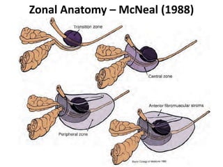
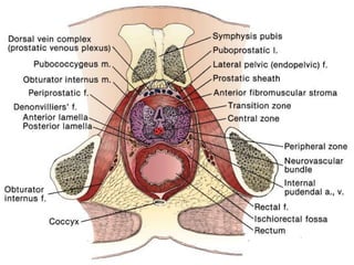
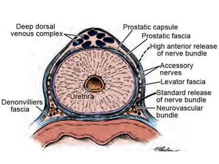
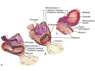
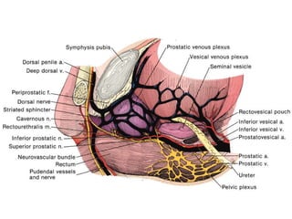
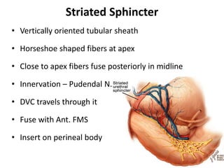
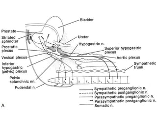
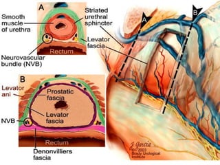
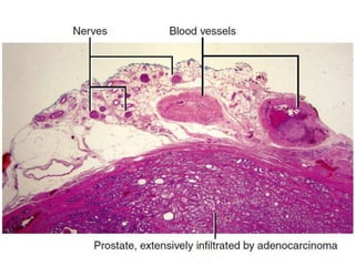
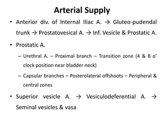
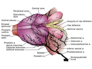
RADICAL PROSTATECTOMY
SURGICAL ANATOMY
- Dr. Abhishek Pandey
Prostatic relations
• Pyramidal shape –
– Base against bladder
– Apex joining membranous urethra
• Deep in pelvis – difficult surgical exposure
• Anterior – Pubic symphysis
• Lateral – Levator ani (wedged b/w levators)
• Posterior – Rectal ampulla separated by
denonvillier’s fascia
• Postero-superior
– Seminal vesicles
– Ampullae of the vasa
• Normal – 18-20g
• 70% glandular
• 30% Fibromuscular
stroma
Zonal Anatomy – McNeal (1988)
Prostatic Capsule
• Surgical capsule – relevant to TURP
– Compressed prostate gland remaining after procedure
• Anatomic Prostatic Capsule – relevant to RP
– Non-glandular fibromuscular stromal portion of prostatic
parenchyma distributed over surface
– Non-discrete with indistinct margin with strands mingling
in periprostatic fascia
– 2-3mm thick
– Microscopically integral part of prostate
Prostaic Sheath
• AKA Peri-Prostatic Fascia –
– Loose areolar tissue encasing prostate
– Derived from intermediate stratum of
retroperitoneal fascia
– Fuses anterolaterally with endopelvic fascia
(lateral pelvic fascia) continuous with obturator &
transversalis fasciae
– Lateral pelvic fascia – derived from outer stratum
Three parts of periprostatic fascia
• Anterior leaf – encloses dorsal venous complex
• Lateral leaf – most important surgically
– Extends from anterolateral to posterolateral area
– Encloses the Neurovascular bundle
• Posterior leaf – fuses with denonvillier’s fascia
Denonvillier’s Fascia
• Between posterior wall of prostate & rectum
• Loose areolar connective tissue – more condensed
than periprostatic fascia
Two Lamellae
• Anterior lamella – single layer fusion-fascia
– Arises by distal-to-proximal fusion of mesothelial layers of
two layers of pelvic peritoneum in rectovesical pouch
– Derived from inner stratum
A
– Adherent to posterior prostatic surface
– Limited laterally – merges with periprostatic fascia
– Shield shaped
• Posterior lamella – rectal fascia
– Overlies anterolateral wall of rectum over lamina propria
– Derived from inner stratum
• Both layers taken with specimen in RP
• No surgically separable plane between them
• Pearly Gates – Hugh Young
– Combined layers of denonvillier’s fascia appearing
glistening white on
perineal exposure
– Signify safe passage
past rectum
• Beware – Rectourethralis
Puboprostatic Ligaments
• AKA median puboprostatic ligaments
• Paired ligaments each 4.5mm wide
• Attached to perichondrium near inferior border of
symphysis pubis lateral to synchondrosis
• Each deviates slightly medially towards prostate
• Becomes continuous with prostatic sheath
• Branches of deep dorsal vein that run beneath & b/w
puboprostatic ligaments
Dorsal Venous Complex
• Deep Dorsal Vein of Penis
– Under Buck’s fascia between corpora cavernosa
– Penetrates Urogenital diaphragm to enter Retzius space
• Three major branches
– Superficial branch – Single median
• Travels between puboprostatic lig.
• Lies outside peri-prostatic fascia
– Lateral branches – Right & Left
• Travel posterolaterally peri-prostatic fascia
Prostatic Venous Plexus of Santorini
• Lies over anterior fibromuscular stroma & lateral
surface
• Embeded in peri-prostatic fascia
• Main input – Deep dorsal vein
• Empty in vesical venous plexus & Inferior Vesical Vein
• Thin-walled valve-less veins
• Intercommunication with all deep pelvic veins
Striated Sphincter
• Vertically oriented tubular sheath
• Horseshoe shaped fibers at apex
• Close to apex fibers fuse posteriorly in midline
• Innervation – Pudendal N.
• DVC travels through it
• Fuse with Ant. FMS
• Insert on perineal body
Prostatic Innervation
• Sympathetic – L1 & L2 → Sup. hypogastric plexus →
Inferior hypogastric plexus
• Parasympathetic - Pelvic Splanchnic nerves from S2,
3, 4 via Inferior hypogastric plexus
• Somatic – from sacral nerves S2, 3, 4 → supply EUS
via Pudendal N.
Inferior Hypogastric (Pelvic) Plexus
• Located retroperitoneally besides rectum
• 5-11cm from the anal verge
• Form a fenestrated rectangular plate in sagittal plane
• Midpoint at the level of tip of seminal vesicles
• Input from sacral centers & hypogastric nerves
• Gives rise to Neurovascular Bundle (NVB)
Neurovascular Bundle of Walsh
• Runs within periprostatic fascia at the postero-lateral
junction
• 1.5mm from prostate at base
• 3.0mm from prostate at apex
• Contains fibres of Prostatic nerves & Cavernous N.
• Prostatic nerve fibres closely associated with
prostatic capsular vessels
• Nerve-sparing – lateral displacement of NVB
Arterial Supply
• Anterior div. of Internal Iliac A. → Gluteo-pudendal
trunk → Prostatovesical A. → Inf. Vesicle & Prostatic A.
• Prostatic A.
– Urethral A. – Proximal branch – Transition zone (4 & 8 o’
clock position near bladder neck)
– Capsular branches – Posterolateral offshoots – Peripheral &
central zones
• Superior vesicle A. → Vesiculodeferential A. →
Seminal vesicles & vasa
Lymphatic Drainage
Three lymphatic routes of nodal spread
• Route 1 – along Prostatic pedicle – major lymphatic
vessels - obturator and internal iliac nodes
• Route 2 – along Vesiculodeferential A. – Central zone
– external iliac nodes
• Route 3 – along capsular vessels – capsular lymphatic
vessels – sacral nodes
• Primary drainage – Obturator & External Iliac nodes
Thank You
A
• A

Radical prostatectomy - Surgical anatomy

  • 1.
  • 2.
    Prostatic relations • Pyramidalshape – – Base against bladder – Apex joining membranous urethra • Deep in pelvis – difficult surgical exposure • Anterior – Pubic symphysis • Lateral – Levator ani (wedged b/w levators) • Posterior – Rectal ampulla separated by denonvillier’s fascia
  • 5.
    • Postero-superior – Seminalvesicles – Ampullae of the vasa • Normal – 18-20g • 70% glandular • 30% Fibromuscular stroma
  • 6.
    Zonal Anatomy –McNeal (1988)
  • 7.
    Prostatic Capsule • Surgicalcapsule – relevant to TURP – Compressed prostate gland remaining after procedure • Anatomic Prostatic Capsule – relevant to RP – Non-glandular fibromuscular stromal portion of prostatic parenchyma distributed over surface – Non-discrete with indistinct margin with strands mingling in periprostatic fascia – 2-3mm thick – Microscopically integral part of prostate
  • 10.
    Prostaic Sheath • AKAPeri-Prostatic Fascia – – Loose areolar tissue encasing prostate – Derived from intermediate stratum of retroperitoneal fascia – Fuses anterolaterally with endopelvic fascia (lateral pelvic fascia) continuous with obturator & transversalis fasciae – Lateral pelvic fascia – derived from outer stratum
  • 11.
    Three parts ofperiprostatic fascia • Anterior leaf – encloses dorsal venous complex • Lateral leaf – most important surgically – Extends from anterolateral to posterolateral area – Encloses the Neurovascular bundle • Posterior leaf – fuses with denonvillier’s fascia
  • 13.
    Denonvillier’s Fascia • Betweenposterior wall of prostate & rectum • Loose areolar connective tissue – more condensed than periprostatic fascia Two Lamellae • Anterior lamella – single layer fusion-fascia – Arises by distal-to-proximal fusion of mesothelial layers of two layers of pelvic peritoneum in rectovesical pouch – Derived from inner stratum
  • 14.
    A – Adherent toposterior prostatic surface – Limited laterally – merges with periprostatic fascia – Shield shaped • Posterior lamella – rectal fascia – Overlies anterolateral wall of rectum over lamina propria – Derived from inner stratum • Both layers taken with specimen in RP • No surgically separable plane between them
  • 16.
    • Pearly Gates– Hugh Young – Combined layers of denonvillier’s fascia appearing glistening white on perineal exposure – Signify safe passage past rectum • Beware – Rectourethralis
  • 17.
    Puboprostatic Ligaments • AKAmedian puboprostatic ligaments • Paired ligaments each 4.5mm wide • Attached to perichondrium near inferior border of symphysis pubis lateral to synchondrosis • Each deviates slightly medially towards prostate • Becomes continuous with prostatic sheath • Branches of deep dorsal vein that run beneath & b/w puboprostatic ligaments
  • 20.
    Dorsal Venous Complex •Deep Dorsal Vein of Penis – Under Buck’s fascia between corpora cavernosa – Penetrates Urogenital diaphragm to enter Retzius space • Three major branches – Superficial branch – Single median • Travels between puboprostatic lig. • Lies outside peri-prostatic fascia – Lateral branches – Right & Left • Travel posterolaterally peri-prostatic fascia
  • 22.
    Prostatic Venous Plexusof Santorini • Lies over anterior fibromuscular stroma & lateral surface • Embeded in peri-prostatic fascia • Main input – Deep dorsal vein • Empty in vesical venous plexus & Inferior Vesical Vein • Thin-walled valve-less veins • Intercommunication with all deep pelvic veins
  • 25.
    Striated Sphincter • Verticallyoriented tubular sheath • Horseshoe shaped fibers at apex • Close to apex fibers fuse posteriorly in midline • Innervation – Pudendal N. • DVC travels through it • Fuse with Ant. FMS • Insert on perineal body
  • 26.
    Prostatic Innervation • Sympathetic– L1 & L2 → Sup. hypogastric plexus → Inferior hypogastric plexus • Parasympathetic - Pelvic Splanchnic nerves from S2, 3, 4 via Inferior hypogastric plexus • Somatic – from sacral nerves S2, 3, 4 → supply EUS via Pudendal N.
  • 28.
    Inferior Hypogastric (Pelvic)Plexus • Located retroperitoneally besides rectum • 5-11cm from the anal verge • Form a fenestrated rectangular plate in sagittal plane • Midpoint at the level of tip of seminal vesicles • Input from sacral centers & hypogastric nerves • Gives rise to Neurovascular Bundle (NVB)
  • 30.
    Neurovascular Bundle ofWalsh • Runs within periprostatic fascia at the postero-lateral junction • 1.5mm from prostate at base • 3.0mm from prostate at apex • Contains fibres of Prostatic nerves & Cavernous N. • Prostatic nerve fibres closely associated with prostatic capsular vessels • Nerve-sparing – lateral displacement of NVB
  • 34.
    Arterial Supply • Anteriordiv. of Internal Iliac A. → Gluteo-pudendal trunk → Prostatovesical A. → Inf. Vesicle & Prostatic A. • Prostatic A. – Urethral A. – Proximal branch – Transition zone (4 & 8 o’ clock position near bladder neck) – Capsular branches – Posterolateral offshoots – Peripheral & central zones • Superior vesicle A. → Vesiculodeferential A. → Seminal vesicles & vasa
  • 36.
    Lymphatic Drainage Three lymphaticroutes of nodal spread • Route 1 – along Prostatic pedicle – major lymphatic vessels - obturator and internal iliac nodes • Route 2 – along Vesiculodeferential A. – Central zone – external iliac nodes • Route 3 – along capsular vessels – capsular lymphatic vessels – sacral nodes • Primary drainage – Obturator & External Iliac nodes
  • 39.
  • 40.