
Recommended
More Related Content
What's hot
What's hot (20)
Similar to Types of Bone cells
Similar to Types of Bone cells (20)
Recently uploaded
Recently uploaded (20)
Types of Bone cells
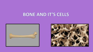
- 2. BONE • Bone is a specialized connective tissue, which makes up the body skeleton and is one of the hardest structures of the animal body. • Bone or osseous tissue represents the highest differentiation among supporting tissues.
- 3. COMPOSITION OF BONE ORGANIC PART 33%-35% • COLLAGEN: 88-90% TYPE 1 • NON COLLAGEN:10-11% A) GYLCOPROTEINS 6-9%(EX.OSTEONECTIN) B) PROTEOGLYCANS 8% C) SIALOPROTEINS 35% D) LIPIDS 4% INORGANIC PART 65%-67% • CALCIUM • PHOSPHATE • MAGNESIUM • TRACE ELEMENTS: Nickel, iron, Fluoride, cadmium, zinc, magnesium
- 4. OSTEOPROGENITOR CELLS • Derived from mesenchyme stem cells in bone marrow. • Undergo mitosis and develop into "osteoblasts“ • Found on inner surface of periosteum and endosteum.
- 5. OSTEOBLASTS • They are mononucleated cells responsible for the synthesis and secretion of the macromolecular organic constituents of bone matrix. • Osteoblasts are cuboidal or slightly elongated cells. • The cells are found on the forming surface of growing or remodeling bone. • They form a protein mixture known as osteoid (primarily type I collagen), which mineralizes to become bone.
- 6. FUNCTIONS OF OSTEOBLAST • Formation of new bone via synthesis of various proteins and polysaccharides. • Regulation of bone remodeling and mineral metabolism. • Osteoblasts also secrete small amount of type I collagen, osteonectin, osteopontin, RANKL, proteoglycans, proteases, growth factors etc. • Osteoblasts recognize the resorptive signal and transmit it to the osteoclast.
- 7. OSTEOCYTES • The number of osteoblasts that becomes osteocytes depends on the rapidity of bone formation. • Within the bone matrix, the osteocyte reduces in size, creating a space around it called the osteocytic lacuna. • Normally the most abundant cells in bone, the almond shaped osteocytes exhibit significantly less RER, smaller Golgi complexes, and more condensed nuclear chromatin than osteoblasts
- 8. FUNCTIONS OF OSTEOCYTES • Maintains the integrity of the lacunae and canaliculi. • Keep open the channels for diffusion of nutrients through bone. • Play role in removal and deposition of matrix and of calcium when required. • They release proteins that inhibit/stimulate/other bone cells- osteoblasts and osteoclasts.
- 9. OSTEOCLASTS • The large size and multinucleated condition are due to their origin from the fusion of bone marrow–derived monocytes. • osteoclasts on the bone surface lie within enzymatically etched depressions or cavities in the matrix known as resorption lacunae (or Howship lacunae). • In an active osteoclast the membrane domain that contacts the bone forms a circular sealing zone which binds the cell tightly to the bone matrix and surrounds an area with many surface projections, called the ruffled border.
- 10. Function of Osteoclasts • Osteoclasts are the cells that degrade bone to initiate normal bone remodeling and mediate bone loss in pathologic conditions by increasing their resorptive activity. • Osteoclast pumps protons to acidify and promote dissolution of the adjacent hydroxyapatite, and releases matrix metalloproteinases and other hydrolytic enzymes from lysosome- related secretory vesicles for the localized digestion of matrix proteins.
- 11. REGULATION OF OSTEOCLAST ACTIVITY