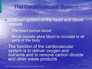
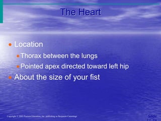
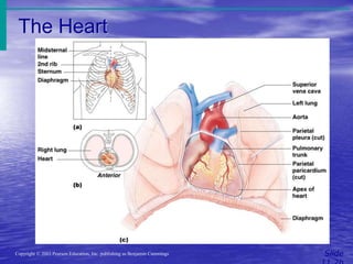
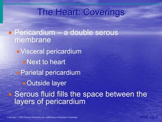
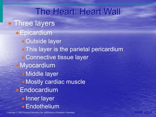
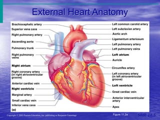
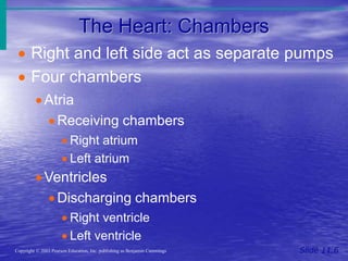
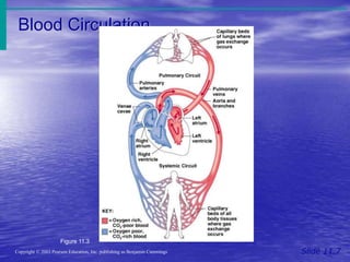
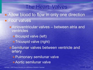
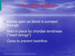
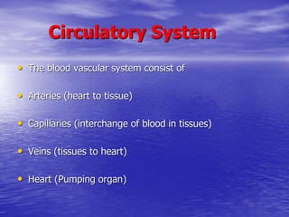
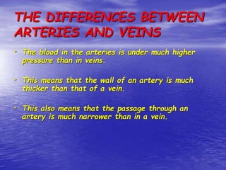
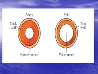
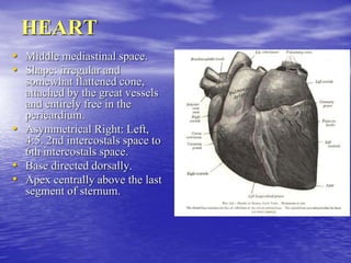
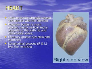
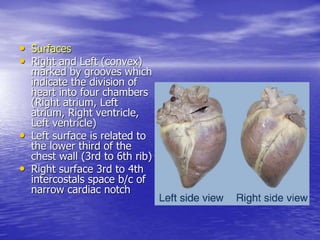
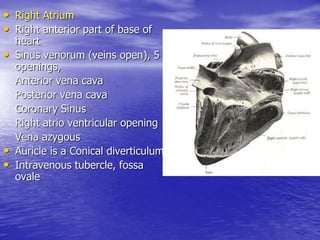
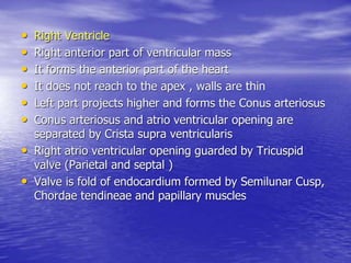
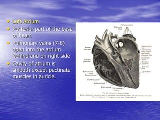
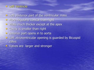
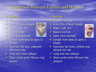
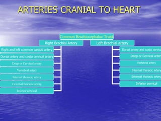
CIRCULATORY SYSTEM
By
Dr.Razia Kausar
Assistant Professor
The Cardiovascular System
Slide 11.1
Copyright © 2003 Pearson Education, Inc. publishing as Benjamin Cummings
ď‚· A closed system of the heart and blood
vessels
ď‚·The heart pumps blood
ď‚·Blood vessels allow blood to circulate to all
parts of the body
ď‚· The function of the cardiovascular
system is to deliver oxygen and
nutrients and to remove carbon dioxide
and other waste products
The Heart
Slide
Copyright © 2003 Pearson Education, Inc. publishing as Benjamin Cummings
ď‚· Location
ď‚·Thorax between the lungs
ď‚·Pointed apex directed toward left hip
ď‚· About the size of your fist
The Heart
Slide
Copyright © 2003 Pearson Education, Inc. publishing as Benjamin Cummings
Figure 11.1
The Heart: Coverings
Slide 11.3
Copyright © 2003 Pearson Education, Inc. publishing as Benjamin Cummings
 Pericardium – a double serous
membrane
ď‚·Visceral pericardium
ď‚·Next to heart
ď‚·Parietal pericardium
ď‚·Outside layer
ď‚· Serous fluid fills the space between the
layers of pericardium
The Heart: Heart Wall
Slide 11.4
Copyright © 2003 Pearson Education, Inc. publishing as Benjamin Cummings
ď‚· Three layers
ď‚·Epicardium
ď‚· Outside layer
ď‚· This layer is the parietal pericardium
ď‚· Connective tissue layer
ď‚·Myocardium
ď‚· Middle layer
ď‚· Mostly cardiac muscle
ď‚·Endocardium
ď‚· Inner layer
ď‚· Endothelium
External Heart Anatomy
Slide 11.5
Copyright © 2003 Pearson Education, Inc. publishing as Benjamin Cummings Figure 11.2a
The Heart: Chambers
Slide 11.6
Copyright © 2003 Pearson Education, Inc. publishing as Benjamin Cummings
ď‚· Right and left side act as separate pumps
ď‚· Four chambers
ď‚·Atria
ď‚· Receiving chambers
ď‚· Right atrium
ď‚· Left atrium
ď‚·Ventricles
ď‚· Discharging chambers
ď‚· Right ventricle
ď‚· Left ventricle
Blood Circulation
Slide 11.7
Copyright © 2003 Pearson Education, Inc. publishing as Benjamin Cummings
Figure 11.3
The Heart: Valves
Slide 11.8
Copyright © 2003 Pearson Education, Inc. publishing as Benjamin Cummings
ď‚· Allow blood to flow in only one direction
ď‚· Four valves
Atrioventricular valves – between atria and
ventricles
ď‚· Bicuspid valve (left)
ď‚· Tricuspid valve (right)
ď‚·Semilunar valves between ventricle and
artery
ď‚· Pulmonary semilunar valve
ď‚· Aortic semilunar valve
The Heart: Valves
Slide 11.9
Copyright © 2003 Pearson Education, Inc. publishing as Benjamin Cummings
ď‚· Valves open as blood is pumped
through
ď‚· Held in place by chordae tendineae
(“heart strings”)
ď‚· Close to prevent backflow
Circulatory System
• The blood vascular system consist of
• Arteries (heart to tissue)
• Capillaries (interchange of blood in tissues)
• Veins (tissues to heart)
• Heart (Pumping organ)
THE DIFFERENCES BETWEEN
ARTERIES AND VEINS
• The blood in the arteries is under much higher
pressure than in veins.
• This means that the wall of an artery is much
thicker than that of a vein.
• This also means that the passage through an
artery is much narrower than in a vein.
Pericardium
• Fibro serous sac which encloses the heart and great vessels
• Fibrous layer, thin, strong and inelastic
• Serous layer, surrounded by fibrous pericardium, smooth,
glistening
• Clear serous fluid liquor pericardii.
• Two parts
• Parietal part: lines the fibrous layer
• Visceral part: covers the heart and great vessels and called
epicardium.
HEART
• Middle mediastinal space.
• Shape: irregular and
somewhat flattened cone,
attached by the great vessels
and entirely free in the
pericardium.
• Asymmetrical Right: Left,
4:5. 2nd intercostals space to
6th intercostals space.
• Base directed dorsally.
• Apex centrally above the last
segment of sternum.
HEART
• Anterior border strongly convex
and parallel with the sternum
• Posterior border is much
shorter, nearly vertical and is
opposite to the sixth rib and
intercostals space.
• Coronary groove b/w atria and
ventricles
• Longitudinal grooves (R & L)
b/w the ventricles
• Surfaces
• Right and Left (convex)
marked by grooves which
indicate the division of
heart into four chambers
(Right atrium, Left
atrium, Right ventricle,
Left ventricle)
• Left surface is related to
the lower third of the
chest wall (3rd to 6th rib)
• Right surface 3rd to 4th
intercostals space b/c of
narrow cardiac notch
• Right Atrium
• Right anterior part of base of
heart
• Sinus venorum (veins open), 5
openings,
Anterior vena cava
Posterior vena cava
Coronary Sinus
Right atrio ventricular opening
Vena azygous
• Auricle is a Conical diverticulum
• Intravenous tubercle, fossa
ovale
• Right Ventricle
• Right anterior part of ventricular mass
• It forms the anterior part of the heart
• It does not reach to the apex , walls are thin
• Left part projects higher and forms the Conus arteriosus
• Conus arteriosus and atrio ventricular opening are
separated by Crista supra ventricularis
• Right atrio ventricular opening guarded by Tricuspid
valve (Parietal and septal )
• Valve is fold of endocardium formed by Semilunar Cusp,
Chordae tendineae and papillary muscles
• Left Atrium
• Posterior part of the base
of heart
• Pulmonary veins (7-8)
open into the atrium
behind and on right side
• Cavity of atrium is
smooth except pectinate
muscles in auricle.
• Left Ventricle
• Left posterior part of the ventricular mass
• More regularly conical than right
• Walls much thicker except at the apex
• Cavity is smaller than right
• Anterior part opens in to aorta
• Left atrioventricular opening is guarded by Bicuspid
valve.
• Valves are larger and stronger
Comparison Between Equines and Bovines
• Equines
• Weight , 4 Kg
• 0.7% of Body Weight
• Right: Left, 4:5
• Base is wider
• Apex less pointed
• Length from base to apex is
shorter
• Coronary fat less, yellowish
and more oily
• Long axis more oblique
• Ossa cordis aortic fibrous ring
absent
• Bovines
• Weight , 2.5 Kg
• 0.4-0.5% of Body Weight
• Right: Left, 3:4
• Base is narrow
• Apex more pointed
• Length from base to apex is
longer
• Coronary fat more, whitish and
almost non oily
• Long axis less oblique
• Ossa cordis aortic fibrous ring
present
ARTERIES CRANIAL TO HEART
Common Brachiocephaluc Trunk
Right Brachial Artery Left Brachial artery
Right and left common carotid artery
Dorsal artery and costo cervical artery
Deep or Cervical artery
Vertebral artery
Internal thoracic artery
External thoracic artery
Inferior cervical
Dorsal artery and costo cervica
Deep or Cervical artery
Vertebral artery
Internal thoracic artery
External thoracic artery
Inferior cervical
Branches of Brachial Artery
1. Suprascapular artery (Anterior deep pectoral, Brachiocephalic, Supraspinatous )
2. Subscapular Artery (Subscapularis, teres major, triceps, tensor fascia antibrachii)
3. Thoraco dorsal (Latisimuss dorsi,triceps)
4. Posterior circumflex artery of humerus (long and lateral head of triceps, joint capsule)
5. Circumflex artery of scapula (Supraspinatous, infraspinatous, teres major)
6. Anterior circumflex artery of humerus (Coraco-brachialis, deep pectoral)
7. Deep Brachial artery (Triceps, tensor fascia antibrachii, Anconeus, brachialis )
8. Muscular branches (Distal part of biceps)
9. Ulnar artery (Superficial pectoral,Flexor carpii ulnaris)
10. Nutrient artery of humerus (Humerus)
11. Anterior radial artery ( Elbow joint, Biceps, brachialis, extensors of carpus and digit)
12. Median artery
a) Articular branches ( Elbow joint)
b) Muscular branches ( Flexors of carpus and digit)
c) Common interosseous artery ( Common and lateral extensors of digit)
Branches of Thoracic Aorta
• Broncho-oesophageal Artery
a) Bronchial artery ( lung tissue, bronchial lymph
nodes)
b) Esophageal artery ( Esophagus, pleura)
• Intercostal Artery (intercostal muscles)
• Phrenic Artery (Curar of Diaphragm)
Branches of Abdominal Aorta
• Cealic artery (Gastric, Hepatic, splenic/ Right ruminal, left
ruminal, omaso-abomasl, ruminoreticular, hepatic, splenic)
• Anterior Mesenteric artery (Small intestine)
• Posterior Mesenteric artery ( Large Intestine)
• Renal artery (Right and Left kidney)
• Internal spermatic arteries (Male)
• Utero-ovarian artery (Female)
• Lumber arteries (6 pairs)
• External iliac ( Circumlflex iliac artery)
• Internal iliac (Last pair of lumber artery, Internal pudic artery)
• Middle coccygeal artery/ Median sacral artery

Circulatory System.ppt

  • 1.
  • 2.
    The Cardiovascular System Slide11.1 Copyright © 2003 Pearson Education, Inc. publishing as Benjamin Cummings  A closed system of the heart and blood vessels The heart pumps blood Blood vessels allow blood to circulate to all parts of the body  The function of the cardiovascular system is to deliver oxygen and nutrients and to remove carbon dioxide and other waste products
  • 3.
    The Heart Slide Copyright ©2003 Pearson Education, Inc. publishing as Benjamin Cummings  Location Thorax between the lungs Pointed apex directed toward left hip  About the size of your fist
  • 4.
    The Heart Slide Copyright ©2003 Pearson Education, Inc. publishing as Benjamin Cummings Figure 11.1
  • 5.
    The Heart: Coverings Slide11.3 Copyright © 2003 Pearson Education, Inc. publishing as Benjamin Cummings  Pericardium – a double serous membrane Visceral pericardium Next to heart Parietal pericardium Outside layer  Serous fluid fills the space between the layers of pericardium
  • 6.
    The Heart: HeartWall Slide 11.4 Copyright © 2003 Pearson Education, Inc. publishing as Benjamin Cummings  Three layers Epicardium  Outside layer  This layer is the parietal pericardium  Connective tissue layer Myocardium  Middle layer  Mostly cardiac muscle Endocardium  Inner layer  Endothelium
  • 7.
    External Heart Anatomy Slide11.5 Copyright © 2003 Pearson Education, Inc. publishing as Benjamin Cummings Figure 11.2a
  • 8.
    The Heart: Chambers Slide11.6 Copyright © 2003 Pearson Education, Inc. publishing as Benjamin Cummings  Right and left side act as separate pumps  Four chambers Atria  Receiving chambers  Right atrium  Left atrium Ventricles  Discharging chambers  Right ventricle  Left ventricle
  • 9.
    Blood Circulation Slide 11.7 Copyright© 2003 Pearson Education, Inc. publishing as Benjamin Cummings Figure 11.3
  • 10.
    The Heart: Valves Slide11.8 Copyright © 2003 Pearson Education, Inc. publishing as Benjamin Cummings  Allow blood to flow in only one direction  Four valves Atrioventricular valves – between atria and ventricles  Bicuspid valve (left)  Tricuspid valve (right) Semilunar valves between ventricle and artery  Pulmonary semilunar valve  Aortic semilunar valve
  • 11.
    The Heart: Valves Slide11.9 Copyright © 2003 Pearson Education, Inc. publishing as Benjamin Cummings  Valves open as blood is pumped through  Held in place by chordae tendineae (“heart strings”)  Close to prevent backflow
  • 12.
    Circulatory System • Theblood vascular system consist of • Arteries (heart to tissue) • Capillaries (interchange of blood in tissues) • Veins (tissues to heart) • Heart (Pumping organ)
  • 13.
    THE DIFFERENCES BETWEEN ARTERIESAND VEINS • The blood in the arteries is under much higher pressure than in veins. • This means that the wall of an artery is much thicker than that of a vein. • This also means that the passage through an artery is much narrower than in a vein.
  • 15.
    Pericardium • Fibro seroussac which encloses the heart and great vessels • Fibrous layer, thin, strong and inelastic • Serous layer, surrounded by fibrous pericardium, smooth, glistening • Clear serous fluid liquor pericardii. • Two parts • Parietal part: lines the fibrous layer • Visceral part: covers the heart and great vessels and called epicardium.
  • 16.
    HEART • Middle mediastinalspace. • Shape: irregular and somewhat flattened cone, attached by the great vessels and entirely free in the pericardium. • Asymmetrical Right: Left, 4:5. 2nd intercostals space to 6th intercostals space. • Base directed dorsally. • Apex centrally above the last segment of sternum.
  • 17.
    HEART • Anterior borderstrongly convex and parallel with the sternum • Posterior border is much shorter, nearly vertical and is opposite to the sixth rib and intercostals space. • Coronary groove b/w atria and ventricles • Longitudinal grooves (R & L) b/w the ventricles
  • 18.
    • Surfaces • Rightand Left (convex) marked by grooves which indicate the division of heart into four chambers (Right atrium, Left atrium, Right ventricle, Left ventricle) • Left surface is related to the lower third of the chest wall (3rd to 6th rib) • Right surface 3rd to 4th intercostals space b/c of narrow cardiac notch
  • 19.
    • Right Atrium •Right anterior part of base of heart • Sinus venorum (veins open), 5 openings, Anterior vena cava Posterior vena cava Coronary Sinus Right atrio ventricular opening Vena azygous • Auricle is a Conical diverticulum • Intravenous tubercle, fossa ovale
  • 20.
    • Right Ventricle •Right anterior part of ventricular mass • It forms the anterior part of the heart • It does not reach to the apex , walls are thin • Left part projects higher and forms the Conus arteriosus • Conus arteriosus and atrio ventricular opening are separated by Crista supra ventricularis • Right atrio ventricular opening guarded by Tricuspid valve (Parietal and septal ) • Valve is fold of endocardium formed by Semilunar Cusp, Chordae tendineae and papillary muscles
  • 21.
    • Left Atrium •Posterior part of the base of heart • Pulmonary veins (7-8) open into the atrium behind and on right side • Cavity of atrium is smooth except pectinate muscles in auricle.
  • 22.
    • Left Ventricle •Left posterior part of the ventricular mass • More regularly conical than right • Walls much thicker except at the apex • Cavity is smaller than right • Anterior part opens in to aorta • Left atrioventricular opening is guarded by Bicuspid valve. • Valves are larger and stronger
  • 23.
    Comparison Between Equinesand Bovines • Equines • Weight , 4 Kg • 0.7% of Body Weight • Right: Left, 4:5 • Base is wider • Apex less pointed • Length from base to apex is shorter • Coronary fat less, yellowish and more oily • Long axis more oblique • Ossa cordis aortic fibrous ring absent • Bovines • Weight , 2.5 Kg • 0.4-0.5% of Body Weight • Right: Left, 3:4 • Base is narrow • Apex more pointed • Length from base to apex is longer • Coronary fat more, whitish and almost non oily • Long axis less oblique • Ossa cordis aortic fibrous ring present
  • 24.
    ARTERIES CRANIAL TOHEART Common Brachiocephaluc Trunk Right Brachial Artery Left Brachial artery Right and left common carotid artery Dorsal artery and costo cervical artery Deep or Cervical artery Vertebral artery Internal thoracic artery External thoracic artery Inferior cervical Dorsal artery and costo cervica Deep or Cervical artery Vertebral artery Internal thoracic artery External thoracic artery Inferior cervical
  • 25.
    Branches of BrachialArtery 1. Suprascapular artery (Anterior deep pectoral, Brachiocephalic, Supraspinatous ) 2. Subscapular Artery (Subscapularis, teres major, triceps, tensor fascia antibrachii) 3. Thoraco dorsal (Latisimuss dorsi,triceps) 4. Posterior circumflex artery of humerus (long and lateral head of triceps, joint capsule) 5. Circumflex artery of scapula (Supraspinatous, infraspinatous, teres major) 6. Anterior circumflex artery of humerus (Coraco-brachialis, deep pectoral) 7. Deep Brachial artery (Triceps, tensor fascia antibrachii, Anconeus, brachialis ) 8. Muscular branches (Distal part of biceps) 9. Ulnar artery (Superficial pectoral,Flexor carpii ulnaris) 10. Nutrient artery of humerus (Humerus) 11. Anterior radial artery ( Elbow joint, Biceps, brachialis, extensors of carpus and digit) 12. Median artery a) Articular branches ( Elbow joint) b) Muscular branches ( Flexors of carpus and digit) c) Common interosseous artery ( Common and lateral extensors of digit)
  • 26.
    Branches of ThoracicAorta • Broncho-oesophageal Artery a) Bronchial artery ( lung tissue, bronchial lymph nodes) b) Esophageal artery ( Esophagus, pleura) • Intercostal Artery (intercostal muscles) • Phrenic Artery (Curar of Diaphragm)
  • 27.
    Branches of AbdominalAorta • Cealic artery (Gastric, Hepatic, splenic/ Right ruminal, left ruminal, omaso-abomasl, ruminoreticular, hepatic, splenic) • Anterior Mesenteric artery (Small intestine) • Posterior Mesenteric artery ( Large Intestine) • Renal artery (Right and Left kidney) • Internal spermatic arteries (Male) • Utero-ovarian artery (Female) • Lumber arteries (6 pairs) • External iliac ( Circumlflex iliac artery) • Internal iliac (Last pair of lumber artery, Internal pudic artery) • Middle coccygeal artery/ Median sacral artery