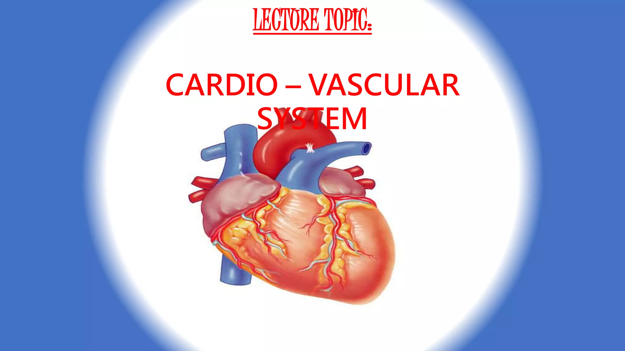
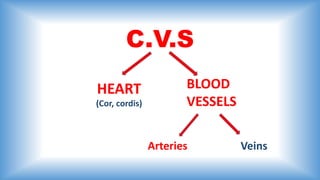
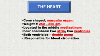
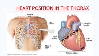
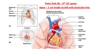
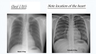
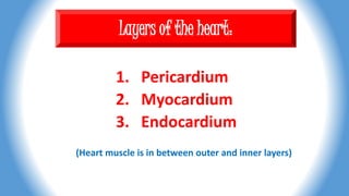
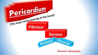
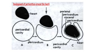
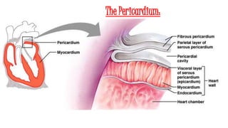
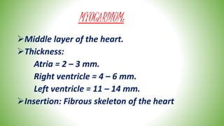
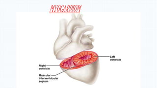
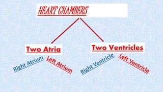
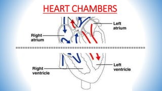
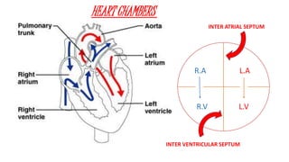
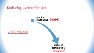
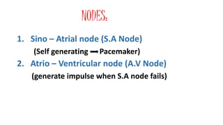
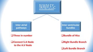
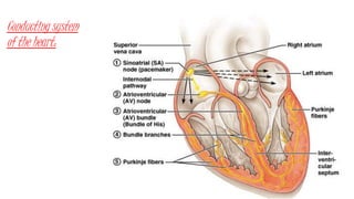
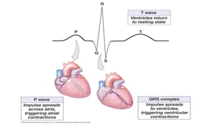
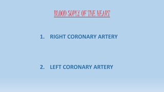
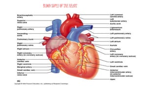
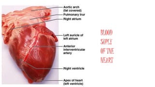
LECTURE TOPIC:
CARDIO – VASCULAR
SYSTEM
C.V.S
HEART BLOOD
VESSELS
Arteries Veins
(Cor, cordis)
 Cone shaped, muscular organ.
 Weight = 250 – 350 gm.
 Located in the middle mediastinum
 Four chambers: two atria, two ventricles
 Both ventricles – double pump
 Responsible for blood circulation
THE HEART
HEART POSITION IN THE THORAX
From 2nd rib – 5th I/C space
Apex – 1 cm inside to left mid clavicular line
Chest X RAY:
Male X Ray
Female X Ray
Note location of the heart
Layers of the heart:
1. Pericardium
2. Myocardium
3. Endocardium
(Heart muscle is in between outer and inner layers)
Fibrous
Serous
(Visceral = Epicardium)
Development of pericardium around the heart
The Pericardium:
MYOCARDIUM:
Middle layer of the heart.
Thickness:
Atria = 2 – 3 mm.
Right ventricle = 4 – 6 mm.
Left ventricle = 11 – 14 mm.
Insertion: Fibrous skeleton of the heart
MYOCARDIUM
Myocardium :
In atria: Pectinate muscles
In ventricles: Papillary muscles
3 Papillary muscles in the RV (attaches to the
tricuspid valve cusps).
 2 Papillary muscles in the LV ( attaches to the
bicuspid value cusps.
(From inside)
MiddlePapillary
Muscles
Chordae
Tendineae
Valve
cusps
ENDOCARDIUM:
•Inner layer of the heart.
•Covers hart chambers from inside.
•Formed by endothelia.
•Participates in the formation of heart valves.
Myocardium
Tricuspid
Mitral
Aortic
Pulmonary
HEART CHAMBERS
Two Atria Two Ventricles
HEART CHAMBERS
=======================================================
R.A
R.V
L.A
L.V
R R
INTER VENTRICULAR SEPTUM
INTER ATRIAL SEPTUM
HEART CHAMBERS
HEART VALVES:
Four Valves in the heart.
Open and close (heart beating)
VALVES OF THE HEART
Two in between
Atria and Ventricles
Two in the start
Of Great vessels
VALVES INSIDE THE HEART:
• RIGHT A.V. VALVE = TRICUSPID VALVE
• LEFT A.V. VALVE = BICUSPID VALVE
(Mitral valve)
R RVALVEVALVE
TRICUSPID BICUSPID
ATRIO – VENTRICULAR VALVES
(TRICUSPID AND BICUSPID VALVES)
PULMONARY AND AORTIC VALVES
(SEMI-LUNAR VALVES)
Conducting system of the heart:
ATYPICAL MYOCARDIUM:
IMPULSE
GENERATING
IMPULSE
CONDECTING
(NODE)
(BUNDLE)
NODES:
1. Sino – Atrial node (S.A Node)
(Self generating Pacemaker)
2. Atrio – Ventricular node (A.V Node)
(generate impulse when S.A node fails)
BUNDLES:
Inter atrial
pathways
Inter ventricular
bundles
Three in number
Connect S.A Node
to the A.V Node
Bundle of Hiss
Right Bundle Branch
Left Bundle Branch
Conducting system
of the heart:
BLOOD SUPLY OF THE HEART
1. RIGHT CORONARY ARTERY
2. LEFT CORONARY ARTERY
BLOOD SUPLY OF THE HEART
BLOOD
SUPLY
OF THE
HEART
thank you for attention.
THE END!

Cvs anatomy