This document provides an overview of the cardiovascular system. It discusses the composition and functions of blood, the structure and layers of the heart, types of circulation including pulmonary and systemic circulation, the cardiac cycle, heart valves and conducting system, blood pressure, electrocardiograms, and some common cardiovascular disorders like hypertension and stroke. The document is intended as an educational resource on the key components and functioning of the cardiovascular system.
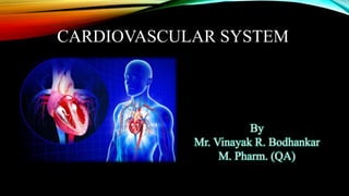
This presentation deals with circulation in human body.
I have made this presentation for H.S.C students. I have tried my level best to simplify the topic. Visual presentation will definitely help the students to understand the topic easily. Please share your opinions regarding this presentation. Thank you.
If you or anyone you know has heart disease, hypertension, disorders of the heart valves, or cholesterol issues then you need this information. In this presentation you will learn about the circulatory system, risk factors, and what supplements you can take to help it achieve optimal health.
Be sure to order any supplements mentioned in this presesntation from NaturesSunshine.com (use sponsor number 2849323 / or search for MCM Products) or from BrilliantNaturalHealth.com.
This presentation deals with circulation in human body.
I have prepared this for H.S.C students of Maharashtra board. The topic is simplified. This presentation will definitely help students to understand circulation in depth. Thank you.
Cancer cell metabolism: special Reference to Lactate PathwayAADYARAJPANDEY1
Normal Cell Metabolism:
Cellular respiration describes the series of steps that cells use to break down sugar and other chemicals to get the energy we need to function.
Energy is stored in the bonds of glucose and when glucose is broken down, much of that energy is released.
Cell utilize energy in the form of ATP.
The first step of respiration is called glycolysis. In a series of steps, glycolysis breaks glucose into two smaller molecules - a chemical called pyruvate. A small amount of ATP is formed during this process.
Most healthy cells continue the breakdown in a second process, called the Kreb's cycle. The Kreb's cycle allows cells to “burn” the pyruvates made in glycolysis to get more ATP.
The last step in the breakdown of glucose is called oxidative phosphorylation (Ox-Phos).
It takes place in specialized cell structures called mitochondria. This process produces a large amount of ATP. Importantly, cells need oxygen to complete oxidative phosphorylation.
If a cell completes only glycolysis, only 2 molecules of ATP are made per glucose. However, if the cell completes the entire respiration process (glycolysis - Kreb's - oxidative phosphorylation), about 36 molecules of ATP are created, giving it much more energy to use.
IN CANCER CELL:
Unlike healthy cells that "burn" the entire molecule of sugar to capture a large amount of energy as ATP, cancer cells are wasteful.
Cancer cells only partially break down sugar molecules. They overuse the first step of respiration, glycolysis. They frequently do not complete the second step, oxidative phosphorylation.
This results in only 2 molecules of ATP per each glucose molecule instead of the 36 or so ATPs healthy cells gain. As a result, cancer cells need to use a lot more sugar molecules to get enough energy to survive.
introduction to WARBERG PHENOMENA:
WARBURG EFFECT Usually, cancer cells are highly glycolytic (glucose addiction) and take up more glucose than do normal cells from outside.
Otto Heinrich Warburg (; 8 October 1883 – 1 August 1970) In 1931 was awarded the Nobel Prize in Physiology for his "discovery of the nature and mode of action of the respiratory enzyme.
WARNBURG EFFECT : cancer cells under aerobic (well-oxygenated) conditions to metabolize glucose to lactate (aerobic glycolysis) is known as the Warburg effect. Warburg made the observation that tumor slices consume glucose and secrete lactate at a higher rate than normal tissues.
Richard's entangled aventures in wonderlandRichard Gill
Since the loophole-free Bell experiments of 2020 and the Nobel prizes in physics of 2022, critics of Bell's work have retreated to the fortress of super-determinism. Now, super-determinism is a derogatory word - it just means "determinism". Palmer, Hance and Hossenfelder argue that quantum mechanics and determinism are not incompatible, using a sophisticated mathematical construction based on a subtle thinning of allowed states and measurements in quantum mechanics, such that what is left appears to make Bell's argument fail, without altering the empirical predictions of quantum mechanics. I think however that it is a smoke screen, and the slogan "lost in math" comes to my mind. I will discuss some other recent disproofs of Bell's theorem using the language of causality based on causal graphs. Causal thinking is also central to law and justice. I will mention surprising connections to my work on serial killer nurse cases, in particular the Dutch case of Lucia de Berk and the current UK case of Lucy Letby.
Observation of Io’s Resurfacing via Plume Deposition Using Ground-based Adapt...Sérgio Sacani
Since volcanic activity was first discovered on Io from Voyager images in 1979, changes
on Io’s surface have been monitored from both spacecraft and ground-based telescopes.
Here, we present the highest spatial resolution images of Io ever obtained from a groundbased telescope. These images, acquired by the SHARK-VIS instrument on the Large
Binocular Telescope, show evidence of a major resurfacing event on Io’s trailing hemisphere. When compared to the most recent spacecraft images, the SHARK-VIS images
show that a plume deposit from a powerful eruption at Pillan Patera has covered part
of the long-lived Pele plume deposit. Although this type of resurfacing event may be common on Io, few have been detected due to the rarity of spacecraft visits and the previously low spatial resolution available from Earth-based telescopes. The SHARK-VIS instrument ushers in a new era of high resolution imaging of Io’s surface using adaptive
optics at visible wavelengths.
This pdf is about the Schizophrenia.
For more details visit on YouTube; @SELF-EXPLANATORY;
https://www.youtube.com/channel/UCAiarMZDNhe1A3Rnpr_WkzA/videos
Thanks...!
Nutraceutical market, scope and growth: Herbal drug technologyLokesh Patil
As consumer awareness of health and wellness rises, the nutraceutical market—which includes goods like functional meals, drinks, and dietary supplements that provide health advantages beyond basic nutrition—is growing significantly. As healthcare expenses rise, the population ages, and people want natural and preventative health solutions more and more, this industry is increasing quickly. Further driving market expansion are product formulation innovations and the use of cutting-edge technology for customized nutrition. With its worldwide reach, the nutraceutical industry is expected to keep growing and provide significant chances for research and investment in a number of categories, including vitamins, minerals, probiotics, and herbal supplements.
This presentation explores a brief idea about the structural and functional attributes of nucleotides, the structure and function of genetic materials along with the impact of UV rays and pH upon them.
Multi-source connectivity as the driver of solar wind variability in the heli...Sérgio Sacani
The ambient solar wind that flls the heliosphere originates from multiple
sources in the solar corona and is highly structured. It is often described
as high-speed, relatively homogeneous, plasma streams from coronal
holes and slow-speed, highly variable, streams whose source regions are
under debate. A key goal of ESA/NASA’s Solar Orbiter mission is to identify
solar wind sources and understand what drives the complexity seen in the
heliosphere. By combining magnetic feld modelling and spectroscopic
techniques with high-resolution observations and measurements, we show
that the solar wind variability detected in situ by Solar Orbiter in March
2022 is driven by spatio-temporal changes in the magnetic connectivity to
multiple sources in the solar atmosphere. The magnetic feld footpoints
connected to the spacecraft moved from the boundaries of a coronal hole
to one active region (12961) and then across to another region (12957). This
is refected in the in situ measurements, which show the transition from fast
to highly Alfvénic then to slow solar wind that is disrupted by the arrival of
a coronal mass ejection. Our results describe solar wind variability at 0.5 au
but are applicable to near-Earth observatories.
Earliest Galaxies in the JADES Origins Field: Luminosity Function and Cosmic ...Sérgio Sacani
We characterize the earliest galaxy population in the JADES Origins Field (JOF), the deepest
imaging field observed with JWST. We make use of the ancillary Hubble optical images (5 filters
spanning 0.4−0.9µm) and novel JWST images with 14 filters spanning 0.8−5µm, including 7 mediumband filters, and reaching total exposure times of up to 46 hours per filter. We combine all our data
at > 2.3µm to construct an ultradeep image, reaching as deep as ≈ 31.4 AB mag in the stack and
30.3-31.0 AB mag (5σ, r = 0.1” circular aperture) in individual filters. We measure photometric
redshifts and use robust selection criteria to identify a sample of eight galaxy candidates at redshifts
z = 11.5 − 15. These objects show compact half-light radii of R1/2 ∼ 50 − 200pc, stellar masses of
M⋆ ∼ 107−108M⊙, and star-formation rates of SFR ∼ 0.1−1 M⊙ yr−1
. Our search finds no candidates
at 15 < z < 20, placing upper limits at these redshifts. We develop a forward modeling approach to
infer the properties of the evolving luminosity function without binning in redshift or luminosity that
marginalizes over the photometric redshift uncertainty of our candidate galaxies and incorporates the
impact of non-detections. We find a z = 12 luminosity function in good agreement with prior results,
and that the luminosity function normalization and UV luminosity density decline by a factor of ∼ 2.5
from z = 12 to z = 14. We discuss the possible implications of our results in the context of theoretical
models for evolution of the dark matter halo mass function.
2. CONTENTS
Blood
Composition of Blood
Functions of blood
Blood group
Coagulation of blood
Disorders of blood
Structure & function of Heart
Types of Circulation
Cardiac cycle, Arteries & Veins
Conducting system of Heart
ECG
Blood pressure & its recording
CV disorders
3. BLOOD
Fluid connective tissue.
Composed of plasma (55%) & blood cells (45%).
Physical properties of blood
Colour – Red coloured, viscous fluid
Thicker and heavier than water.
Constitute 8% of body weight.
Temp. – 380C.
Total amount in human body – 5 to5.5 litre.
pH – 7.4 to 7.5
4. BLOOD PLASMA
Slightly yellow coloured fluid.
Composition
Water – 90 to 92%
Plasma protein – 6 to 8%
Nutrients – Amino acids, glucose, etc.
Organic waste – Urea, uric acid, etc.
Mineral salts – Na, K, Ca, etc.
Gases – Oxygen, carbon dioxide, etc.
Hormones and enzymes – Antibodies and enzymes.
5. FUNCTIONS OF BLOOD
Transport of oxygen from lung to the body.
Return of carbon dioxide from cell to lungs.
Drain out waste material in the body.
Transport of enzymes and hormones.
Transport system for nourishment.
Help to maintain body temp.
Help in defensive mechanism of the body.
Prevent the loss of body fluid and blood cells.
Maintain acid base balance of the body.
6. COMPOSITION OF BLOOD
1. Blood cells
a) RBC’s
b) WBC’s
i) Granulocytes: Neutrophils (70%)
Basophils (1%)
Eosinophils (4%)
ii) Agranulocytes: Lymphocytes (20%)
Monocytes (5%)
c) Thrombocytes
2. Plasma
7. RBC’s
Small circular, disc shaped.
Suspended in blood plasma.
In female: 4 to 4.5 millions per cubic mm
In male: 5 to 5.5 millions per cubic mm
Production
o In foetal life: liver
o After birth: Red bone marrow
Process of production is called erythropoiesis.
Megacarocytes: from which RBC’s developed.
Life span – 115 to 120 days.
8. WBC’s
Irregular shaped.
Normal count: 6000 to 10000 per cubic mm
1. Granulocytes
A. Neutrophils
70% of total WBC.
Stained with neutral dye – purple
Responsible for defence mechanism.
B. Eosinophils
4% of total WBC
Stain red with eosin.
C. Basophils
1% of total WBC.
Stain with basic dye and appear blue colour.
9. 2. Agranulocytes
Produced in lymph glands.
Constitute 25% of all WBC.
i) Lymphocytes
Constitute 23%.
Responsible for development of immunity.
ii) Monocytes
Constitute 2%
Larger than lymphocytes.
Phagocytic in action.
C. Thrombocytes
Smaller fragments of megakaryocytes.
Normal count: 3 to 4 lakh per cubic mm.
Help in blood clotting.
10. BLOOD GROUP
Discovered by Karl Landsteiner in 1900.
Why Blood group..?
Blood donor
Universal donor
Universal acceptor
Agglutinogen: Antigen in the membrane of RBC.
Agglutinin: Natural antibodies.
Agglutinins in plasma are opposite type.
Rh antigen
Important in case of pregnancy.
Blood group Receive from Donate to
A A, O A, AB
B B, O B, AB
AB A, B, AB, O AB
O O A, B, AB, O
11. BLOOD CLOTTING
Blood vessel + Injury = Rough surface.
Platelet + rough surface = thromboplastin.
In presence of thromboplastin & Ca in plasma
Prothrombin – Thrombin (which helps) in fibrinogen - fibrin
Thread of fibrin + blood cell = blood clot.
P + Ca + Tp = T
T + Fg = F
F + Blood cell = clot
Bleeding time: 1 to 4 min
Clotting time: 3 to 6 min
12. Factor hastening blood clotting
Temp. excess than body temp.
Contact of blood with rough suface.
Slowness of blood flow.
Factor retarding blood clotting
13. DISORDERS OF BLOOD
Anaemia: Decrease in oxygen carrying capacity of blood.
Types of anaemia:
A. Pernicious anaemia
Factor responsible for absorption of vitamin B12 is absent.
B. Microcytic anaemia
Diameter of RBC is smaller, due to deficiency of iron.
C. Haemolytic anaemia
Life span of RBC is shortened.
D. Sickle cell anaemia
Defect in haemoglobin. Life span of RBC is shortened.
E. Thalassemia
F. Megaloblastic anaemia
Polycythemia: Increase in RBC count.
Bleeding disease.
Leukemia.
Agranulocytosis.
14. STRUCTURE OF THE HEART
The heart is divided into the left and right side by partitions called septa (singular septum).
The interatrial septum separates the two upper chambers, called atria (from atri/o, meaning “upper chambers”).
The interventricular septum separates the two lower chambers, called ventricles (from ventricul/o, meaning
“lower chamber).
Interventricular Septum
Interatrial Septum
15. LAYERS OF HEART
The endocardium (from endo- + cardi/o + -ium, meaning “inner layer of the heart”) is formed by
endothelial cells, and it lines the interior of the heart chambers and valves.
The myocardium (from my/o + cardi/o + -ium, meaning “heart muscle”) is the muscular middle
layer of the heart that consists of heart muscle cells.
The epicardium (from epi- + cardi/o + -ium, meaning “outer layer of the heart”) is formed by
epithelial cells, and forms the outer cell layer of the heart.
The pericardium (from peri- + cardi/o + -ium, meaning “surrounding the heart”) is a membranous
sac that surrounds the heart. It consist of two layers called the visceral pericardium (adheres to the
epicardium) and parietal pericardium (the outer coat). The space between these two layers is called
pericardial cavity and it contains pericardial fluid.
16. HEART CHAMBERS AND VALVES
The human heart has four chambers, which are responsible for pumping blood and
maintaining blood circulation throughout the body.
The four chambers are name:
The right atrium
The left atrium
The right ventricle
The left ventricle
Blood is only pumped to one direction.
Four heart valves ensure that blood does not flow backward within the heart.
17. HEART VALVES
The tricuspid valve (from tri- + cuspid, meaning “having three points”) located between right atrium and
ventricle.
The pulmonary valve (from pulmon/o, meaning “lungs”) located between right ventricle and pulmonary artery.
Also called semilunar valve.
The mitral valve, also called bicuspid valve ( from bi- + cuspid, meaning “having two points”) located
between left atrium and ventricle.
The aortic valve located between left ventricle and aorta.
The tricuspid and bicuspid valves are also called atrioventricular valves (meaning “located between the atrium and
ventricle”).
18. FUNCTIONS OF THE HEART
The heart functions to circulate blood around the body. The right
and left side of the heart pump blood into two different
circulations.
The right side pumps deoxygenated blood into the pulmonary
circulation, while the left side pumps oxygenated blood into the
systemic circulation.
The right atrium receives deoxygenated blood from the body
tissues via the superior and inferior vena cava.
The blood enters the right atrium, which pumps the blood into
the right ventricle. The tricuspid valve prevents blood from
flowing backward into the right atrium. The right ventricle
pumps the blood into the pulmonary artery via the pulmonary
valve.
19. The pulmonary artery will deliver the deoxygenated blood to the lungs, where gas exchange occurs.
Oxygen is taken from the air into the blood (now called oxygenated blood), while carbon dioxide is
expelled from the blood into the air. The oxygenated blood returns to the left side of the heart via the
pulmonary veins.
The oxygenated blood enters the left atrium.
The left atrium pumps blood into the left ventricle. The mitral valve prevents blood from flowing
backward into the left atrium.
The left ventricle pumps the blood into the aorta and systemic circulation. The oxygenated
blood is delivered everywhere in the body (besides the lungs).
20. PULMONARY CIRCULATION
Pulmonary circulation begins at the right ventricle, where the deoxygenated blood from the body
tissues is pumped into the pulmonary arteries and to the lungs.
In the lungs, the blood exchanges carbon dioxide (waste product of cellular respiration) to oxygen.
The oxygenated blood them travels back to the heart and the left atrium, via the pulmonary vein.
21. SYSTEMIC CIRCULATION
The systemic circulation begins at the left ventricle that pumps oxygenated blood into the
aorta.
Aorta branches out into smaller arteries, which carry the oxygenated blood to the rest of the
body (with the exception of lungs).
Oxygen is delivered to the body tissues and exchanged to carbon dioxide. The now deoxygenated
blood is carried back to the heart and the right atrium via veins.
22. PORTAL CIRCULATION
Collection by portal vein – digestive organ.
Pour into liver.
Liver – already supplied with oxy. blood by hepatic artery.
Mixing take place.
Collected by hepatic vein.
Pour into inferior venacava.
23. CARDIAC CYCLE
One complete sequence of atrial and ventricular systole and diastole.
Cycle of events that occurs as the heart contract and relaxes.
Contraction of atria – short
Contraction of ventricle – long.
Time required for one cardiac cycle is 0.8 sec.
SA node – near the opening of superior vena cava in RA.
AV node – On the atrioventricular septum.
Stroke volume: Amount of blood ejected from heart at each contraction of ventricle.
Cardiac output: Amount of blood ejected each minute.
Stroke volume * heart rate
24. ARTERIES VS VEINS
The blood vessels that carry blood AWAY from heart are called arteries.
The blood vessels that carry blood TOWARD the heart are called veins.
Only in systemic circulation arteries carry oxygenated blood, while in the pulmonary circulation arteries carry
deoxygenated blood.
25. CONDUCTION SYSTEM OF THE HEART
The conduction system of the heart controls the rate and pattern of your heartbeat.
26. SINOATRIAL(SA) NODE
Myocardium contracts after it receives an electrical impulse generated by a specialized tissue located within
the right atrium.
This is called the sinoatrial node (SA node), also called the pacemaker of the heart. The SA node is a
bundle of neurons that triggers the contraction of the atria during the cardiac cycle.
The electrical currents next reach the ventricles, which contract after the atria.
The SA node initiates approximately 75 electrical impulses each minute, with variation between individuals’
age and general health.
27. THE PURKINJE FIBERS
The Purkinje fibers are cells in the inner ventricle walls, just beneath the endocardium. These fibers run
between the ventricles to the apex (bottom) of the heart. The Purkinje fibers play a crucial role in the cardiac
cycle.
When an electrical stimulus leaves the AV node, it travels via the bundle of His and branches to the Purkinje
fibers. These fibers then carry the impulse through the inner wall of each ventricle. This causes the ventricles to
contract after the atria contract.
The ventricle contraction forces blood from the right ventricle to the lungs (pulmonary circulation) and
from the left ventricle to the body (systemic circulation).
These three elements generate a healthy heart rhythm known as sinus rhythm. The rhythm, or contraction of
the heart pumps blood throughout the body. In roughly a minute’s time, blood travels from the heart to the
body and back.
28. ECG AND THE P-QRS-T SYSTEM
An electrocardiogram (from electr/o + cardi/o + -gram), also called an EKG or ECG, is a diagnostic
test used to record and trace the electrical activity of the heart.
29. THE ELECTRICALACTIVITY OF THE HEART
The electrical activity in the heart is displayed as a P wave, QRS interval and T wave.
The P wave correlates to atrial depolarization (systole) and atrial contractions. There is not a wave
associated with atrial repolarization as it occurs during ventricular depolarization (during the QRS
interval).
The QRS complex correlates to ventricular depolarization (systole) as the ventricles contract. The Q
wave is the beginning, the R wave the middle of the contraction, and the S wave is the end of
ventricular depolarization, and beginning of ventricular repolarization (diastole).
The T wave correlates to ventricular repolarization (diastole).
30. BLOOD PRESSURE
Types: Systolic & diastolic.
Measured in terms of mm of Hg.
Normal systolic: 120 mm of Hg (100-150)
Normal diastolic: 90 mm of Hg (60-90)
Pulse pressure: Difference between systolic & diastolic B.P.
Measured with the help of instrument “Sphygmomanometer”
Factor affecting blood pressure
Physiological conditions.
Emotional conditions.
Age of individual.
Body weight.
31. DISORDERS OF HEART
Rheumatic heart disease
Streptococcal infection. (Haemolytic)
Bacterial endocarditis
Non haemolytic infection.
Coronary artery occlusion
Myocardial infaract
Congenital heart disease
Defect in formation of septum.
Arrhythmia
Disturbance in rhythm
Atherosclerosis
Deposition of fatty material.
32. HEALTH CONDITIONS
Hypertension:
Hypertension is an abnormal condition that is primarily caused by high blood cholesterol.
Excess cholesterol is deposited on the arterial walls as plaques.
These plaques make the lumen of the artery narrower, which causes the blood to flow with higher
pressure.
If an artery becomes completely blocked, the cells supported by that artery will suffer from lack of oxygen and die.
If this happens in the coronary arteries, which provide blood to the heart, the result can be myocardial
infarction (heart attack).
33. STROKE
An artery leading to the brain can become blocked. This can cause a cerebral vascular accident, known as a
stroke.
34. HYPOTENSION
Hypotension is also an abnormal condition, in which the blood flows with low pressure.
Hypotension occurs when a large volume of water or blood is lost from the body.
The body’s loss of water, dehydration, can occur during diarrhea or vomiting.
The body’s loss of blood, hemorrhage, can occur due to blood disorders or injury to the blood vessels
(trauma).
Hypotension can result in shock.