Downloaded 159 times
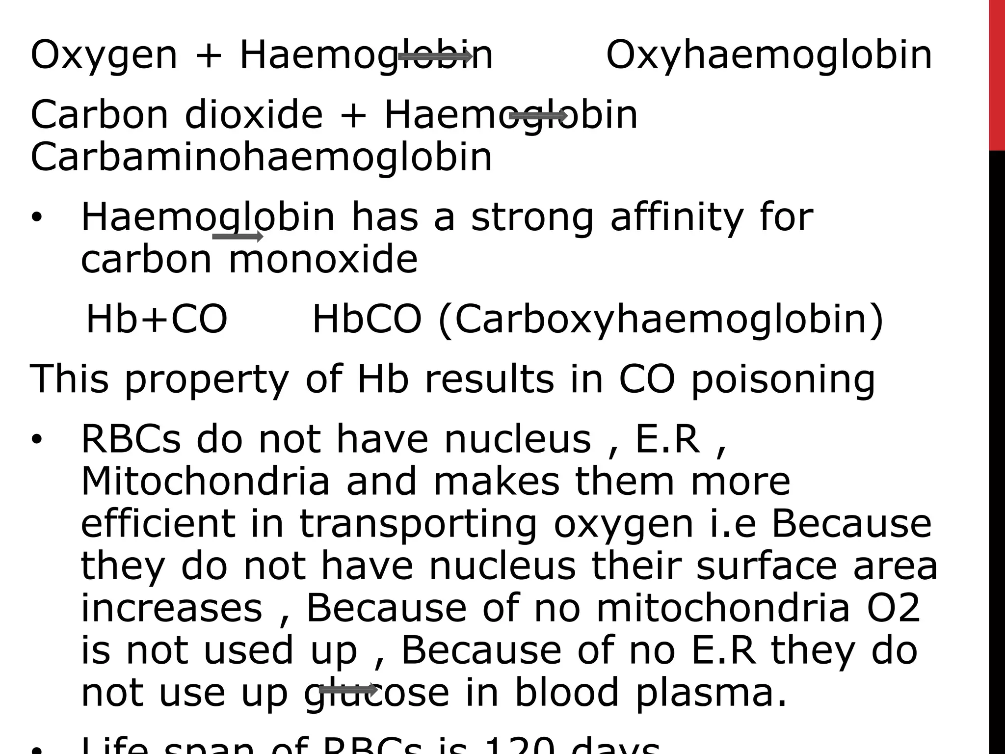
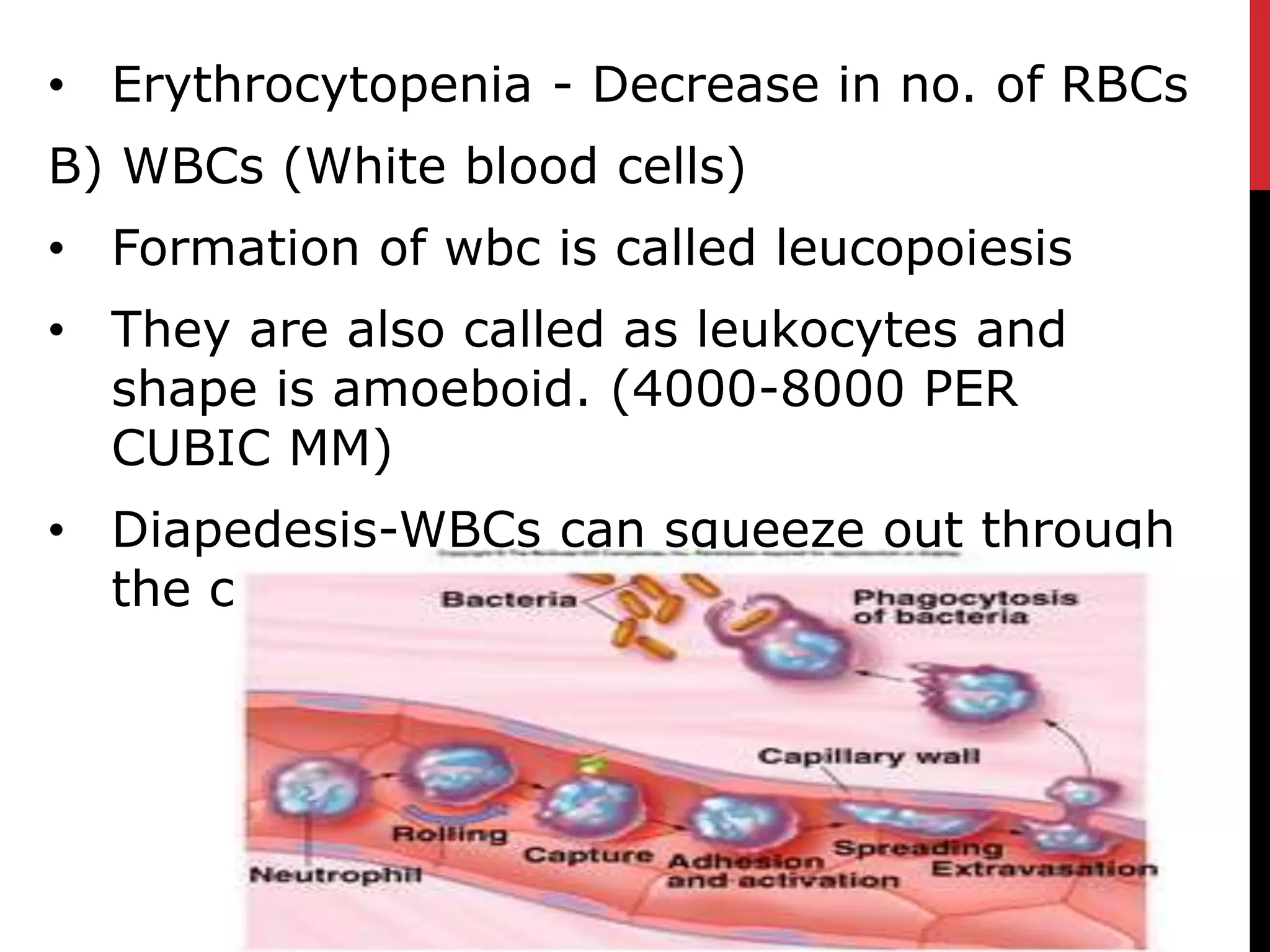
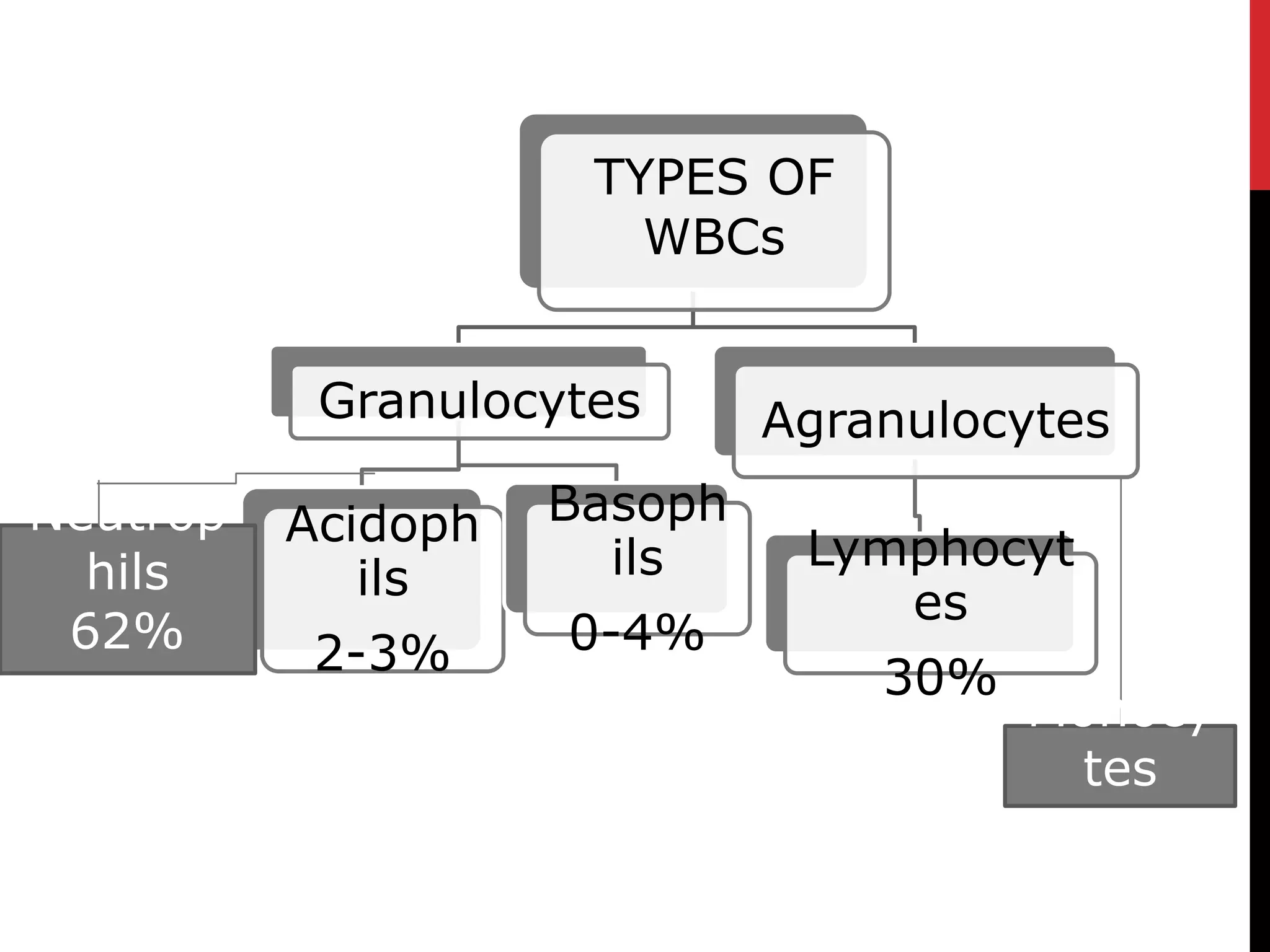
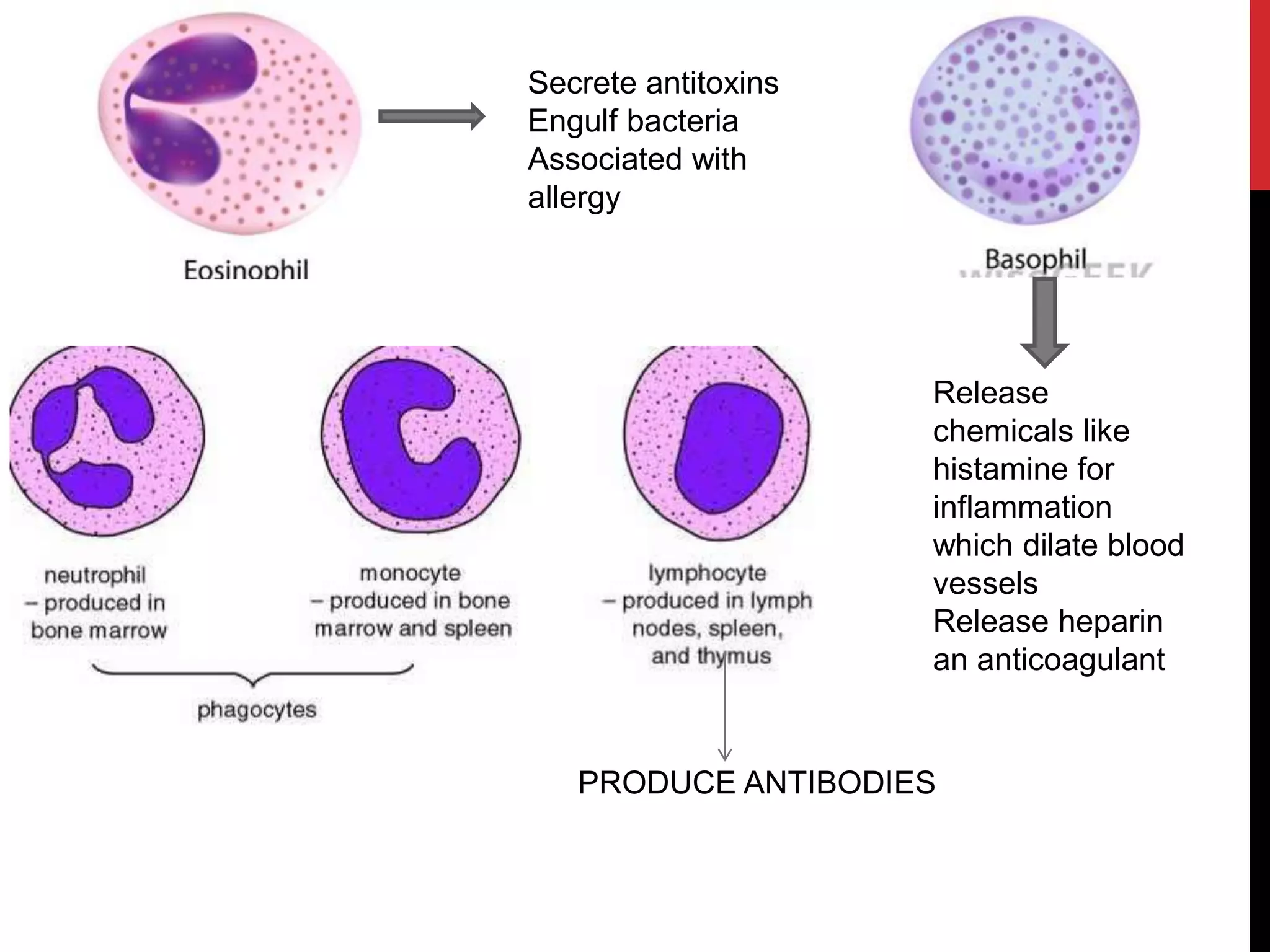
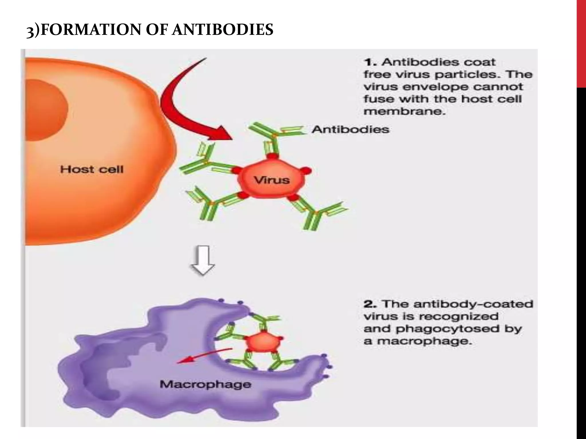
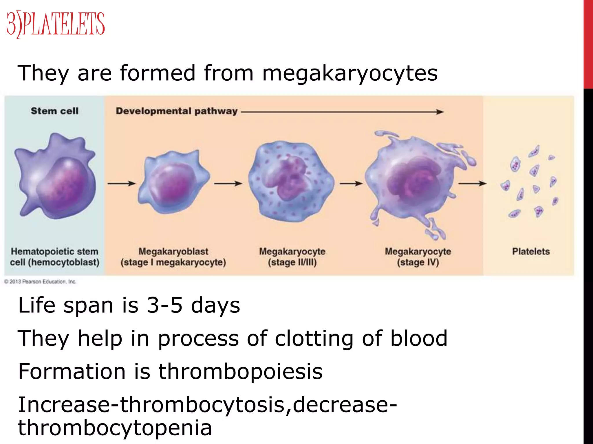
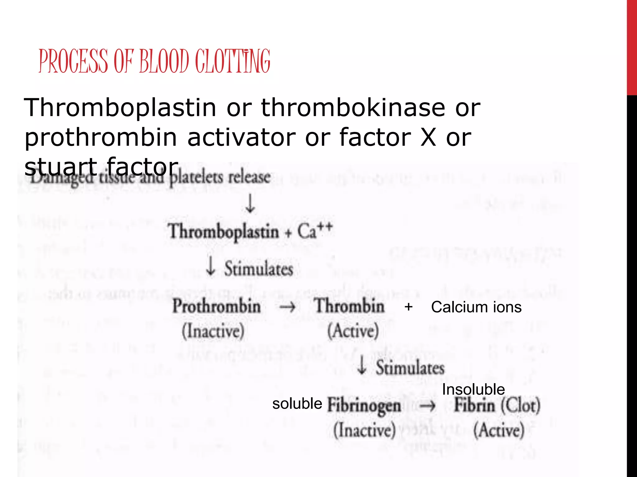
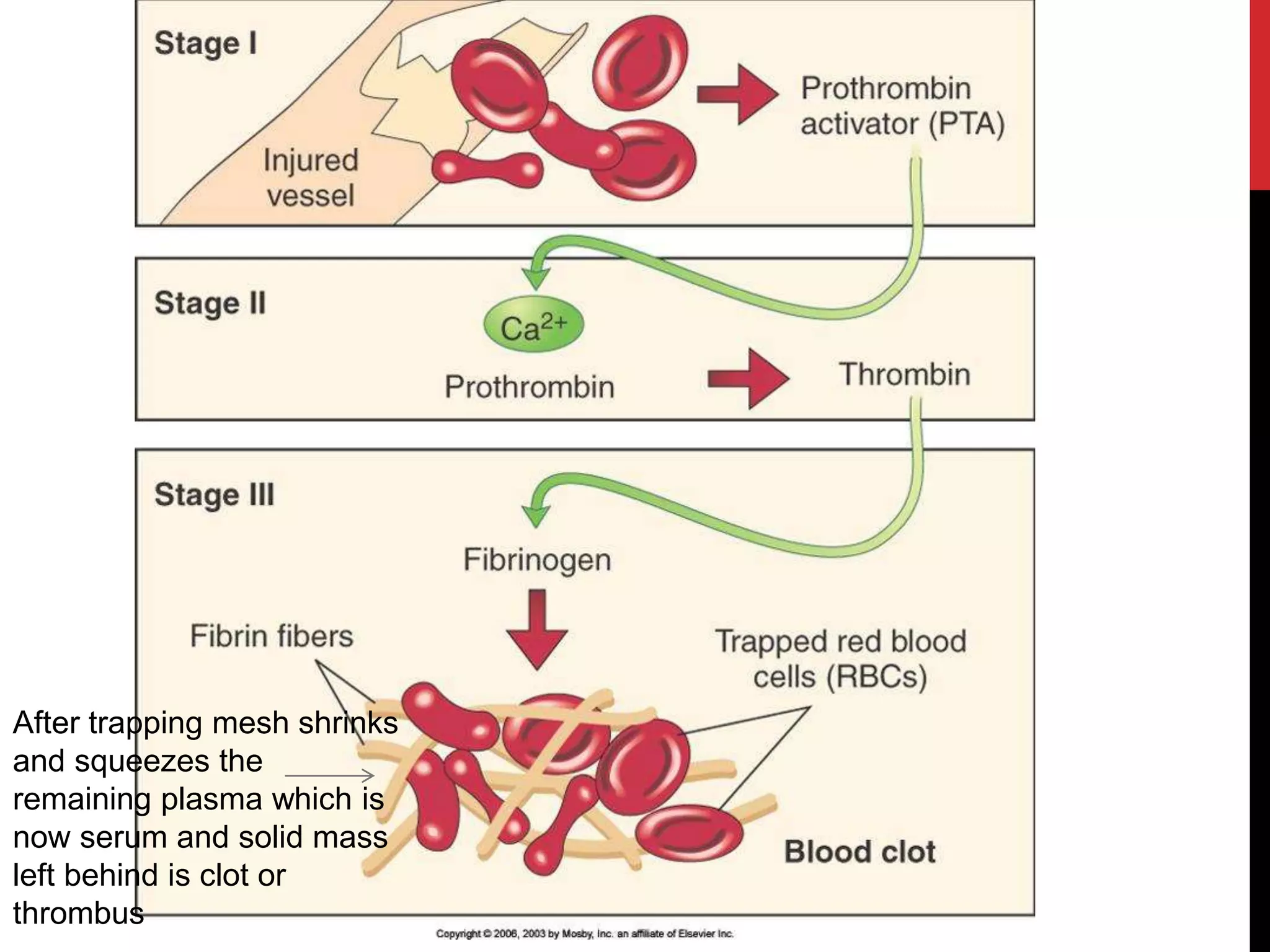

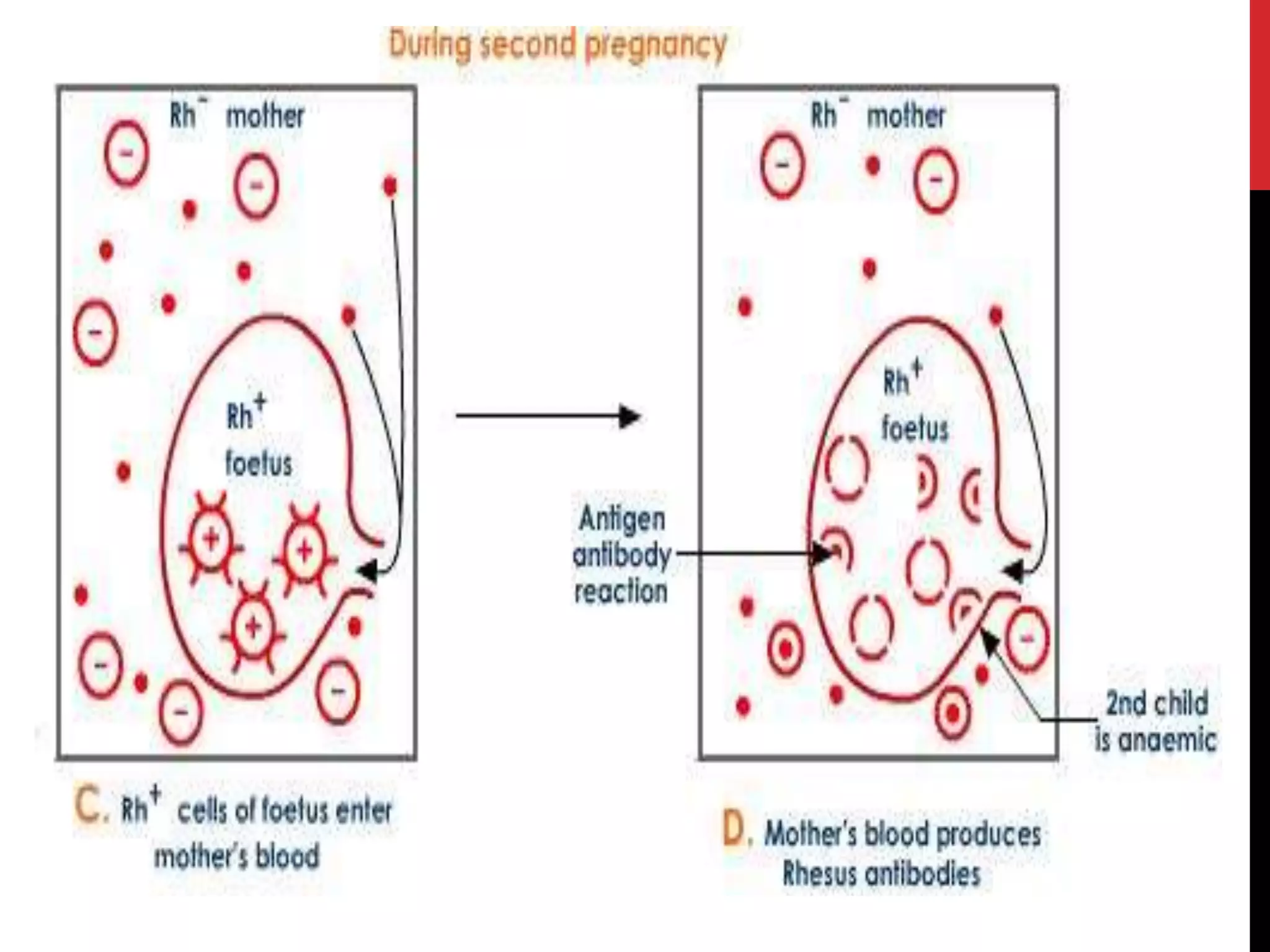
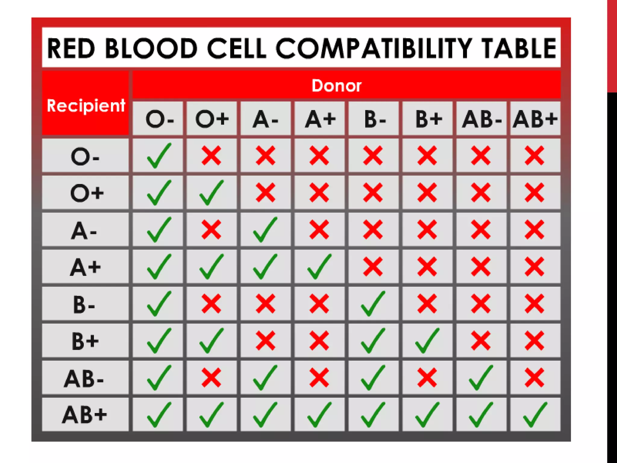
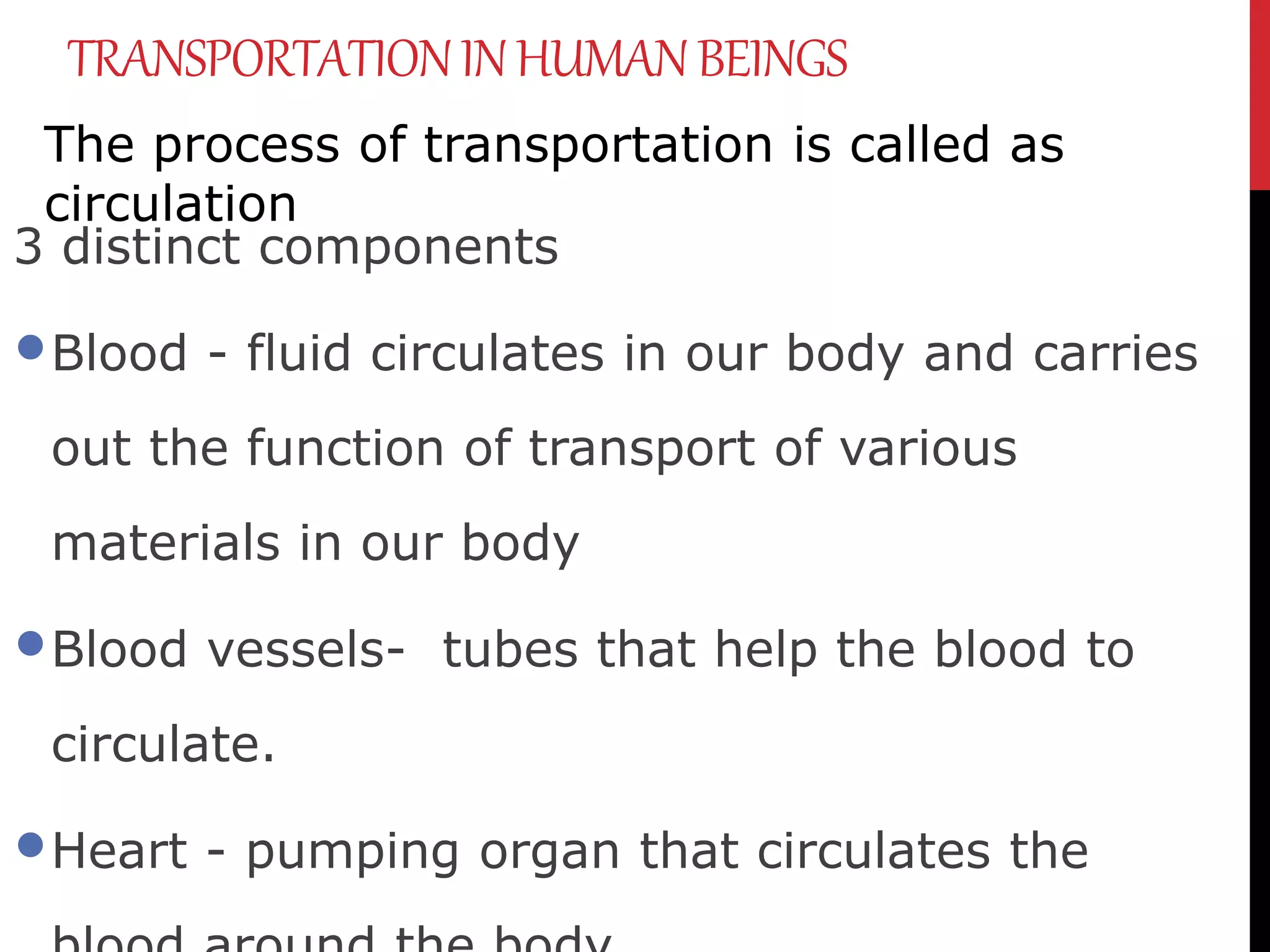
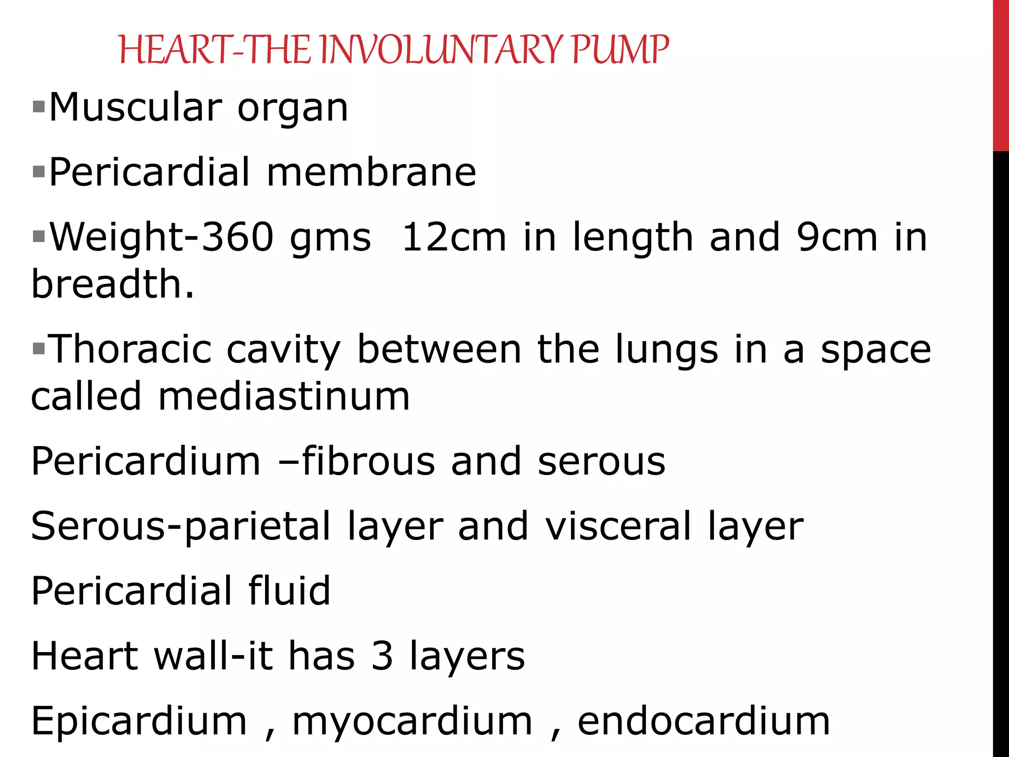

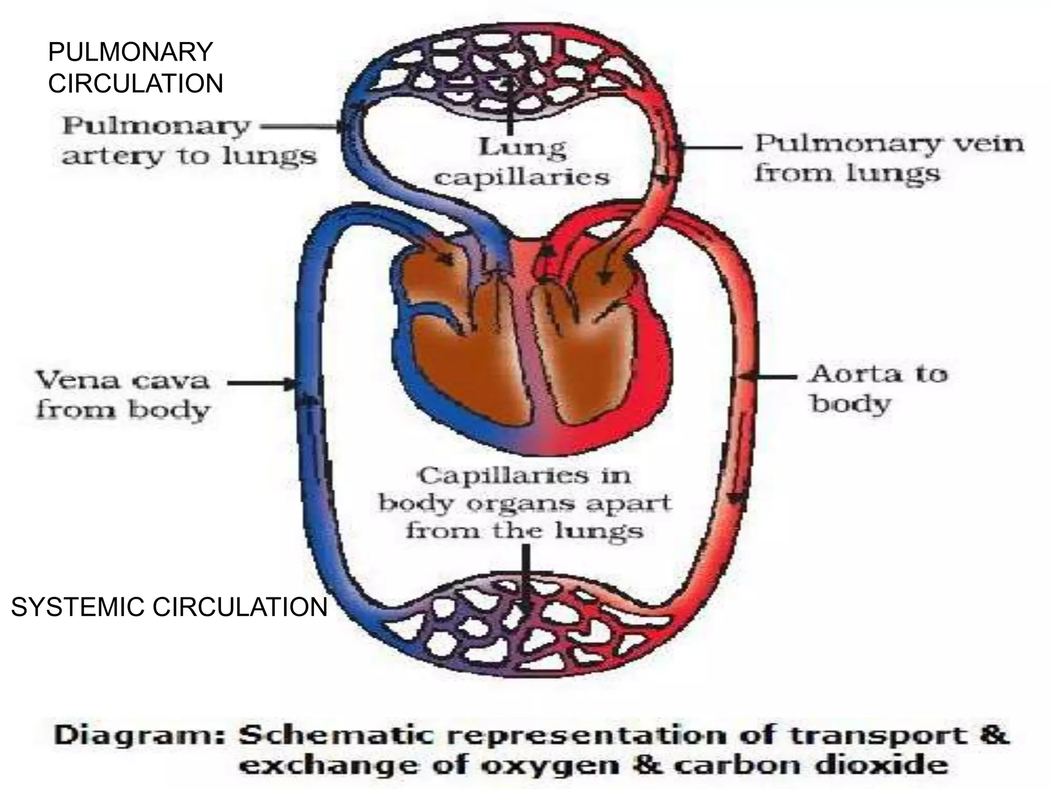
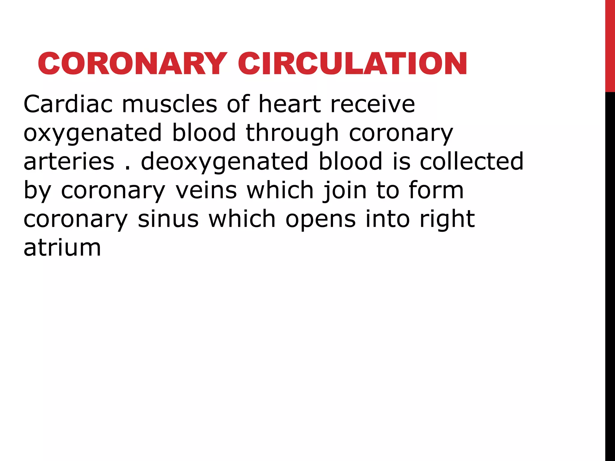
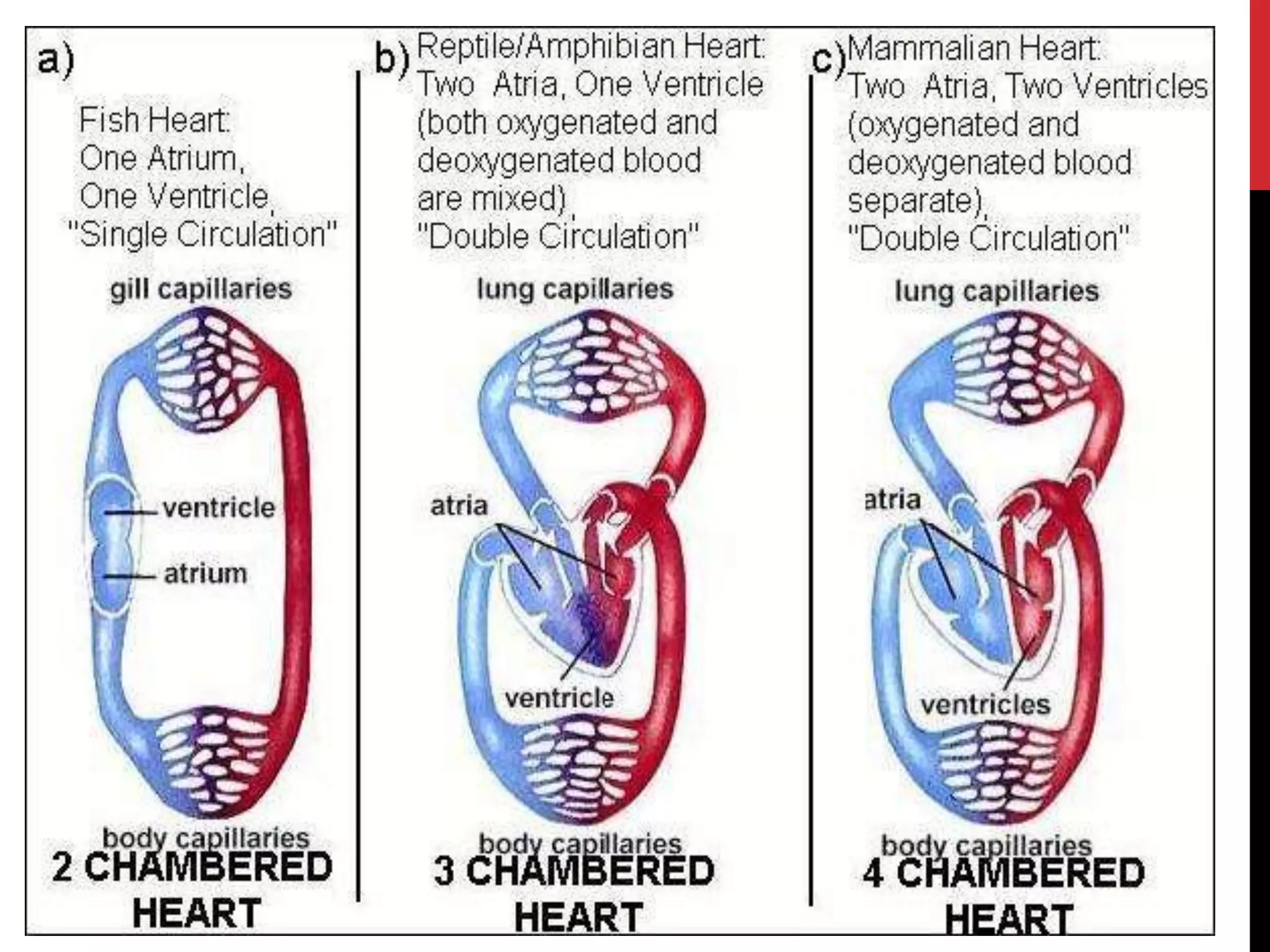
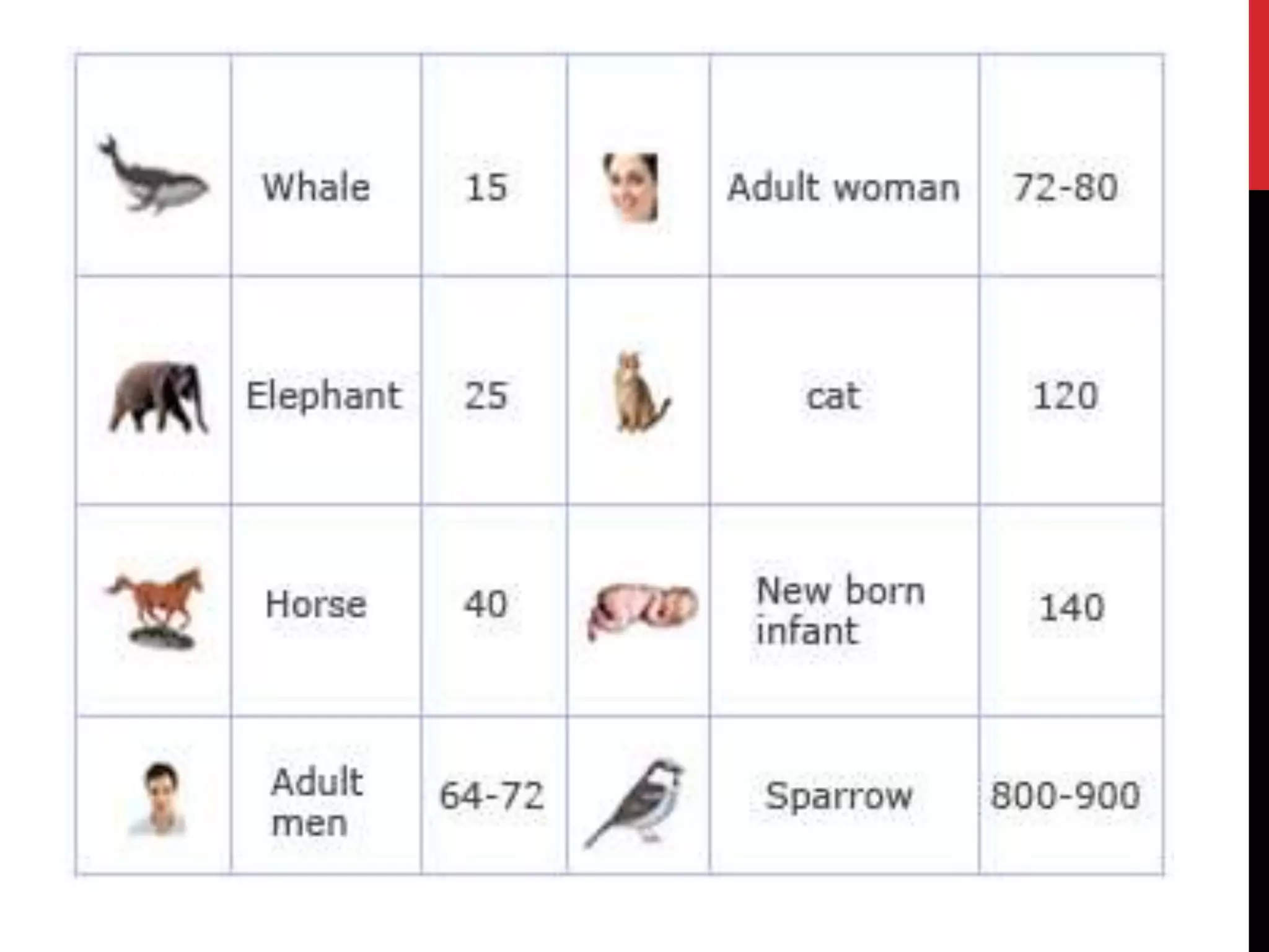
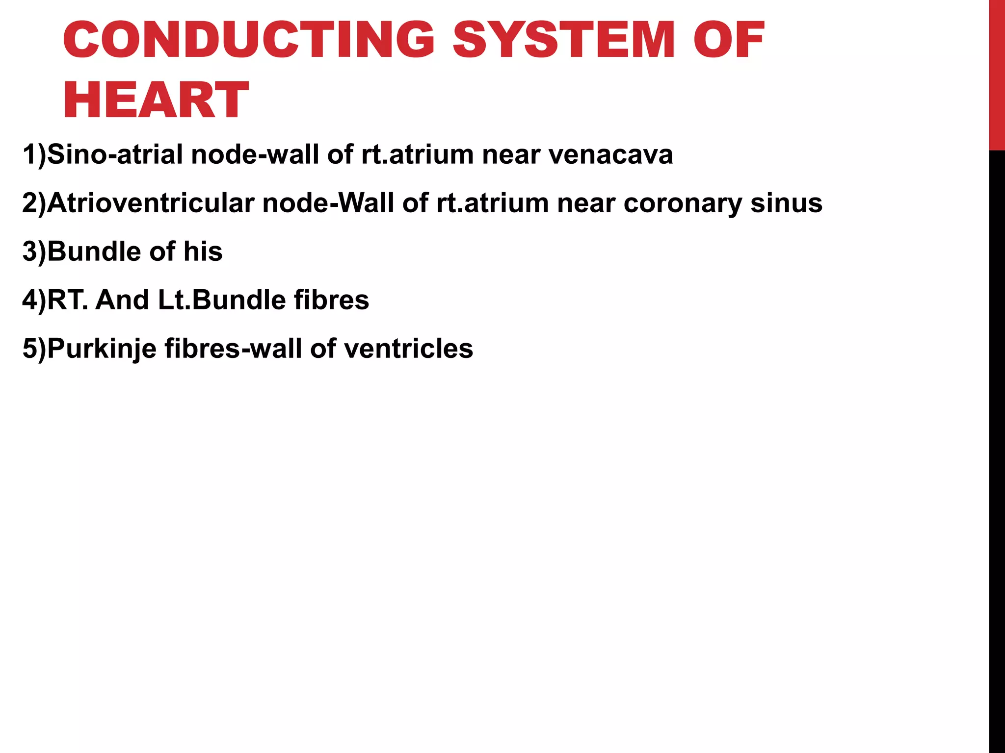
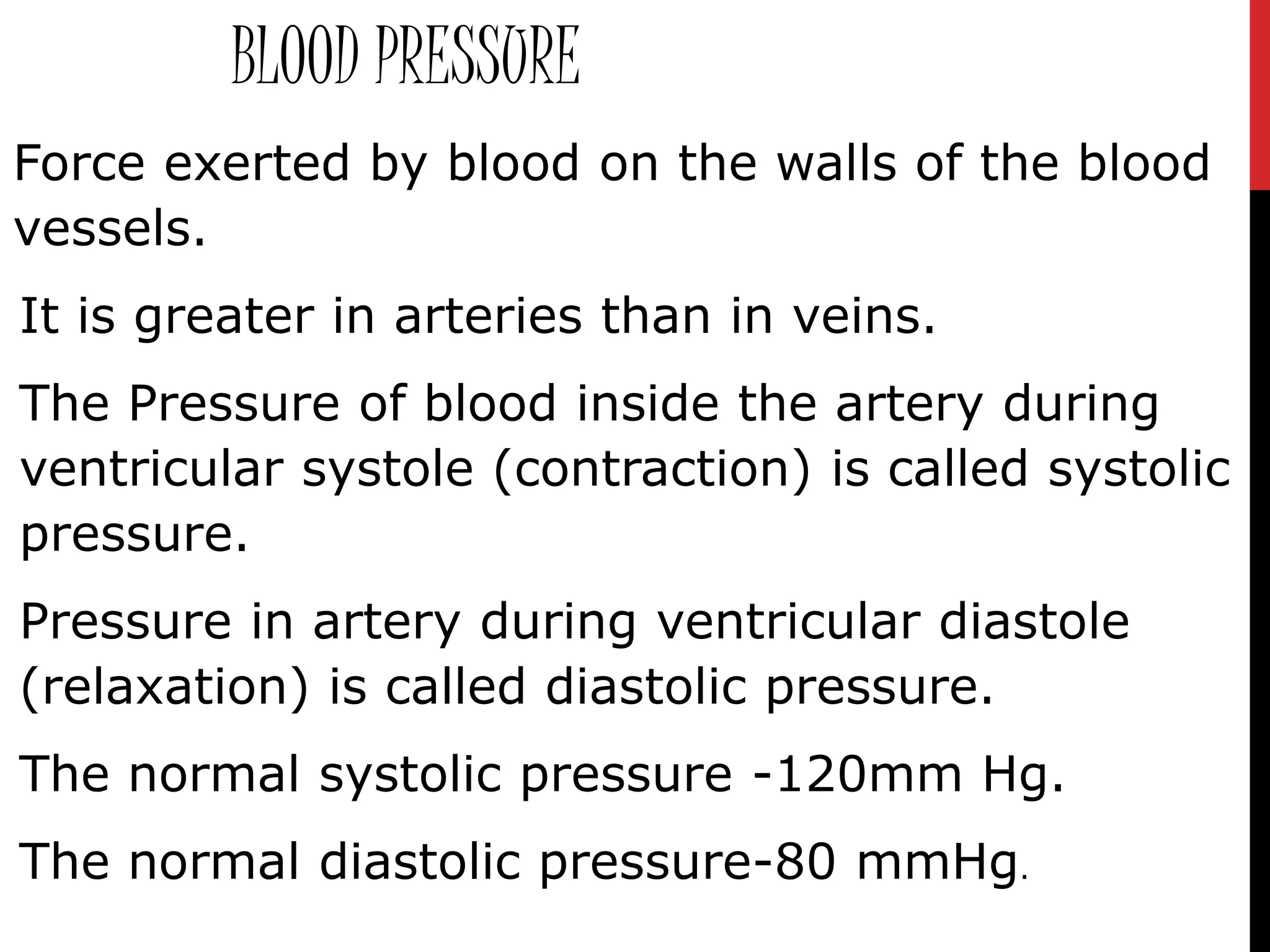
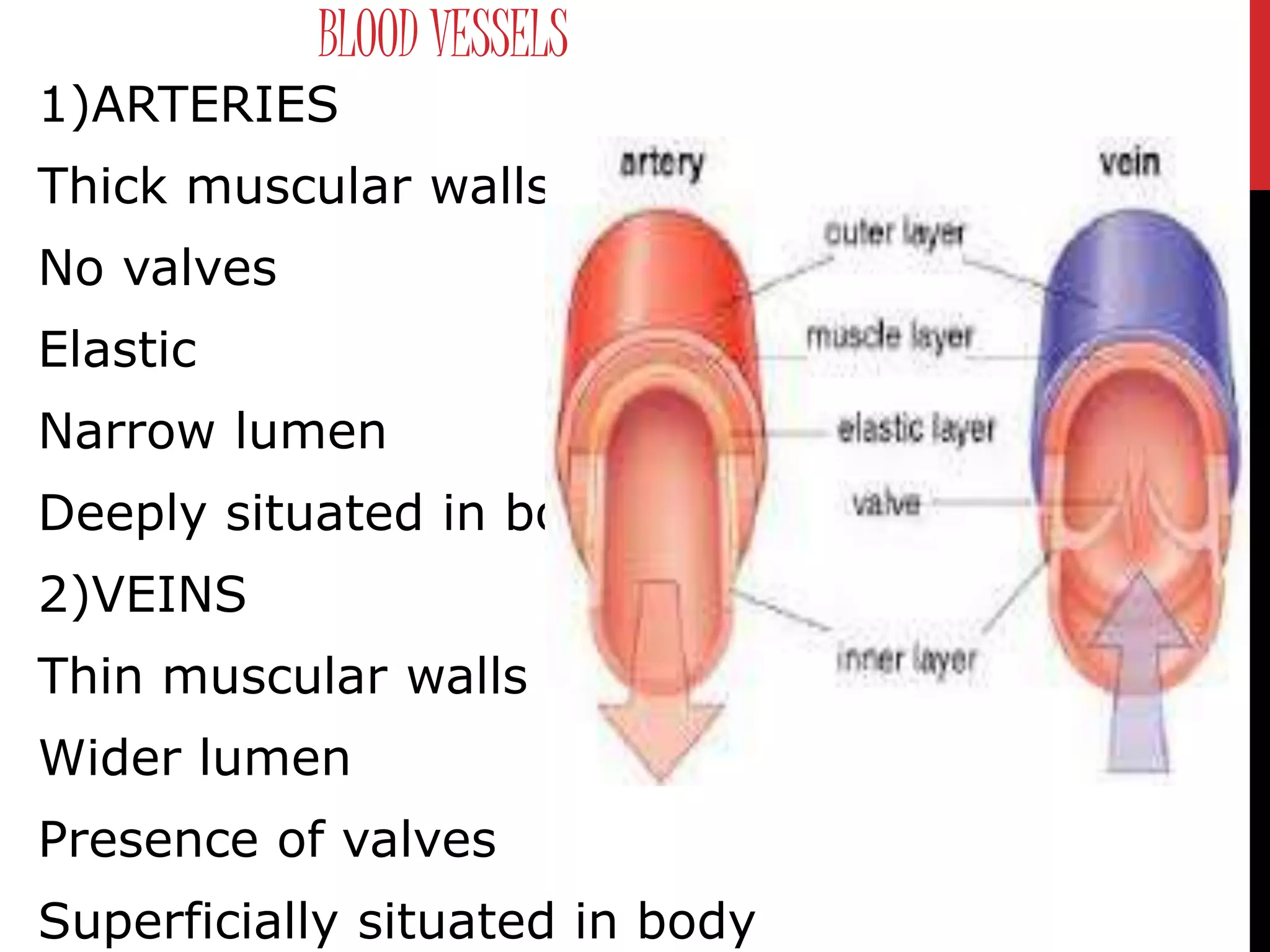
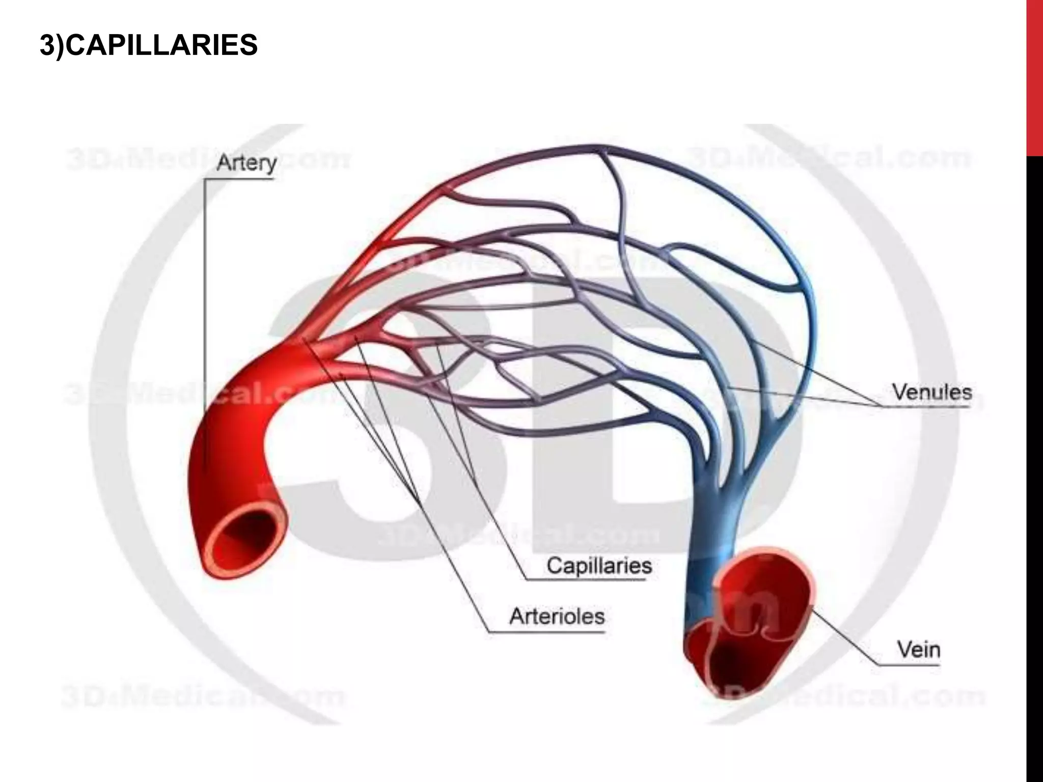
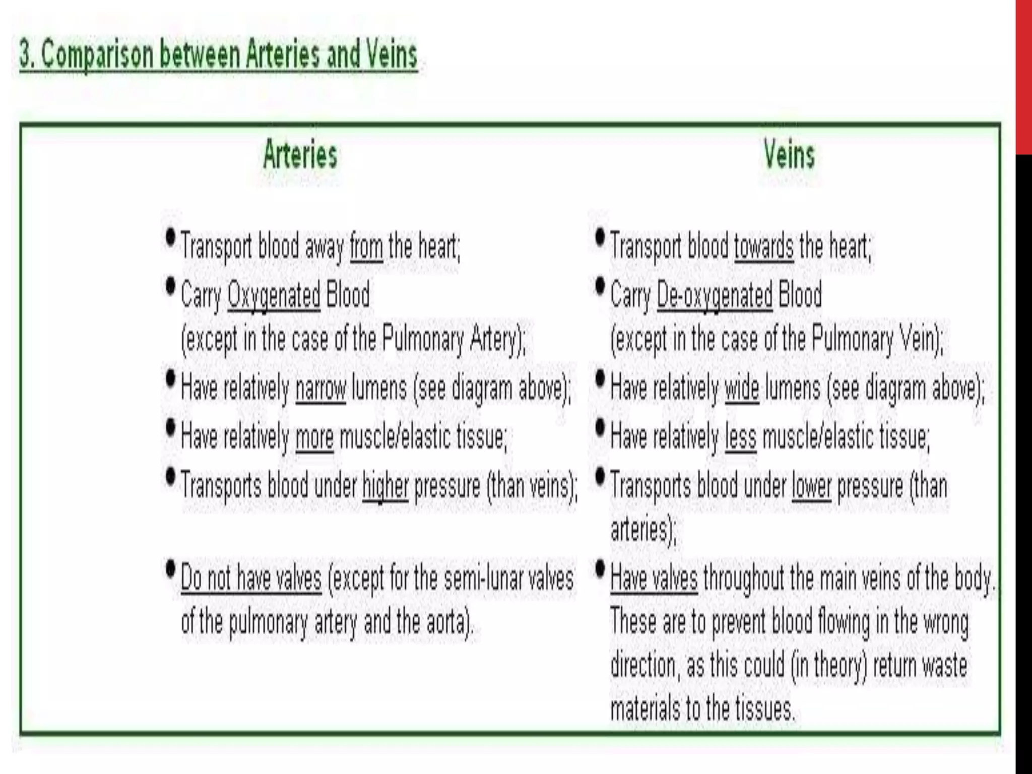
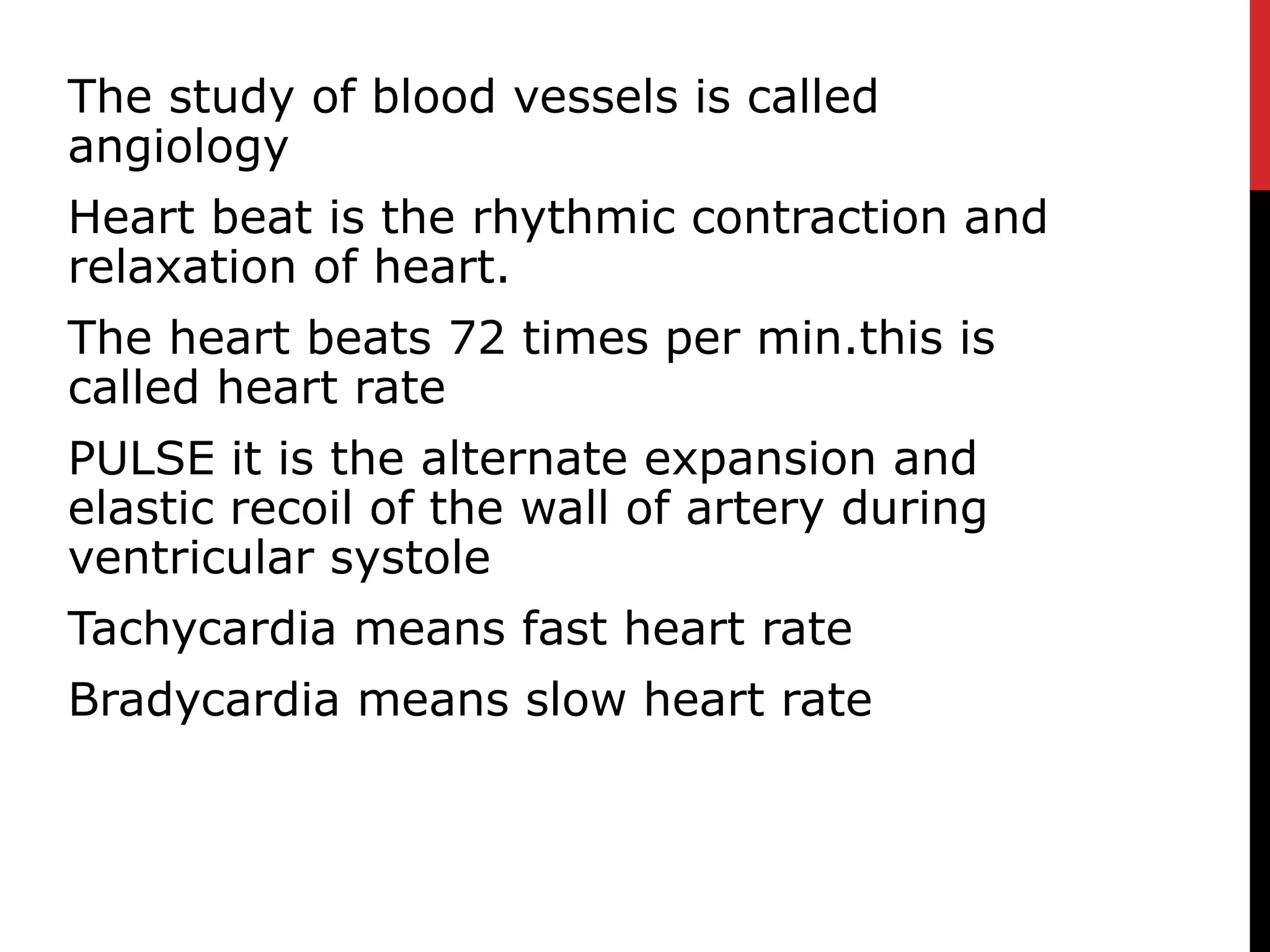
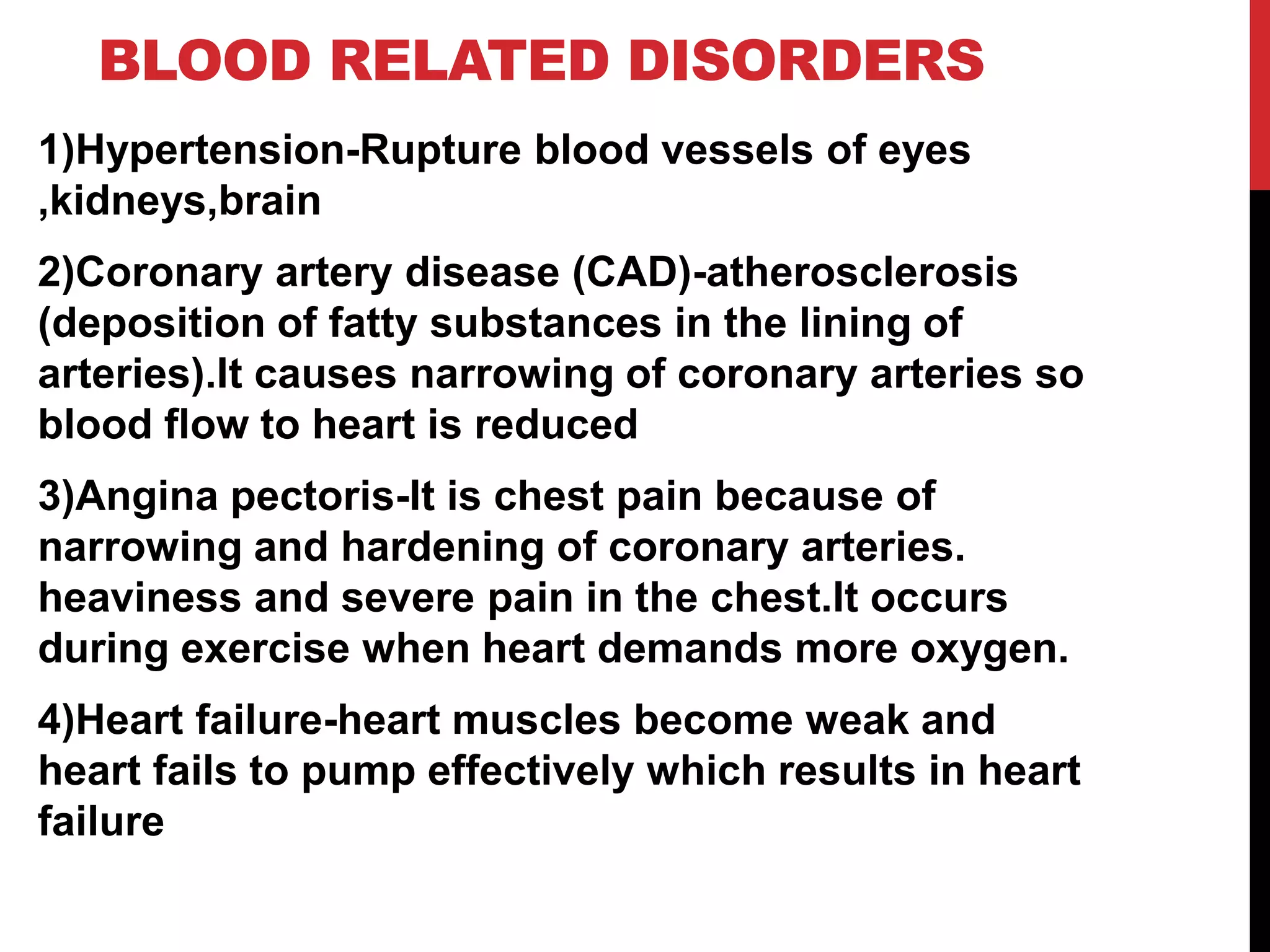
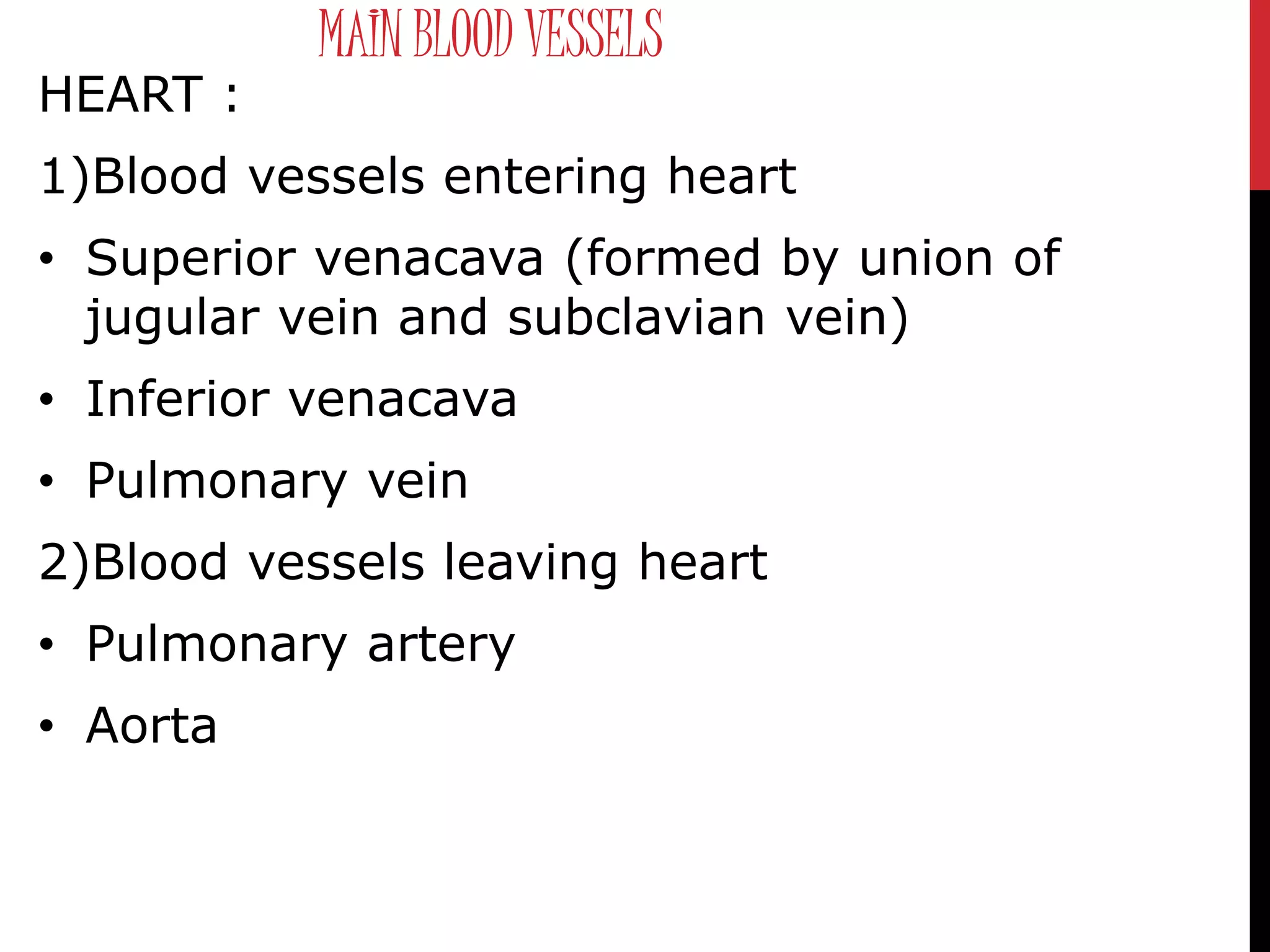
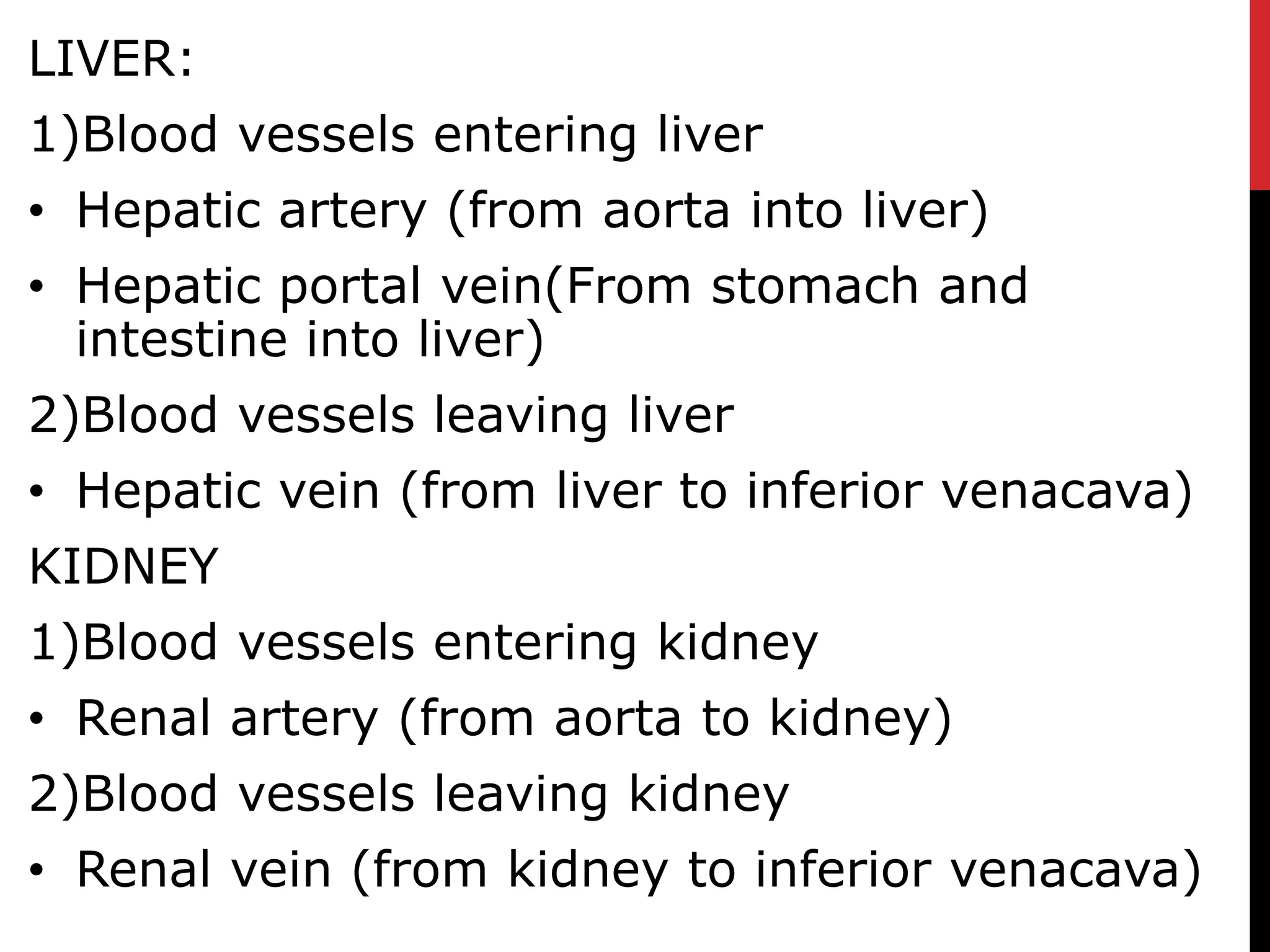
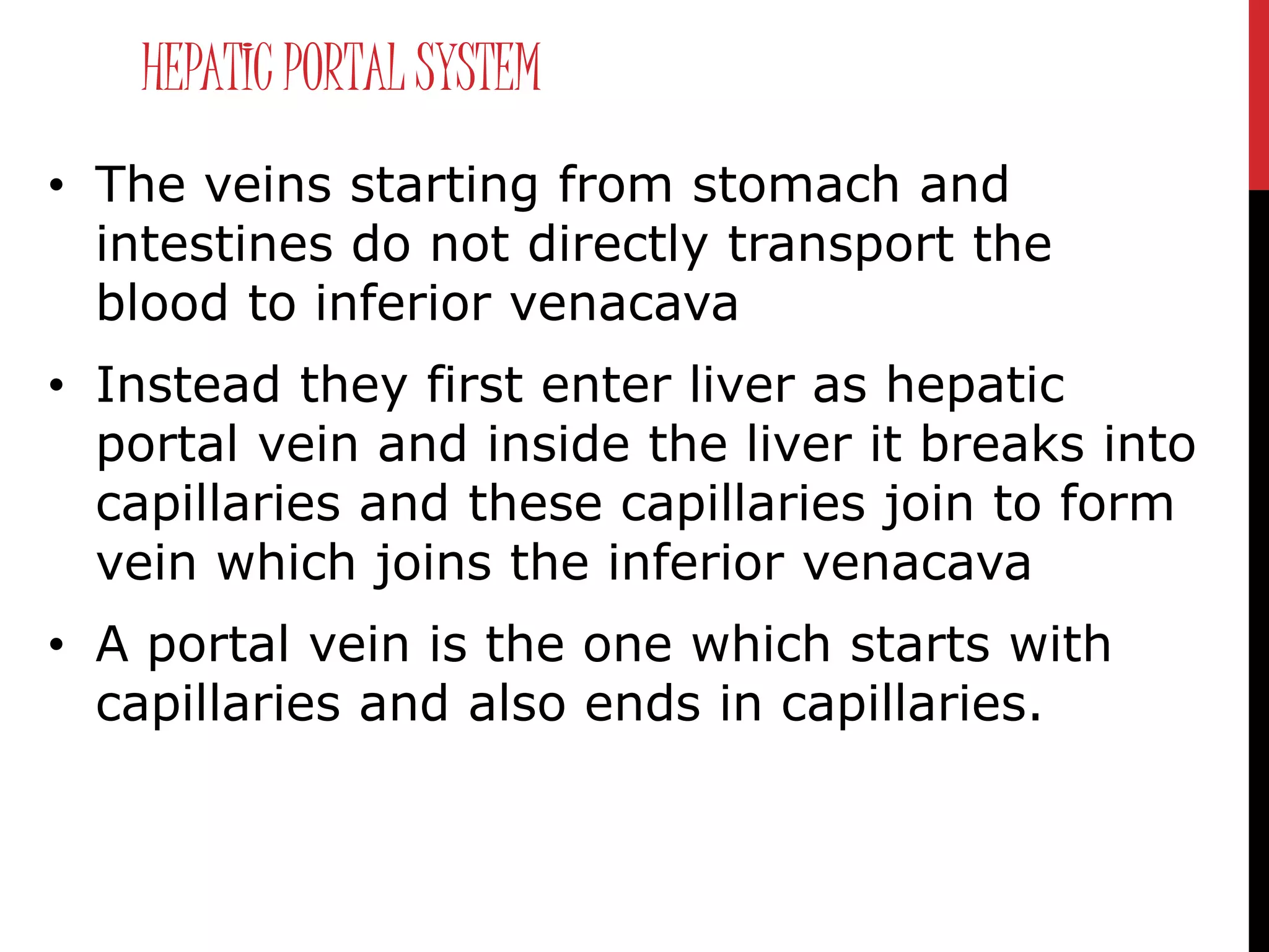

This document provides an overview of the human circulatory system. It describes the components of blood and their functions, including red blood cells, white blood cells, platelets, and plasma. It explains the need for transportation within the body to distribute oxygen, nutrients, waste, hormones, etc. It details the structure and function of the heart as a pump that circulates blood through two circuits: pulmonary and systemic circulation. It also discusses blood vessels, blood pressure, cardiac cycle, conducting system of the heart, and some blood disorders.


