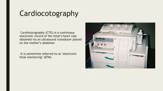
CARDIOTOCOGRAPHY
Cardiocotography
Cardiotocography (CTG) is a continuous
electronic record of the fetal’s heart rate
obtained via an ultrasound transducer placed
on the mother’s abdomen
It is sometimes referred to as ‘electronic
fetal monitoring’ (EFM)
TCG should be offered and recommended for high-risk
pregnancies where there is an increased risk of perinatal death, cerebral palsy
or neonatal encephalopathy.
Continuous TCG should be used where oxytocin is being used for induction or
augmentation of labour.
The indications for continuous CTG
monitoring in labour include:
• Sepsis
• Maternal tachycardia (> 120)
• Significant meconium
• Pre-eclampsia (particularly blood pressure > 160 / 110)
• Fresh antepartum haemorrhage
• Delay in labour
• Use of oxytocin
• Disproportionate maternal pain
There are five key features to look for on a CTG:
Contractions – the number of uterine contractions per 10 minutes
Baseline rate – the baseline fetal heart rate
Variability – how the fetal heart rate varies up and down around the
baseline
Accelerations – periods where the fetal heart rate spikes
Decelerations – periods where the fetal heart rate drops
Contractions are used to gauge the activity of labour. Too few
contractions indicate labour is not progressing. Too many
contractions can mean uterine hyperstimulation, which can lead to
fetal compromise. It is also important to interpret the fetal heart
rate in the context of the uterine contractions
Variability
The duration of the cardiac cycle is different for each fetus and is
constantly changing. These changes are termed instantaneous
frequency variability or simply “variability”.
Variability is composed of two components: short-term and long-
term variability.
Baseline rate and variability can be described as reassuring, non-reassuring and abnormal
Accelerations
They are defined as a short and rapid increase in fetal HR.
It is the cardiac response to fetal movements, also known as the
“Myocardial Reflex”, characterized by an increased HR in response
to physical activity.
Decelerations
They are defined as a short-term decline in fetal HR.
Given two main factors - deceleration form and relationship between
decelerations and the onset and the end of the uterine contraction, There are
four types of decelerations to be aware of:
Early decelerations
Late decelerations
Variable decelerations
Prolonged decelerations
Early decelerations are gradual dips and recoveries in heart rate that correspond with uterine contractions. The lowest point of the declaration
corresponds to the peak of the contraction. Early decelerations are normal and not considered pathological. They are caused by the uterus
compressing the head the fetus, stimulating the vagus nerve of the fetus, slowing the heart rate.
Late decelerations are gradual falls in heart rate that starts after the uterine contraction has already begun. There is a delay between the uterine
contraction and the deceleration. The lowest point of the declaration occurs after the peak of the contraction. Late decelerations are caused
by hypoxia in the fetus, and are a more concerning finding. They may be caused by excessive uterine contractions, maternal hypotension or
maternal hypoxia.
Variable decelerations are abrupt decelerations that may be unrelated to uterine contractions. There is a fall of more than 15 bpm from the
baseline. The lowest point of the declaration occurs within 30 seconds, and the deceleration lasts less than 2 minutes in total. Variable
decelerations often indicate intermittent compression of the umbilical cord, causing fetal hypoxia. Brief accelerations before and after the
deceleration are known as shoulders, and are a reassuring sign that the fetus is coping.
Prolonged decelerations last between 2 and 10 minutes with a drop of more than 15 bpm from baseline. This often indicates compression of the
umbilical cord, causing fetal hypoxia. These are abnormal and concerning.
Types
External cardiotocography can be used for continuous or
intermittent monitoring.
The fetal heart rate and the activity of the uterine muscle are
detected by two transducers placed on the mother's abdomen, with
one above the fetal heart to monitor heart rate, and the other at
the fundus of the uterus to measure frequency of contractions.
Internal cardiotocography uses an electronic transducer connected directly to the
fetus.
A wire electrode, sometimes called a spiral or scalp electrode, is attached to the
fetal scalp through the cervical opening and is connected to the monitor. Internal
monitoring provides a more accurate and consistent transmission of the fetal
heart rate, as unlike external monitoring, it is not affected by factors such as
movement.
Criteria for Internal Monitoring:
Amniotic membranes must be ruptured
Cervix dilated 2 cm.
Presentation must be cephalic.
Presenting part down against the cervix
There are several pathological variants of CTG:
Silent fetal CTG is characterized by the absence of accelerations or decelerations of the rhythm, while the basal rhythm may be in the normal
range. Sometimes such a cardiotocogram is called monotonous, the graphic image of the heartbeat looks like a straight line.
Sinusoidal CTG has a characteristic form of a sinusoid. In this case, the amplitude is small, equal to 6-10 beats. in min. This type of CTG is very
unfavorable and indicates severe fetal hypoxia. In rare cases, this type of CTG may appear when a pregnant woman takes narcotic or psychotropic
drugs.
lambda rhythm is the alternation of accelerations and decelerations immediately after them. In 95% of cases, this type of CTG is the result of
compression (compression) of the umbilical cord.
In addition, there are many types of CTG that are considered conditionally pathological . They are characterized by the following features:
■ Presence of decelerations after accelerations;
■ Reduced motor activity of the fetus;
■ Insufficient amplitude and rhythm variability.
We can see these signs if there is :
■ entanglement of the umbilical cord;
■ The presence of the umbilical cord;
■ Violation of placental blood flow;
■ fetal hypoxia;
■ Kid's heart defects;
■ The presence of diseases in the mother. For example, in pregnant women with hyperthyroidism, thyroid hormones can cross the placental
barrier and cause arrhythmias in the fetus;
■ Anemia of the baby (for example, with hemolytic disease associated with immunological incompatibility of the blood of the mother and fetus);
■ Inflammation of the membranes (amnionitis);
■ Taking certain medications. For example, Ginipral, widely used in obstetrics, can cause an increase in the baby's rhythm.
CTG during child birth
The study of the heart rate is necessary both in the first (opening of the cervix)
and in the second (pulling) period of labor. This is necessary in order to prevent
acute intrauterine hypoxia, which threatens the life of the fetus and is an
indication for an emergency caesarean section.
It is for this reason that the CTG recording must be started already at the first
signs of labor. With a normal birth, it is enough to register CTG every hour.
This study also shows:
After the outflow of amniotic fluid;
When conducting epidural anesthesia in childbirth (after the introduction of an
anesthetic).
Permanent recording of CTG is necessary for conditions such as:
1. Prolapse of the umbilical cord;
2. The pelvic position of the fetus;
3. Multiple pregnancy;
4. Double and triple entanglement of the umbilical cord around the neck of the
fetus;
5. Signs of preeclampsia;
6. Diabetes;
7. yellow or green amniotic fluid;
8. Hemolytic disease of the fetus;
9. intrauterine growth retardation;
10. premature birth;
11. Scar on the uterus after previous operations;
12. With weak or excessively strong labor activity.
13. When labor is stimulated with drugs, such as Oxytocin or prostaglandins.

ctd pregnancy

  • 1.
  • 2.
    Cardiocotography Cardiotocography (CTG) isa continuous electronic record of the fetal’s heart rate obtained via an ultrasound transducer placed on the mother’s abdomen It is sometimes referred to as ‘electronic fetal monitoring’ (EFM)
  • 3.
    TCG should beoffered and recommended for high-risk pregnancies where there is an increased risk of perinatal death, cerebral palsy or neonatal encephalopathy. Continuous TCG should be used where oxytocin is being used for induction or augmentation of labour.
  • 4.
    The indications forcontinuous CTG monitoring in labour include: • Sepsis • Maternal tachycardia (> 120) • Significant meconium • Pre-eclampsia (particularly blood pressure > 160 / 110) • Fresh antepartum haemorrhage • Delay in labour • Use of oxytocin • Disproportionate maternal pain
  • 5.
    There are fivekey features to look for on a CTG: Contractions – the number of uterine contractions per 10 minutes Baseline rate – the baseline fetal heart rate Variability – how the fetal heart rate varies up and down around the baseline Accelerations – periods where the fetal heart rate spikes Decelerations – periods where the fetal heart rate drops
  • 6.
    Contractions are usedto gauge the activity of labour. Too few contractions indicate labour is not progressing. Too many contractions can mean uterine hyperstimulation, which can lead to fetal compromise. It is also important to interpret the fetal heart rate in the context of the uterine contractions
  • 7.
    Variability The duration ofthe cardiac cycle is different for each fetus and is constantly changing. These changes are termed instantaneous frequency variability or simply “variability”. Variability is composed of two components: short-term and long- term variability.
  • 8.
    Baseline rate andvariability can be described as reassuring, non-reassuring and abnormal
  • 9.
    Accelerations They are definedas a short and rapid increase in fetal HR. It is the cardiac response to fetal movements, also known as the “Myocardial Reflex”, characterized by an increased HR in response to physical activity.
  • 10.
    Decelerations They are definedas a short-term decline in fetal HR. Given two main factors - deceleration form and relationship between decelerations and the onset and the end of the uterine contraction, There are four types of decelerations to be aware of: Early decelerations Late decelerations Variable decelerations Prolonged decelerations
  • 11.
    Early decelerations aregradual dips and recoveries in heart rate that correspond with uterine contractions. The lowest point of the declaration corresponds to the peak of the contraction. Early decelerations are normal and not considered pathological. They are caused by the uterus compressing the head the fetus, stimulating the vagus nerve of the fetus, slowing the heart rate. Late decelerations are gradual falls in heart rate that starts after the uterine contraction has already begun. There is a delay between the uterine contraction and the deceleration. The lowest point of the declaration occurs after the peak of the contraction. Late decelerations are caused by hypoxia in the fetus, and are a more concerning finding. They may be caused by excessive uterine contractions, maternal hypotension or maternal hypoxia. Variable decelerations are abrupt decelerations that may be unrelated to uterine contractions. There is a fall of more than 15 bpm from the baseline. The lowest point of the declaration occurs within 30 seconds, and the deceleration lasts less than 2 minutes in total. Variable decelerations often indicate intermittent compression of the umbilical cord, causing fetal hypoxia. Brief accelerations before and after the deceleration are known as shoulders, and are a reassuring sign that the fetus is coping. Prolonged decelerations last between 2 and 10 minutes with a drop of more than 15 bpm from baseline. This often indicates compression of the umbilical cord, causing fetal hypoxia. These are abnormal and concerning.
  • 12.
    Types External cardiotocography canbe used for continuous or intermittent monitoring. The fetal heart rate and the activity of the uterine muscle are detected by two transducers placed on the mother's abdomen, with one above the fetal heart to monitor heart rate, and the other at the fundus of the uterus to measure frequency of contractions.
  • 13.
    Internal cardiotocography usesan electronic transducer connected directly to the fetus. A wire electrode, sometimes called a spiral or scalp electrode, is attached to the fetal scalp through the cervical opening and is connected to the monitor. Internal monitoring provides a more accurate and consistent transmission of the fetal heart rate, as unlike external monitoring, it is not affected by factors such as movement. Criteria for Internal Monitoring: Amniotic membranes must be ruptured Cervix dilated 2 cm. Presentation must be cephalic. Presenting part down against the cervix
  • 14.
    There are severalpathological variants of CTG: Silent fetal CTG is characterized by the absence of accelerations or decelerations of the rhythm, while the basal rhythm may be in the normal range. Sometimes such a cardiotocogram is called monotonous, the graphic image of the heartbeat looks like a straight line. Sinusoidal CTG has a characteristic form of a sinusoid. In this case, the amplitude is small, equal to 6-10 beats. in min. This type of CTG is very unfavorable and indicates severe fetal hypoxia. In rare cases, this type of CTG may appear when a pregnant woman takes narcotic or psychotropic drugs. lambda rhythm is the alternation of accelerations and decelerations immediately after them. In 95% of cases, this type of CTG is the result of compression (compression) of the umbilical cord. In addition, there are many types of CTG that are considered conditionally pathological . They are characterized by the following features: ■ Presence of decelerations after accelerations; ■ Reduced motor activity of the fetus; ■ Insufficient amplitude and rhythm variability. We can see these signs if there is : ■ entanglement of the umbilical cord; ■ The presence of the umbilical cord; ■ Violation of placental blood flow; ■ fetal hypoxia; ■ Kid's heart defects; ■ The presence of diseases in the mother. For example, in pregnant women with hyperthyroidism, thyroid hormones can cross the placental barrier and cause arrhythmias in the fetus; ■ Anemia of the baby (for example, with hemolytic disease associated with immunological incompatibility of the blood of the mother and fetus); ■ Inflammation of the membranes (amnionitis); ■ Taking certain medications. For example, Ginipral, widely used in obstetrics, can cause an increase in the baby's rhythm.
  • 15.
    CTG during childbirth The study of the heart rate is necessary both in the first (opening of the cervix) and in the second (pulling) period of labor. This is necessary in order to prevent acute intrauterine hypoxia, which threatens the life of the fetus and is an indication for an emergency caesarean section. It is for this reason that the CTG recording must be started already at the first signs of labor. With a normal birth, it is enough to register CTG every hour. This study also shows: After the outflow of amniotic fluid; When conducting epidural anesthesia in childbirth (after the introduction of an anesthetic).
  • 16.
    Permanent recording ofCTG is necessary for conditions such as: 1. Prolapse of the umbilical cord; 2. The pelvic position of the fetus; 3. Multiple pregnancy; 4. Double and triple entanglement of the umbilical cord around the neck of the fetus; 5. Signs of preeclampsia; 6. Diabetes; 7. yellow or green amniotic fluid; 8. Hemolytic disease of the fetus; 9. intrauterine growth retardation; 10. premature birth; 11. Scar on the uterus after previous operations; 12. With weak or excessively strong labor activity. 13. When labor is stimulated with drugs, such as Oxytocin or prostaglandins.