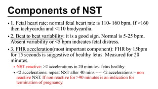
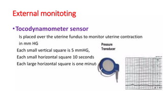
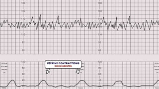
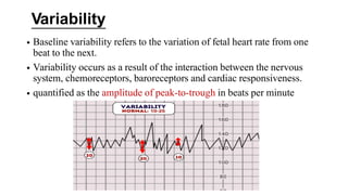
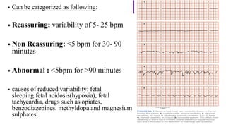
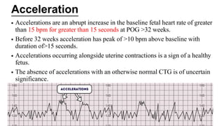
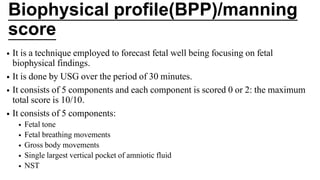
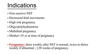
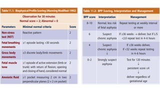
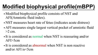
NST, Biophysical Profile
And Modified Biophysical
Profile
Presented by Shivang Verma
20092
Dr. RKGMCH
Introduction
•non stress test and biophysical profile are part
of antepartum fetal monitoring.
•Non stress test(NST) includes monitoring of
fetal heart rate in response to fetal
movements to assess fetal well being.
•Biophysical profile is a screening test for
uteroplacental insufficiency.
• Fetal biophysical activities are initiated,
modulated and regulated through fetal nervous
system.
•Fetal CNS in sensitive to hypoxia and changes
occurs in fetal biophysical activity
Non Stress Test(NST)
•non-stress test, a continuous electronic
monitoring of the fetal heart rate along with
recording of fetal movements
(cardiotocography) is undertaken.
•We observe association of FHR acceleration
with fetal movements, which when present,
indicates a healthy fetus.
Indications of Non Stress
Test
• Premature rupture of membrane
• Chronic renal disease
• Gestational diabetes mellitus
• High blood pressure/gestational
hypertension
• Trauma
• Vaginal bleeding in 2nd / 3rd trimester
• H/O stillbirth
• Rh sensitization
• Decreased fetal movements
• Intrauterine growth
restriction(IUGR)
• Oligo/polyhydramnios
Procedure of performing NST
• Explain the mother procedure and its purpose.
• Make sure woman has eaten food and empty
bladder.
• Monitor is turned on and pre checked.
• Perform abdominal palpation.
• Confirm FHR with stethoscope or doppler and
note area of maximum intensity.
• Place woman in semi fowler’s position and place
monitor belt under back
• Connect ultrasound transducer and
tocotransducer and apply gel on ultrasound.
• Place ultrasound transducer on fetal back and
move till clear, audible FHS heard.
• Secure device in place with belt.
• Place tocotransducer on fundus of uterus and
secure with belt.
• Give the hand button to woman and ask her to
press the button every time she feels the fetal
movements.
• Run for 20 minutes, on completion take out stripe
of paper.
• Remove abdominal straps, clear gel and make
woman comfortable.
Components of NST
• 1. Fetal heart rate: normal fetal heart rate is 110- 160 bpm. If >160
then tachycardia and <110 bradycardia.
• 2. Beat to beat variability: it is a good sign. Normal is 5-25 bpm.
Absent variability or <5 bpm indicates fetal distress.
• 3. FHR acceleration(most important component): FHR by 15bpm
for 15 seconds is suggestive of healthy fetus. Measured for 20
minutes.
• NST reactive: >2 accelerations in 20 minutes- fetus healthy
• <2 accelerations: repeat NST after 40 mins ---- <2 accelerations – non
reactive NST. If non reactive for >90 minutes is an indication for
termination of pregnancy.
Cardiotocography(CTG)
•CTG is defined as graphical recoding of
fetal heart rate and uterine contractions by
the use of electronic devices indicated for
assesment of fetal, condition.
•It is also known as electronic fetal
monitoring(EFM)
Indications
• Maternal
–Hypertension
–previous cesarian
section
–induced labour
–APH
–PROM
• Fetal
–Small fetus(FGR)
–oligohydroamnios
–multiple pregnancy
–abnormal FHR on
auscultation
Methods of doing CTG
External :Continuous tracing of FHR can be obtained using
ultrasound Doppler . The transducers are placed on the
maternal abdomen, one over the fundus and the other at a
site where the fetal heart sound is best audible. Frequency of
uterine contractions and uterine pressure are recorded
simultaneously by tocodynamometer.
Internal: Fetal ECG tracing is made by applying an
electrode to the fetal scalp after rupturing the membranes.
Not done now a days.
Interpretation of CTG
• Remembered using the acronym DR C BRAVADO
• DR: Define risk
• C: Contractions
• BRa: Baseline rate
• V: Variability
• A: Accelerations
• D: Decelerations
• O: Overall impression
•When performing CTG interpretation, determine if
the pregnancy is high or low risk. This is important
as it gives more context to the CTG reading (e.g. if
the pregnancy categorised as high-risk, the threshold
for intervention may be lower).
Defining risk
Indications
• Maternal medical illness
like
–Gestational diabetes
–Hypertension
–Asthma
• Obstetric complications like
– Multiple gestation
– Post-dated gestation
– Previous cesarean section
– IUGR
– PROM
– congenital malformations
– induction/augmentation of
labour
– Pre-eclampsia.
Contractions
•Record the number of contractions present in a 10
minute period.
•Each. big square on CTG represents one minute
and no. of contractions in 10 big squares(10
minutes )
•Contractions are seen as peaks on the part of CTG.
•Assess Duration, Intensity and no. of contractions.
Baseline rate
• Is the mean level of FHR excluding accelerations
and decelerations. It is expressed in beats per
minute (bpm). Normal baseline FHR is 110–160
bpm.
• FHR >160 is defined
as tachycardia.
Causes include:
• hypoxia
• chorioamninoitis
• fetal anemia
• maternal anmeia
• FHR <110 is defined
as bradycardia.
causes include:
• post dated gestation
• OP/transverse
position
• cord compression
• cord prolapse
• epi./spinal anasthesia.
Variability
• Baseline variability refers to the variation of fetal heart rate from one
beat to the next.
• Variability occurs as a result of the interaction between the nervous
system, chemoreceptors, baroreceptors and cardiac responsiveness.
• quantified as the amplitude of peak-to-trough in beats per minute
• Can be categorized as following:
• Reassuring: variability of 5- 25 bpm
• Non Reassuring: <5 bpm for 30- 90
minutes
• Abnormal : <5bpm for >90 minutes
• causes of reduced variability: fetal
sleeping,fetal acidosis(hypoxia), fetal
tachycardia, drugs such as opiates,
benzodiazepines, methyldopa and magnesium
sulphates
• Accelerations are an abrupt increase in the baseline fetal heart rate of greater
than 15 bpm for greater than 15 seconds at POG >32 weeks.
• Before 32 weeks acceleration has peak of >10 bpm above baseline with
duration of>15 seconds.
• Accelerations occurring alongside uterine contractions is a sign of a healthy
fetus.
• The absence of accelerations with an otherwise normal CTG is of uncertain
significance.
Acceleration
Decelerations
• Decelerations are an abrupt decrease in the baseline fetal heart
rate of greater than 15 bpm for greater than 15 seconds.
• In response to hypoxic stress, the fetus reduces its heart rate to
preserve myocardial oxygenation and perfusion.
• This reduction in heart rate to reduce myocardial demand is
referred to as a deceleration.
• There are a number of different types of decelerations, each
with varying significance.
1. Early deceleration
• This physiological response shows a
gradual FHR decline and return to
baseline that mirrors the contraction.
This occurs due to head compression
during active labour when the cervix
is 4 to 7 cm dilated.
• early deceleration are considered
benign and are not associated with
fatal hypoxia , acidemia or low apgar
score
2.Late Deceleration
• this deceleration is a smooth gradual
symmetrical decline in FHR that
begins at or after the contraction peak
and returns to baseline only after
contraction has ended.
• This type of deceleration indicates
there is insufficient blood flow to the
uterus and placenta. As a result, blood
flow to the fetus is significantly
reduced causing fetal hypoxia and
acidosis.
3.Variable Deceleration
• Variable decelerations are observed as a
rapid fall in baseline fetal heart rate with a
variable recovery phase.
• Variable decelerations are usually caused
by umbilical cord compression which
may occur during labour process.
• The presence of persistent variable
decelerations indicates the need for close
monitoring.
Biophysical profile(BPP)/manning
score
• It is a technique employed to forecast fetal well being focusing on fetal
biophysical findings.
• It is done by USG over the period of 30 minutes.
• It consists of 5 components and each component is scored 0 or 2: the maximum
total score is 10/10.
• It consists of 5 components:
• Fetal tone
• Fetal breathing movements
• Gross body movements
• Single largest vertical pocket of amniotic fluid
• NST
Indications
• Non reactive NST
• Decreased fetal movements
• High risk pregnancy
• Oligo/polyhydramnios
• Multifetal pregnancy
• Mother>35 yr at time of pregnancy
• Frequency: done weekly after NST is normal, twice to thrice
weekly if abnormal ; ≥28 weeks of pregnancy.
Modified biophysical profile(mBPP)
•Modified biophysical profile consists of NST and
AFI(Amniotic fluid index).
•NST measures heart rate of fetus (indicates acute distress)
•AFI measures single largest vertical pocket of amniotic fluid
>2 cm.
•It is considered as normal when NST is reassuring and/or
AFI>5cm
•It is considered as abnormal when NST is non reactive
and/or AFI is<5cm
NST, biophysical score, cardiotocography(CTG).pdf

NST, biophysical score, cardiotocography(CTG).pdf

  • 1.
    NST, Biophysical Profile AndModified Biophysical Profile Presented by Shivang Verma 20092 Dr. RKGMCH
  • 2.
    Introduction •non stress testand biophysical profile are part of antepartum fetal monitoring. •Non stress test(NST) includes monitoring of fetal heart rate in response to fetal movements to assess fetal well being.
  • 3.
    •Biophysical profile isa screening test for uteroplacental insufficiency. • Fetal biophysical activities are initiated, modulated and regulated through fetal nervous system. •Fetal CNS in sensitive to hypoxia and changes occurs in fetal biophysical activity
  • 4.
    Non Stress Test(NST) •non-stresstest, a continuous electronic monitoring of the fetal heart rate along with recording of fetal movements (cardiotocography) is undertaken. •We observe association of FHR acceleration with fetal movements, which when present, indicates a healthy fetus.
  • 5.
    Indications of NonStress Test • Premature rupture of membrane • Chronic renal disease • Gestational diabetes mellitus • High blood pressure/gestational hypertension • Trauma • Vaginal bleeding in 2nd / 3rd trimester • H/O stillbirth • Rh sensitization • Decreased fetal movements • Intrauterine growth restriction(IUGR) • Oligo/polyhydramnios
  • 6.
    Procedure of performingNST • Explain the mother procedure and its purpose. • Make sure woman has eaten food and empty bladder. • Monitor is turned on and pre checked. • Perform abdominal palpation. • Confirm FHR with stethoscope or doppler and note area of maximum intensity. • Place woman in semi fowler’s position and place monitor belt under back • Connect ultrasound transducer and tocotransducer and apply gel on ultrasound.
  • 7.
    • Place ultrasoundtransducer on fetal back and move till clear, audible FHS heard. • Secure device in place with belt. • Place tocotransducer on fundus of uterus and secure with belt. • Give the hand button to woman and ask her to press the button every time she feels the fetal movements. • Run for 20 minutes, on completion take out stripe of paper. • Remove abdominal straps, clear gel and make woman comfortable.
  • 8.
    Components of NST •1. Fetal heart rate: normal fetal heart rate is 110- 160 bpm. If >160 then tachycardia and <110 bradycardia. • 2. Beat to beat variability: it is a good sign. Normal is 5-25 bpm. Absent variability or <5 bpm indicates fetal distress. • 3. FHR acceleration(most important component): FHR by 15bpm for 15 seconds is suggestive of healthy fetus. Measured for 20 minutes. • NST reactive: >2 accelerations in 20 minutes- fetus healthy • <2 accelerations: repeat NST after 40 mins ---- <2 accelerations – non reactive NST. If non reactive for >90 minutes is an indication for termination of pregnancy.
  • 9.
    Cardiotocography(CTG) •CTG is definedas graphical recoding of fetal heart rate and uterine contractions by the use of electronic devices indicated for assesment of fetal, condition. •It is also known as electronic fetal monitoring(EFM)
  • 10.
    Indications • Maternal –Hypertension –previous cesarian section –inducedlabour –APH –PROM • Fetal –Small fetus(FGR) –oligohydroamnios –multiple pregnancy –abnormal FHR on auscultation
  • 11.
    Methods of doingCTG External :Continuous tracing of FHR can be obtained using ultrasound Doppler . The transducers are placed on the maternal abdomen, one over the fundus and the other at a site where the fetal heart sound is best audible. Frequency of uterine contractions and uterine pressure are recorded simultaneously by tocodynamometer. Internal: Fetal ECG tracing is made by applying an electrode to the fetal scalp after rupturing the membranes. Not done now a days.
  • 15.
    Interpretation of CTG •Remembered using the acronym DR C BRAVADO • DR: Define risk • C: Contractions • BRa: Baseline rate • V: Variability • A: Accelerations • D: Decelerations • O: Overall impression
  • 16.
    •When performing CTGinterpretation, determine if the pregnancy is high or low risk. This is important as it gives more context to the CTG reading (e.g. if the pregnancy categorised as high-risk, the threshold for intervention may be lower). Defining risk
  • 17.
    Indications • Maternal medicalillness like –Gestational diabetes –Hypertension –Asthma • Obstetric complications like – Multiple gestation – Post-dated gestation – Previous cesarean section – IUGR – PROM – congenital malformations – induction/augmentation of labour – Pre-eclampsia.
  • 18.
    Contractions •Record the numberof contractions present in a 10 minute period. •Each. big square on CTG represents one minute and no. of contractions in 10 big squares(10 minutes ) •Contractions are seen as peaks on the part of CTG. •Assess Duration, Intensity and no. of contractions.
  • 19.
    Baseline rate • Isthe mean level of FHR excluding accelerations and decelerations. It is expressed in beats per minute (bpm). Normal baseline FHR is 110–160 bpm.
  • 20.
    • FHR >160is defined as tachycardia. Causes include: • hypoxia • chorioamninoitis • fetal anemia • maternal anmeia • FHR <110 is defined as bradycardia. causes include: • post dated gestation • OP/transverse position • cord compression • cord prolapse • epi./spinal anasthesia.
  • 21.
    Variability • Baseline variabilityrefers to the variation of fetal heart rate from one beat to the next. • Variability occurs as a result of the interaction between the nervous system, chemoreceptors, baroreceptors and cardiac responsiveness. • quantified as the amplitude of peak-to-trough in beats per minute
  • 22.
    • Can becategorized as following: • Reassuring: variability of 5- 25 bpm • Non Reassuring: <5 bpm for 30- 90 minutes • Abnormal : <5bpm for >90 minutes • causes of reduced variability: fetal sleeping,fetal acidosis(hypoxia), fetal tachycardia, drugs such as opiates, benzodiazepines, methyldopa and magnesium sulphates
  • 23.
    • Accelerations arean abrupt increase in the baseline fetal heart rate of greater than 15 bpm for greater than 15 seconds at POG >32 weeks. • Before 32 weeks acceleration has peak of >10 bpm above baseline with duration of>15 seconds. • Accelerations occurring alongside uterine contractions is a sign of a healthy fetus. • The absence of accelerations with an otherwise normal CTG is of uncertain significance. Acceleration
  • 24.
    Decelerations • Decelerations arean abrupt decrease in the baseline fetal heart rate of greater than 15 bpm for greater than 15 seconds. • In response to hypoxic stress, the fetus reduces its heart rate to preserve myocardial oxygenation and perfusion. • This reduction in heart rate to reduce myocardial demand is referred to as a deceleration. • There are a number of different types of decelerations, each with varying significance.
  • 25.
    1. Early deceleration •This physiological response shows a gradual FHR decline and return to baseline that mirrors the contraction. This occurs due to head compression during active labour when the cervix is 4 to 7 cm dilated. • early deceleration are considered benign and are not associated with fatal hypoxia , acidemia or low apgar score
  • 26.
    2.Late Deceleration • thisdeceleration is a smooth gradual symmetrical decline in FHR that begins at or after the contraction peak and returns to baseline only after contraction has ended. • This type of deceleration indicates there is insufficient blood flow to the uterus and placenta. As a result, blood flow to the fetus is significantly reduced causing fetal hypoxia and acidosis.
  • 27.
    3.Variable Deceleration • Variabledecelerations are observed as a rapid fall in baseline fetal heart rate with a variable recovery phase. • Variable decelerations are usually caused by umbilical cord compression which may occur during labour process. • The presence of persistent variable decelerations indicates the need for close monitoring.
  • 28.
    Biophysical profile(BPP)/manning score • Itis a technique employed to forecast fetal well being focusing on fetal biophysical findings. • It is done by USG over the period of 30 minutes. • It consists of 5 components and each component is scored 0 or 2: the maximum total score is 10/10. • It consists of 5 components: • Fetal tone • Fetal breathing movements • Gross body movements • Single largest vertical pocket of amniotic fluid • NST
  • 29.
    Indications • Non reactiveNST • Decreased fetal movements • High risk pregnancy • Oligo/polyhydramnios • Multifetal pregnancy • Mother>35 yr at time of pregnancy • Frequency: done weekly after NST is normal, twice to thrice weekly if abnormal ; ≥28 weeks of pregnancy.
  • 31.
    Modified biophysical profile(mBPP) •Modifiedbiophysical profile consists of NST and AFI(Amniotic fluid index). •NST measures heart rate of fetus (indicates acute distress) •AFI measures single largest vertical pocket of amniotic fluid >2 cm. •It is considered as normal when NST is reassuring and/or AFI>5cm •It is considered as abnormal when NST is non reactive and/or AFI is<5cm