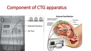
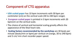
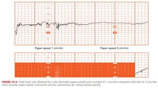
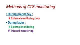
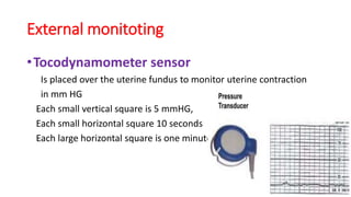
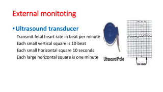
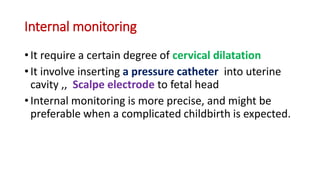
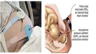
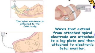
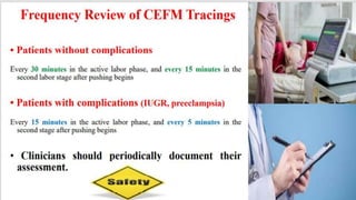
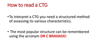
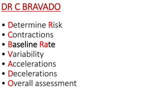
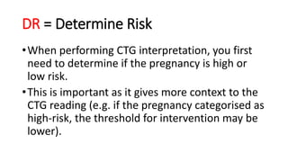
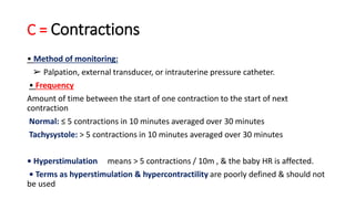
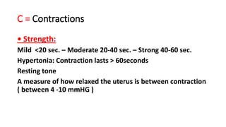
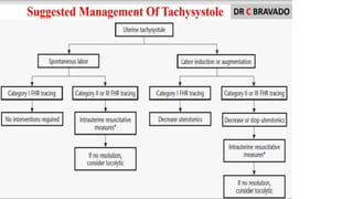
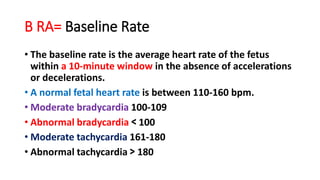
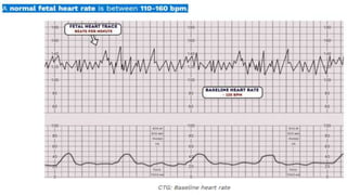
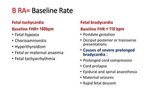
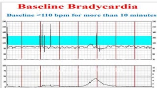
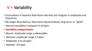
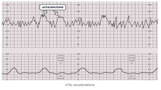
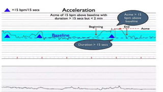
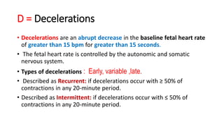
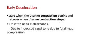
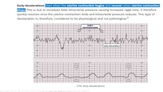
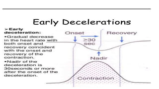
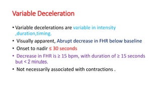
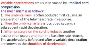
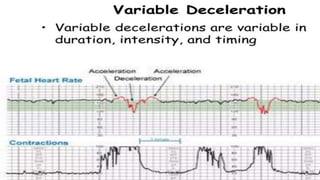
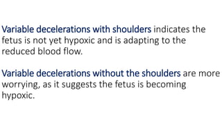
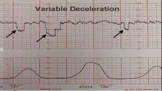
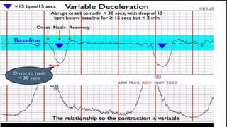
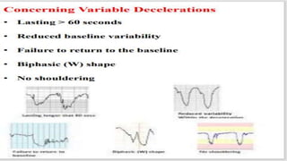
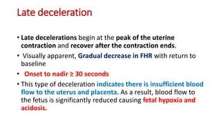
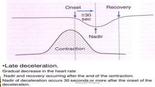
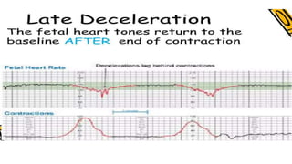
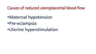
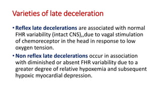
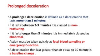
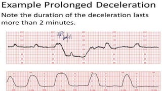
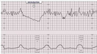
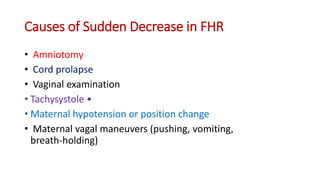
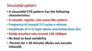
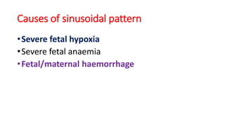
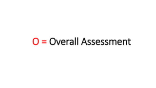
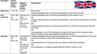
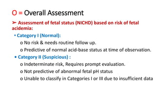
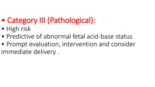
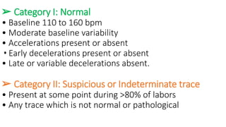
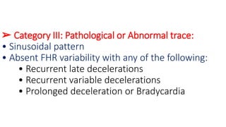
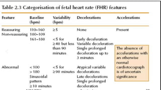
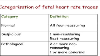
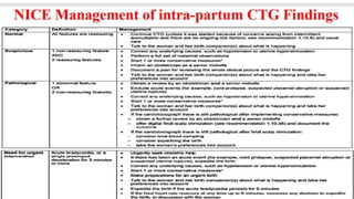
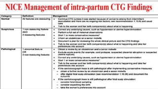
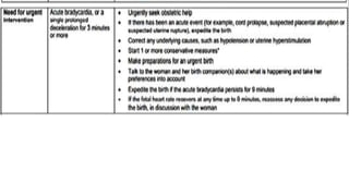
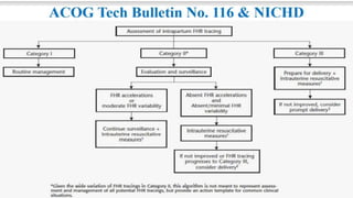
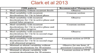
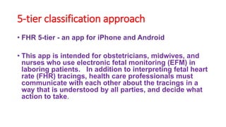
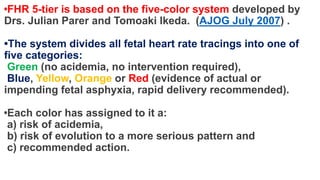
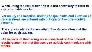
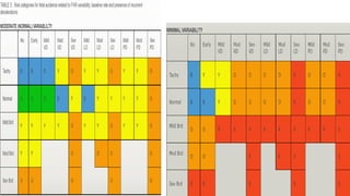
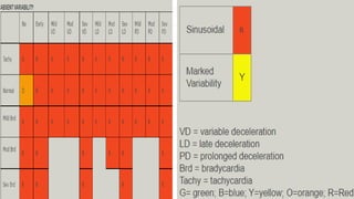
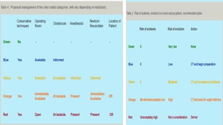
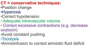
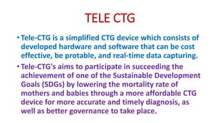
How to read a CTG
(CARDIOTOCOGRAPHY)
D / SALAH ELDEEN REZK
MD, COSULTANT OF OBS&GYN
HEAD OF OBS&GYN ,,SHERBEEN
HOSPITAL
CARDIOTOCOGRAPHY ( DEFINITION)
•Is defined as the graphic recording of fetal
heart rate and uterine contractions by the
use of electronic devices indicated for the
assessment of fetal condition.
•It is also refered to as electronic fetal
monitoring ( EFM )
Component of CTG apparatus
• ultrasonic transducer/probe.
Capable of measuring FHR in the 50-240 beats per minute (bpm), Can
monitor twins (i.e., has two transducers).
• pressure sensitive transducer.
Capable of measuring relative uterine contractions (UCs) in the range
of 0-100 units .
Component of CTG apparatus
• Fetal movement counter.
• PRINTER/RECORDER .
Integrated thermal printer with automatic and manual
print-out modes.
Prints FHR1, FHR2, UCs, marked events and parameters
and relevant alarms.
Print speeds include 1, 2 and 3 cm/min.
Component of CTG apparatus
• Paper and Paper tray:
Paper is heat sensitive with two distinct sections or
channels. The top is referred to as the FHR channel and
the bottom is the UA (uterine activity) channel. Some
monitors also record fetal movement in the UA
channel. The FHR channel may be printed in 5 or 10
beats per minute (bpm) increments on the vertical
scale.
Component of CTG apparatus
• USA scaled paper has 10 bpm increments with 30 bpm per
centimeter (cm) on the vertical scale (30 to 240 bpm range).
• European scaled paper is printed in 5 bpm increments with 20
bpm/cm on the vertical scale.
• The choice of vertical and horizontal scaling greatly affects the
appearance of the fetal heart rate.
• Scaling factors recommended by the workshop are 30 beats per
minute (beats/min or bpm) per vertical cm (range, 30 to 240 bpm)
and 3 cm/ min chart recorder paper speed.
Methods of CTG monitoring
• During pregnancy :
# External monitoring only
• During labor :
# External monitoring
# Internal monitoring
External monitoting
•Tocodynamometer sensor
Is placed over the uterine fundus to monitor uterine contraction
in mm HG
Each small vertical square is 5 mmHG,
Each small horizontal square 10 seconds
Each large horizontal square is one minute
External monitoting
•Ultrasound transducer
Transmit fetal heart rate in beat per minute
Each small vertical square is 10 beat
Each small horizontal square 10 seconds
Each large horizontal square is one minute
Internal monitoring
• It require a certain degree of cervical dilatation
• It involve inserting a pressure catheter into uterine
cavity ,, Scalpe electrode to fetal head
• Internal monitoring is more precise, and might be
preferable when a complicated childbirth is expected.
Preparation for CTG
• Explain the indication of the test to the patient .
• Empty bladder,,supine position.
• Uncover the abdomen.
• Place the tocosensor to the fundus of the uterus, and US
transduser to the site of fetal heart and fixation of them with
abdominal belt.
• Assure the recording of FHS and uterine contraction
• Report the patient to push the bottom when she feel any
movement
High risk indications of fetal monitoring
• Maternal medical illness :
Gestational diabetes,, Hypertension ,,Asthma
• Obstetric complications :
Multiple gestation,,post date gestation,,previous cesarean
section,,intrauterine growth restriction,,oligohydramnios ,,
premature rupture of membranes,, congenital
malformations,,third trimester bleeding,, preeclampsia ,,
meconium stained liquor,, oxytocin induction and
augmentation of labor ,,drug abuse ,,absence of prenatal
care..
Continuous electronic fetal monitoring
( CEFM )
• Introduced in the 1960’s
• Goal to improve neonatal outcomes has been unfulfilled despite
widespread use .
• Strengths:
1- High sensitivity 2-Category I (Normal) tracing predictive of good
outcome
• Limitations:
1- Low specificity 2- Category III (Pathological) tracing not
predictive of poor outcome
Outcomes with CEFM
• ➢ Evidence from 13 RCTs randomized with over 37,000 women:
• ➢ CEFM versus SIA:
• No difference in 1minute Apgar scores < 7 or cord Ph.
• No difference in NICU admission rates or CP rates.
• Increase in cesarean deliveries, especially in low‐risk pregnancies
(NNH = 20).
• Increase in operative vaginal deliveries (NNH = 30).
• Decrease in neonatal seizures (NNT = 661).
How to read a CTG
•To interpret a CTG you need a structured method
of assessing its various characteristics.
• The most popular structure can be remembered
using the acronym DR C BRAVADO:
DR C BRAVADO
• Determine Risk
• Contractions
• Baseline Rate
• Variability
• Accelerations
• Decelerations
• Overall assessment
DR = Determine Risk
•When performing CTG interpretation, you first
need to determine if the pregnancy is high or
low risk.
•This is important as it gives more context to the
CTG reading (e.g. if the pregnancy categorised as
high-risk, the threshold for intervention may be
lower).
C = Contractions
• Method of monitoring:
➢ Palpation, external transducer, or intrauterine pressure catheter.
• Frequency
Amount of time between the start of one contraction to the start of next
contraction
Normal: ≤ 5 contractions in 10 minutes averaged over 30 minutes
Tachysystole: > 5 contractions in 10 minutes averaged over 30 minutes
• Hyperstimulation means > 5 contractions / 10m , & the baby HR is affected.
• Terms as hyperstimulation & hypercontractility are poorly defined & should not
be used
C = Contractions
• Strength:
Mild <20 sec. – Moderate 20-40 sec. – Strong 40-60 sec.
Hypertonia: Contraction lasts > 60seconds
Resting tone
A measure of how relaxed the uterus is between contraction
( between 4 -10 mmHG )
B RA= Baseline Rate
• The baseline rate is the average heart rate of the fetus
within a 10-minute window in the absence of accelerations
or decelerations.
• A normal fetal heart rate is between 110-160 bpm.
• Moderate bradycardia 100-109
• Abnormal bradycardia < 100
• Moderate tachycardia 161-180
• Abnormal tachycardia > 180
B RA= Baseline Rate
Fetal tachycardia
Baseline FHR> 160bpm
• Fetal hypoxia
• Chorioamnionitis
• Hyperthyroidism
• Fetal or maternal anaemia
• Fetal tachyarrhythmia
Fetal bradycardia
Baseline FHR < 110 bpm
• Postdate gestation
• Occiput posterior or transverse
presentations
• Causes of severe prolonged
bradycardia :
• Prolonged cord compression
• Cord prolapse
• Epidural and spinal anaesthesia
• Maternal seizures
• Rapid fetal descent
V = Variability
• Fluctuations in baseline fetal heart rate that are irregular in amplitude and
frequency.
• No longer described as, Short‐term (beat‐to‐beat), long‐term, or “good” .
• Normal variability is between 5-25 bpm.
• Variability categorization.
• Absent: amplitude range undetectable.
• Minimal: amplitude range ≤ 5 bpm.
• Moderate: 6 to 25 bpm .
• Marked: >25 bpm
Causes of Decreased Variability
Maternal causes
• Fever
• Drugs: opiates, benzodiazepines,
methyldopa and magnesium
sulphate.
• General anethesia,,Alcohol.
• Corticosteroids.
• Parasympatholytics
Fetal causes
• Fetal sleeping: this should last no
longer than 40 minutes (this is the
most common cause).
• Fetal acidosis (due to hypoxia) The
lack of oxygen depress the fetal heart
and nervous system
• Prematurity. The fetal nervous
system in a premature baby cannot
effectively control the heart rate.
• Cardiac and CNS anomalies
A = Accelerations
• Accelerations are an abrupt increase in the baseline fetal
heart rate of greater than 15 bpm for greater than 15
seconds.
• Prolonged acceleration ≥ 2 min., but < 10 min.
• Before 32 wks accelerations peek ≥ 10 bpm and a duration ≥
10 seconds .
• The presence of accelerations is reassuring.
• Accelerations occurring alongside uterine contractions is a
sign of a healthy fetus.
D = Decelerations
• Decelerations are an abrupt decrease in the baseline fetal heart rate
of greater than 15 bpm for greater than 15 seconds.
• The fetal heart rate is controlled by the autonomic and somatic
nervous system.
• Types of decelerations : Early, variable ,late.
• Described as Recurrent: if decelerations occur with ≥ 50% of
contractions in any 20‐minute period.
• Described as Intermittent: if decelerations occur with ≤ 50% of
contractions in any 20‐minute period.
Early Deceleration
• start when the uterine contraction begins and
recover when uterine contraction stops.
• Onset to nadir ≥ 30 seconds.
Due to increased vagal tone due to fetal head
compression
Variable Deceleration
• Variable decelerations are variable in intensity
,duration,timing.
• Visually apparent, Abrupt decrease in FHR below baseline
• Onset to nadir ≤ 30 seconds
• Decrease in FHR is ≥ 15 bpm, with duration of ≥ 15 seconds
but < 2 minutes.
• Not necessarily associated with contractions .
Variable decelerations are usually caused by umbilical cord
compression.
The mechanism is as follows:
1. The umbilical vein is often occluded first causing an
acceleration of the fetal heart rate in response.
2. Then the umbilical artery is occluded causing a
subsequent rapid deceleration.
3. When pressure on the cord is reduced another
acceleration occurs and then the baseline rate returns.
The accelerations before and after a variable deceleration
are known as the shoulders of deceleration.
Variable decelerations with shoulders indicates the
fetus is not yet hypoxic and is adapting to the
reduced blood flow.
Variable decelerations without the shoulders are more
worrying, as it suggests the fetus is becoming
hypoxic.
Late deceleration
• Late decelerations begin at the peak of the uterine
contraction and recover after the contraction ends.
• Visually apparent, Gradual decrease in FHR with return to
baseline
• Onset to nadir ≥ 30 seconds
• This type of deceleration indicates there is insufficient blood
flow to the uterus and placenta. As a result, blood flow to
the fetus is significantly reduced causing fetal hypoxia and
acidosis.
Causes of reduced uteroplacental blood flow
•Maternal hypotension
•Pre-eclampsia
•Uterine hyperstimulation
Varieties of late deceleration
• Reflex late decelerations are associated with normal
FHR variability (intact CNS),,due to vagal stimulation
of chemoreceptor in the head in response to low
oxygen tension.
• Non reflex late decelerations occur in association
with diminished or absent FHR variability due to a
greater degree of relative hypoxemia and subsequent
hypoxic myocardial depression.
Prolonged deceleration
• A prolonged deceleration is defined as a deceleration that
lasts more than 2 minutes:
• If it lasts between 2-3 minutes it is classed as non-
reassuring.
• If it lasts longer than 3 minutes it is immediately classed as
abnormal.
• Action must be taken quickly as fetal blood sampling or
emergency C-section.
• A deceleration that last greater than or equal to 10 minute is
a baseline change .
Causes of Sudden Decrease in FHR
• Amniotomy
• Cord prolapse
• Vaginal examination
• Tachysystole •
• Maternal hypotension or position change
• Maternal vagal maneuvers (pushing, vomiting,
breath‐holding)
Sinusoidal pattern
• A sinusoidal CTG pattern has the following
characteristics:
• A smooth, regular, sine wave-like pattern
• Frequency of around 2-5 cycles a minute.
• Amplitude of 5-15 bpm above and below base line
• Stable baseline rate around 120-160bpm
• No beat to beat variability
• Persists for ≥ 20 minutes (Rules out narcotic
induced).
Causes of sinusoidal pattern
•Severe fetal hypoxia
•Severe fetal anaemia
•Fetal/maternal haemorrhage
O = Overall Assessment
➢ Assessment of fetal status (NICHD) based on risk of fetal
acidemia:
• Category I (Normal):
o No risk & needs routine follow up.
o Predictive of normal acid‐base status at time of observation.
• Category II (Suspicious) :
o Indeterminate risk, Requires prompt evaluation.
o Not predictive of abnormal fetal pH status
o Unable to classify in Categories I or III due to insufficient data
• Category III (Pathological):
• High risk
• Predictive of abnormal fetal acid‐base status
• Prompt evaluation, intervention and consider
immediate delivery .
➢ Category I: Normal
• Baseline 110 to 160 bpm
• Moderate baseline variability
• Accelerations present or absent
• Early decelerations present or absent
• Late or variable decelerations absent.
➢ Category II: Suspicious or Indeterminate trace
• Present at some point during >80% of labors
• Any trace which is not normal or pathological
➢ Category III: Pathological or Abnormal trace:
• Sinusoidal pattern
• Absent FHR variability with any of the following:
• Recurrent late decelerations
• Recurrent variable decelerations
• Prolonged deceleration or Bradycardia
Recent proposals for Category II
management:
•Clark algorithm with specific definitions
http://cat2.perigen.com/cat2
•5‐tier classification approach, rather than a
3‐tiered approach
5‐tier classification approach
• FHR 5-tier - an app for iPhone and Android
• This app is intended for obstetricians, midwives, and
nurses who use electronic fetal monitoring (EFM) in
laboring patients. In addition to interpreting fetal heart
rate (FHR) tracings, health care professionals must
communicate with each other about the tracings in a
way that is understood by all parties, and decide what
action to take.
•FHR 5-tier is based on the five-color system developed by
Drs. Julian Parer and Tomoaki Ikeda. (AJOG July 2007) .
•The system divides all fetal heart rate tracings into one of
five categories:
Green (no acidemia, no intervention required),
Blue, Yellow, Orange or Red (evidence of actual or
impending fetal asphyxia, rapid delivery recommended).
•Each color has assigned to it a:
a) risk of acidemia,
b) risk of evolution to a more serious pattern and
c) recommended action.
•When using the FHR 5-tier app it is not necessary to refer to
any other table or chart.
•Variability and baseline, and the shape, nadir, and duration of
decelerations are entered with buttons on the consecutive
screens.
•The app calculates the severity of the deceleration and the
color for each tracing.
• All aspects of the tracing are summarized on the colored
results screen, so that the user can quickly communicate with
others.
CT = conservative techniques:
•Position change
•Hyperoxia
•Correct hypotension
• Adequate intravascular volume
• Correct excessive contractions (e.g. decrease
oxytocin)
•Avoid constant pushing
•Tocolysis
•Amnioinfusion to correct amniotic fluid deficit
TELE CTG
• Tele-CTG is a simplified CTG device which consists of
developed hardware and software that can be cost
effective, be protable, and real-time data capturing.
• Tele-CTG's aims to participate in succeeding the
achievement of one of the Sustainable Development
Goals (SDGs) by lowering the mortality rate of
mothers and babies through a more affordable CTG
device for more accurate and timely diagnosis, as
well as better governance to take place.
My great thanks

How to read a CTG دكتور صلاح رزق.pptx

  • 1.
    How to reada CTG (CARDIOTOCOGRAPHY) D / SALAH ELDEEN REZK MD, COSULTANT OF OBS&GYN HEAD OF OBS&GYN ,,SHERBEEN HOSPITAL
  • 2.
    CARDIOTOCOGRAPHY ( DEFINITION) •Isdefined as the graphic recording of fetal heart rate and uterine contractions by the use of electronic devices indicated for the assessment of fetal condition. •It is also refered to as electronic fetal monitoring ( EFM )
  • 3.
  • 4.
    Component of CTGapparatus • ultrasonic transducer/probe. Capable of measuring FHR in the 50-240 beats per minute (bpm), Can monitor twins (i.e., has two transducers). • pressure sensitive transducer. Capable of measuring relative uterine contractions (UCs) in the range of 0-100 units .
  • 5.
    Component of CTGapparatus • Fetal movement counter. • PRINTER/RECORDER . Integrated thermal printer with automatic and manual print-out modes. Prints FHR1, FHR2, UCs, marked events and parameters and relevant alarms. Print speeds include 1, 2 and 3 cm/min.
  • 6.
    Component of CTGapparatus • Paper and Paper tray: Paper is heat sensitive with two distinct sections or channels. The top is referred to as the FHR channel and the bottom is the UA (uterine activity) channel. Some monitors also record fetal movement in the UA channel. The FHR channel may be printed in 5 or 10 beats per minute (bpm) increments on the vertical scale.
  • 7.
    Component of CTGapparatus • USA scaled paper has 10 bpm increments with 30 bpm per centimeter (cm) on the vertical scale (30 to 240 bpm range). • European scaled paper is printed in 5 bpm increments with 20 bpm/cm on the vertical scale. • The choice of vertical and horizontal scaling greatly affects the appearance of the fetal heart rate. • Scaling factors recommended by the workshop are 30 beats per minute (beats/min or bpm) per vertical cm (range, 30 to 240 bpm) and 3 cm/ min chart recorder paper speed.
  • 10.
    Methods of CTGmonitoring • During pregnancy : # External monitoring only • During labor : # External monitoring # Internal monitoring
  • 11.
    External monitoting •Tocodynamometer sensor Isplaced over the uterine fundus to monitor uterine contraction in mm HG Each small vertical square is 5 mmHG, Each small horizontal square 10 seconds Each large horizontal square is one minute
  • 12.
    External monitoting •Ultrasound transducer Transmitfetal heart rate in beat per minute Each small vertical square is 10 beat Each small horizontal square 10 seconds Each large horizontal square is one minute
  • 13.
    Internal monitoring • Itrequire a certain degree of cervical dilatation • It involve inserting a pressure catheter into uterine cavity ,, Scalpe electrode to fetal head • Internal monitoring is more precise, and might be preferable when a complicated childbirth is expected.
  • 16.
    Preparation for CTG •Explain the indication of the test to the patient . • Empty bladder,,supine position. • Uncover the abdomen. • Place the tocosensor to the fundus of the uterus, and US transduser to the site of fetal heart and fixation of them with abdominal belt. • Assure the recording of FHS and uterine contraction • Report the patient to push the bottom when she feel any movement
  • 17.
    High risk indicationsof fetal monitoring • Maternal medical illness : Gestational diabetes,, Hypertension ,,Asthma • Obstetric complications : Multiple gestation,,post date gestation,,previous cesarean section,,intrauterine growth restriction,,oligohydramnios ,, premature rupture of membranes,, congenital malformations,,third trimester bleeding,, preeclampsia ,, meconium stained liquor,, oxytocin induction and augmentation of labor ,,drug abuse ,,absence of prenatal care..
  • 18.
    Continuous electronic fetalmonitoring ( CEFM ) • Introduced in the 1960’s • Goal to improve neonatal outcomes has been unfulfilled despite widespread use . • Strengths: 1- High sensitivity 2-Category I (Normal) tracing predictive of good outcome • Limitations: 1- Low specificity 2- Category III (Pathological) tracing not predictive of poor outcome
  • 19.
    Outcomes with CEFM •➢ Evidence from 13 RCTs randomized with over 37,000 women: • ➢ CEFM versus SIA: • No difference in 1minute Apgar scores < 7 or cord Ph. • No difference in NICU admission rates or CP rates. • Increase in cesarean deliveries, especially in low‐risk pregnancies (NNH = 20). • Increase in operative vaginal deliveries (NNH = 30). • Decrease in neonatal seizures (NNT = 661).
  • 22.
    How to reada CTG •To interpret a CTG you need a structured method of assessing its various characteristics. • The most popular structure can be remembered using the acronym DR C BRAVADO:
  • 23.
    DR C BRAVADO •Determine Risk • Contractions • Baseline Rate • Variability • Accelerations • Decelerations • Overall assessment
  • 24.
    DR = DetermineRisk •When performing CTG interpretation, you first need to determine if the pregnancy is high or low risk. •This is important as it gives more context to the CTG reading (e.g. if the pregnancy categorised as high-risk, the threshold for intervention may be lower).
  • 25.
    C = Contractions •Method of monitoring: ➢ Palpation, external transducer, or intrauterine pressure catheter. • Frequency Amount of time between the start of one contraction to the start of next contraction Normal: ≤ 5 contractions in 10 minutes averaged over 30 minutes Tachysystole: > 5 contractions in 10 minutes averaged over 30 minutes • Hyperstimulation means > 5 contractions / 10m , & the baby HR is affected. • Terms as hyperstimulation & hypercontractility are poorly defined & should not be used
  • 26.
    C = Contractions •Strength: Mild <20 sec. – Moderate 20-40 sec. – Strong 40-60 sec. Hypertonia: Contraction lasts > 60seconds Resting tone A measure of how relaxed the uterus is between contraction ( between 4 -10 mmHG )
  • 28.
    B RA= BaselineRate • The baseline rate is the average heart rate of the fetus within a 10-minute window in the absence of accelerations or decelerations. • A normal fetal heart rate is between 110-160 bpm. • Moderate bradycardia 100-109 • Abnormal bradycardia < 100 • Moderate tachycardia 161-180 • Abnormal tachycardia > 180
  • 30.
    B RA= BaselineRate Fetal tachycardia Baseline FHR> 160bpm • Fetal hypoxia • Chorioamnionitis • Hyperthyroidism • Fetal or maternal anaemia • Fetal tachyarrhythmia Fetal bradycardia Baseline FHR < 110 bpm • Postdate gestation • Occiput posterior or transverse presentations • Causes of severe prolonged bradycardia : • Prolonged cord compression • Cord prolapse • Epidural and spinal anaesthesia • Maternal seizures • Rapid fetal descent
  • 33.
    V = Variability •Fluctuations in baseline fetal heart rate that are irregular in amplitude and frequency. • No longer described as, Short‐term (beat‐to‐beat), long‐term, or “good” . • Normal variability is between 5-25 bpm. • Variability categorization. • Absent: amplitude range undetectable. • Minimal: amplitude range ≤ 5 bpm. • Moderate: 6 to 25 bpm . • Marked: >25 bpm
  • 34.
    Causes of DecreasedVariability Maternal causes • Fever • Drugs: opiates, benzodiazepines, methyldopa and magnesium sulphate. • General anethesia,,Alcohol. • Corticosteroids. • Parasympatholytics Fetal causes • Fetal sleeping: this should last no longer than 40 minutes (this is the most common cause). • Fetal acidosis (due to hypoxia) The lack of oxygen depress the fetal heart and nervous system • Prematurity. The fetal nervous system in a premature baby cannot effectively control the heart rate. • Cardiac and CNS anomalies
  • 39.
    A = Accelerations •Accelerations are an abrupt increase in the baseline fetal heart rate of greater than 15 bpm for greater than 15 seconds. • Prolonged acceleration ≥ 2 min., but < 10 min. • Before 32 wks accelerations peek ≥ 10 bpm and a duration ≥ 10 seconds . • The presence of accelerations is reassuring. • Accelerations occurring alongside uterine contractions is a sign of a healthy fetus.
  • 42.
    D = Decelerations •Decelerations are an abrupt decrease in the baseline fetal heart rate of greater than 15 bpm for greater than 15 seconds. • The fetal heart rate is controlled by the autonomic and somatic nervous system. • Types of decelerations : Early, variable ,late. • Described as Recurrent: if decelerations occur with ≥ 50% of contractions in any 20‐minute period. • Described as Intermittent: if decelerations occur with ≤ 50% of contractions in any 20‐minute period.
  • 43.
    Early Deceleration • startwhen the uterine contraction begins and recover when uterine contraction stops. • Onset to nadir ≥ 30 seconds. Due to increased vagal tone due to fetal head compression
  • 46.
    Variable Deceleration • Variabledecelerations are variable in intensity ,duration,timing. • Visually apparent, Abrupt decrease in FHR below baseline • Onset to nadir ≤ 30 seconds • Decrease in FHR is ≥ 15 bpm, with duration of ≥ 15 seconds but < 2 minutes. • Not necessarily associated with contractions .
  • 47.
    Variable decelerations areusually caused by umbilical cord compression. The mechanism is as follows: 1. The umbilical vein is often occluded first causing an acceleration of the fetal heart rate in response. 2. Then the umbilical artery is occluded causing a subsequent rapid deceleration. 3. When pressure on the cord is reduced another acceleration occurs and then the baseline rate returns. The accelerations before and after a variable deceleration are known as the shoulders of deceleration.
  • 49.
    Variable decelerations withshoulders indicates the fetus is not yet hypoxic and is adapting to the reduced blood flow. Variable decelerations without the shoulders are more worrying, as it suggests the fetus is becoming hypoxic.
  • 53.
    Late deceleration • Latedecelerations begin at the peak of the uterine contraction and recover after the contraction ends. • Visually apparent, Gradual decrease in FHR with return to baseline • Onset to nadir ≥ 30 seconds • This type of deceleration indicates there is insufficient blood flow to the uterus and placenta. As a result, blood flow to the fetus is significantly reduced causing fetal hypoxia and acidosis.
  • 56.
    Causes of reduceduteroplacental blood flow •Maternal hypotension •Pre-eclampsia •Uterine hyperstimulation
  • 57.
    Varieties of latedeceleration • Reflex late decelerations are associated with normal FHR variability (intact CNS),,due to vagal stimulation of chemoreceptor in the head in response to low oxygen tension. • Non reflex late decelerations occur in association with diminished or absent FHR variability due to a greater degree of relative hypoxemia and subsequent hypoxic myocardial depression.
  • 58.
    Prolonged deceleration • Aprolonged deceleration is defined as a deceleration that lasts more than 2 minutes: • If it lasts between 2-3 minutes it is classed as non- reassuring. • If it lasts longer than 3 minutes it is immediately classed as abnormal. • Action must be taken quickly as fetal blood sampling or emergency C-section. • A deceleration that last greater than or equal to 10 minute is a baseline change .
  • 61.
    Causes of SuddenDecrease in FHR • Amniotomy • Cord prolapse • Vaginal examination • Tachysystole • • Maternal hypotension or position change • Maternal vagal maneuvers (pushing, vomiting, breath‐holding)
  • 62.
    Sinusoidal pattern • Asinusoidal CTG pattern has the following characteristics: • A smooth, regular, sine wave-like pattern • Frequency of around 2-5 cycles a minute. • Amplitude of 5-15 bpm above and below base line • Stable baseline rate around 120-160bpm • No beat to beat variability • Persists for ≥ 20 minutes (Rules out narcotic induced).
  • 63.
    Causes of sinusoidalpattern •Severe fetal hypoxia •Severe fetal anaemia •Fetal/maternal haemorrhage
  • 66.
    O = OverallAssessment
  • 68.
    O = OverallAssessment ➢ Assessment of fetal status (NICHD) based on risk of fetal acidemia: • Category I (Normal): o No risk & needs routine follow up. o Predictive of normal acid‐base status at time of observation. • Category II (Suspicious) : o Indeterminate risk, Requires prompt evaluation. o Not predictive of abnormal fetal pH status o Unable to classify in Categories I or III due to insufficient data
  • 69.
    • Category III(Pathological): • High risk • Predictive of abnormal fetal acid‐base status • Prompt evaluation, intervention and consider immediate delivery .
  • 70.
    ➢ Category I:Normal • Baseline 110 to 160 bpm • Moderate baseline variability • Accelerations present or absent • Early decelerations present or absent • Late or variable decelerations absent. ➢ Category II: Suspicious or Indeterminate trace • Present at some point during >80% of labors • Any trace which is not normal or pathological
  • 71.
    ➢ Category III:Pathological or Abnormal trace: • Sinusoidal pattern • Absent FHR variability with any of the following: • Recurrent late decelerations • Recurrent variable decelerations • Prolonged deceleration or Bradycardia
  • 78.
    Recent proposals forCategory II management: •Clark algorithm with specific definitions http://cat2.perigen.com/cat2 •5‐tier classification approach, rather than a 3‐tiered approach
  • 81.
    5‐tier classification approach •FHR 5-tier - an app for iPhone and Android • This app is intended for obstetricians, midwives, and nurses who use electronic fetal monitoring (EFM) in laboring patients. In addition to interpreting fetal heart rate (FHR) tracings, health care professionals must communicate with each other about the tracings in a way that is understood by all parties, and decide what action to take.
  • 82.
    •FHR 5-tier isbased on the five-color system developed by Drs. Julian Parer and Tomoaki Ikeda. (AJOG July 2007) . •The system divides all fetal heart rate tracings into one of five categories: Green (no acidemia, no intervention required), Blue, Yellow, Orange or Red (evidence of actual or impending fetal asphyxia, rapid delivery recommended). •Each color has assigned to it a: a) risk of acidemia, b) risk of evolution to a more serious pattern and c) recommended action.
  • 83.
    •When using theFHR 5-tier app it is not necessary to refer to any other table or chart. •Variability and baseline, and the shape, nadir, and duration of decelerations are entered with buttons on the consecutive screens. •The app calculates the severity of the deceleration and the color for each tracing. • All aspects of the tracing are summarized on the colored results screen, so that the user can quickly communicate with others.
  • 87.
    CT = conservativetechniques: •Position change •Hyperoxia •Correct hypotension • Adequate intravascular volume • Correct excessive contractions (e.g. decrease oxytocin) •Avoid constant pushing •Tocolysis •Amnioinfusion to correct amniotic fluid deficit
  • 88.
    TELE CTG • Tele-CTGis a simplified CTG device which consists of developed hardware and software that can be cost effective, be protable, and real-time data capturing. • Tele-CTG's aims to participate in succeeding the achievement of one of the Sustainable Development Goals (SDGs) by lowering the mortality rate of mothers and babies through a more affordable CTG device for more accurate and timely diagnosis, as well as better governance to take place.
  • 90.