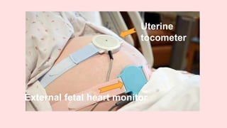
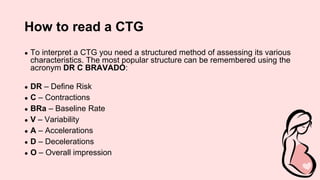
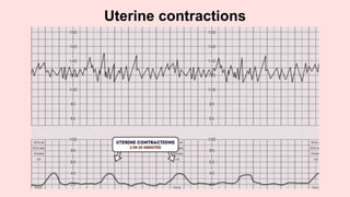
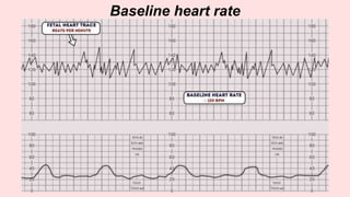
Cardiotocography
(CTG)
Anjala Nizam
&
Sarah Bouka
Contents
1.Cardiotocography
2.How CTG Works
3.How to read CTG
4.Define Risk
5.Contractions
6.Baseline Rate
7.Variability
8.Accelerations
9.Decelerations
10.Overall impression
11.References
Cardiotocography
● Cardiotocography(CTG) or electronic fetal monitoring (EFM) is the most
widely used technique for assessing fetal wellbeing in labour in the
developed world.
● CTGs have a high degree of sensitivity but a low level of specificity which
means that they are very good at telling us which fetuses are well but are
poor at identifying which fetuses are unwell.
● It is most commonly used in the third trimester.
● An abnormal CTG may indicate the need for further investigations and
potential intervention.
Uterine
tocometer
External fetal heart monitor
How CTG works
● The device used in cardiotocography is known as a cardiotocograph.
● It involves the placement of two transducers onto the abdomen of a
pregnant woman.
● One transducer records the fetal heart rate using ultrasound.
● The other transducer monitors the contractions of the uterus.
● It does this by measuring the tension of the maternal abdominal wall.
● This provides an indirect indication of intrauterine pressure.
Remember:
1. Confirm that the CTG belongs to the correct patient (patient’s name,
medical record number/ MRN, and date of birth).
2. Check the date and time.
3. Check the gestational age/ patient history.
4. Comment on the fetal heart 4 features, each feature will be either
reassuring, non-reassuring, or abnormal.
5. Accordingly decide if CTG is normal, suspicious, or pathological .
6. If any deceleration is there decide:
The type of the deceleration
Recurrence of the deceleration
Duration
7. Check the frequency and regularity of uterine contractions, and if any
abnormal uterine contraction patterns.
How to read a CTG
● To interpret a CTG you need a structured method of assessing its various
characteristics. The most popular structure can be remembered using the
acronym DR C BRAVADO:
● DR – Define Risk
● C – Contractions
● BRa – Baseline Rate
● V – Variability
● A – Accelerations
● D – Decelerations
● O – Overall impression
Define risk
● You first need to assess if the pregnancy is high or low risk.
● This is important as it gives more context to the CTG reading
● Some reasons a pregnancy may be considered high risk:
○ Maternal medical illness
○ Obstetric complications
○ Other risk factors
Maternal medical illness
● Gestational diabetes
● Hypertension
● Asthma
Obstetric complications
● Multiple gestation
● Post-date gestation
● Previous cesarean section
● Intrauterine growth restriction
● Premature rupture of membranes
● Congenital malformations
● Oxytocin induction/augmentation of labour
● Pre-eclampsia
Other risk factors
● Absence of prenatal care
● Smoking
● Drug abuse
Contractions
● Record the number of contractions present in a 10 minute period.
● Individual contractions are seen as peaks on the part of the CTG
monitoring uterine activity.
Assess contractions for the following:
● Duration – How long do the contractions last?
● Intensity – How strong are the contractions (assessed using palpation)?
Uterine contractions
Uterine Tachysystole, Uterine hypertonus,
and Hyperstimulation
• Uterine Tachysystole: More than five uterine contractions in 10
minutes without FHR abnormalities.
• Uterine hypertonus: Contractions lasting >2 minutes in duration
or contractions occurring within 60 seconds of each other, without
FHR abnormalities.
• Hyperstimulation: Tachysystole or uterine hypertonus in the
presence of FHR abnormalities
Baseline rate of the fetal heart
● The baseline rate is the average heart rate of the fetus within a 10-
minute window.
● Ignore any accelerations or decelerations.
● A normal fetal heart rate is between 110-160 bpm.
Baseline heart rate
Fetal tachycardia
Baseline heart rate greater than 160 bpm.
Causes of fetal tachycardia include:
● Fetal hypoxia
● Chorioamnionitis
● Hyperthyroidism
● Fetal or maternal anaemia
● Fetal tachyarrhythmia
Fetal bradycardia
● Baseline heart rate of less than 100 bpm.
● It is common to have a baseline heart rate of between 100-120 bpm in
the following situations:
○ Postdate gestation
○ Occiput posterior or transverse presentations
● Severe prolonged bradycardia (less than 80 bpm for more than 3
minutes) indicates severe hypoxia.
Causes of prolonged severe bradycardia
include
● Prolonged cord compression
● Cord prolapse
● Epidural and spinal anaesthesia
● Maternal seizures
● Rapid fetal descent
Variability
● Baseline variability refers to the variation of fetal heart rate from one beat
to the next.
● Variability occurs as a result of the interaction between
the nervous system, chemoreceptors, baroreceptors and cardiac
responsiveness.
● It is, therefore, a good indicator of how healthy a fetus is at that particular
moment in time.
● Normal variability indicates an intact neurological system in the fetus.
Variability can be categorised as follows
● Reassuring (normal): 5 – 25 bpm
● Non-reassuring:
○ less than 5 bpm for between 30-50 minutes
○ more than 25 bpm for 15-25 minutes
● Abnormal:
○ less than 5 bpm for more than 50 minutes
○ more than 25 bpm for more than 25 minutes
○ sinusoidal
Variability
Causes of reduced variability:
● Fetal sleeping- most common cause
● Fetal acidosis- due to hypoxia
● Fetal tachycardia
● Drugs– opiates, benzodiazepines, methyldopa, magnesium sulphate
● Prematurity– <28 weeks
● Congenital heart abnormalities
Reduced variability
Accelerations
● Accelerations are transient increases in FHR of 15bpm or more above
the baseline and lasting 15 seconds.
● The presence of accelerations is reassuring.
● Accelerations occurring alongside uterine contractions is a sign of a
healthy fetus.
Accelerations
Decelerations
● Decelerations are transient episodes of decrease of FHR below the
baseline of more than 15 bpm lasting at least 15 seconds.
● Uterine activity, even in its mildest form, will result in decelerations in a
fetus whose oxygenation is already compromised.
● The fetal heart rate is controlled by the autonomic and somatic nervous
system. In response to hypoxic stress, the fetus reduces its heart rate to
preserve myocardial oxygenation and perfusion. Unlike an adult, a fetus
cannot increase its respiration depth and rate. This reduction in heart
rate to reduce myocardial demand is referred to as a deceleration.
● Recurrent/ repetitive deceleration:
when they are associated with more than (>) 50% of uterine contractions
Types of decceleration:
● Early deccelerations
● Variable deccelerations
● Late deccelerations
Early decelerations
● Early decelerations start when the uterine contraction
begins and recover when uterine contraction stops. (Mirror images)
●Benign/physiological not pathological.
●They are caused by head compression and in general are a normal
physiological response to a mild increase in intracranial pressure.
Early decelerations
Reassuring
Variable decelerations
● Variable decelerations are observed as a rapid fall in baseline fetal
heart rate with a variable recovery phase.
● Decelerations that are variable in shape, depth, duration and timing with
the contractions.
● Time relationships with contraction cycle may be variable but most
commonly occur simultaneously with contractions.
● They are most often seen during labour and in patients’ with reduced
amniotic fluid volume.
Variable decelerations
Variable decelerations are usually caused by
umbilical cord compression:
● The umbilical vein is often occluded first causing an acceleration in
response.
● Then the umbilical artery is occluded causing a subsequent rapid
deceleration.
● When pressure on the cord is reduced another acceleration occurs and
then the baseline rate returns.
● Accelerations before and after a variable deceleration are known as the
“shoulders of deceleration”.
● Their presence indicates the fetus is not yet hypoxic and is adapting to the
reduced blood flow.
● Variable decelerations can sometimes resolve if the mother changes
position.
● The presence of persistent variable decelerations indicates the need for
close monitoring.
● Variable decelerations without the shoulders are more worrying, as it
suggests the fetus is becoming hypoxic.
1.Reassuring
●(Typical) variable decelerations < 90 minutes
2.Non-reassuring
●(Typical) variable decelerations > 90 minutes
●Non-recurrent (atypical) variable decelerations for 30 minutes or more
●Recurrent (atypical) variable decelerations < 30 minutes
3.Abnormal
●Recurrent (atypical) variable decelerations for 30 minutes (or less if any
maternal or fetal clinical risk factors)
Late decelerations
● Uniform, repetitive decreasing of FHR.
● They start after the start of the contraction and the bottom of the
deceleration is more than 20 seconds after the peak of the contraction.
Importantly, they return to the baseline after the contraction has finished
● This type of deceleration indicates there is insufficient blood flow to the
uterus and placenta.
● As a result, blood flow to the fetus is significantly reduced causing fetal
hypoxia and acidosis (contractions in the presence of hypoxia).
Reduced uteroplacental blood flow can
occur due to:
● Maternal hypotension
● Pre-eclampsia
● Uterine hyperstimulation
Late decelerations
1.Non-reassuring
●Recurrent late decelerations for < 30
minutes (with no maternal or fetal clinical
risk factors such as vaginal bleeding or
significant meconium)
2.Abnormal
●Late decelerations for 30 minutes (or less
if any maternal or fetal clinical risk factors)
Prolonged deceleration
● A prolonged deceleration is defined as a deceleration that lasts more
than 2 minutes:
1.If it lasts between 2-3 minutes it is classed as non-reassuring.
2.If it lasts longer than 3 minutes it is immediately classed as abnormal.
Prolonged deceleration
Sinusoidal pattern
Sinusoidal pattern
● This type of pattern is rare, however, if present it is very concerning.
● It is associated with high rates of fetal morbidity and mortality.
● A sinusoidal CTG pattern has the following characteristics:
1. A smooth, regular, wave-like pattern
2. Frequency of around 2-5 cycles a minute
3. Stable baseline rate around 120-160bpm
4. No beat to beat variability
5. No accelerations
● A sinusoidal pattern usually indicates one or more of the following:
1. Severe fetal hypoxia
2. Severe fetal anaemia
3. Fetal/maternal haemorrhage
Overall impression
● The overall impression can be described as either:
○ Reassuring
○ Non-reassuring/Suspicious
○ Abnormal
● The overall impression is determined by how many of the CTG features
are either reassuring, non-reassuring or abnormal. The NICE guideline
demonstrates how to decide which category a CTG falls into.
CTG in labour
1. Normal= all reassuring
2. Suspicious= one non-reassuring
3. Pathological= 2 non-reassuring OR one abnormal
CTG antenatal (not in labour)
1. Normal= all reassuring
2. Abnormal, any abnormality
Reassuring
Baseline heart rate
● 110 to 160 bpm
Baseline variability
● 5 to 25 bpm
Decelerations
● None or early
● Variable decelerations with no concerning characteristics for less than
90 minutes
Non-reassuring
Baseline heart rate
● Either of the below would be classed as non-reassuring:
1. 100 to 109 bpm
2. 161 to 180 bpm
Baseline variability
● Either of the below would be classed as non-reassuring:
1. Less than 5 for 30 to 50 minutes
2. More than 25 for 15 to 25 minutes
Decelerations
● Any of the below would be classed as non-reassuring:
1. Variable decelerations with no concerning characteristics for 90
minutes or more
2. Variable decelerations with any concerning characteristics in up to
50% of contractions for 30 minutes or more
3. Variable decelerations with any concerning characteristics in over
50% of contractions for less than 30 minutes
4. Late decelerations in over 50% of contractions for less than 30
minutes, with no maternal or fetal clinical risk factors such as vaginal
bleeding or significant meconium
Abnormal
Baseline heart rate
● Either of the below would be classed as abnormal:
1. Below 100bpm
2. Above 180 bpm
Baseline variability
● Any of the below would be classed as abnormal:
1. Less than 5 for more than 50 minutes
2. More than 25 for more than 25 minutes
3. Sinusoidal
Decelerations
● Any of the below would be classed as abnormal:
1. Variable decelerations with any concerning characteristics in over
50% of contractions for 30 minutes (or less if any maternal or fetal
clinical risk factors)
2. Late decelerations for 30 minutes (or less if any maternal or fetal
clinical risk factors)
3. Acute bradycardia, or a single prolonged deceleration lasting 3
minutes or more
Concerning characteristics of variable
decelerations
● Lasting more than 60 seconds
● Reduced baseline variability within the deceleration
● Failure to return to baseline
● Biphasic (W) shape
● No shouldering
References
1. AMIR SWEHA, M.D. Interpretation of the Electronic Fetal Heart Rate
During Labor. Am Fam Physician. 1999 May 1;59(9):2487-2500.
2. Clinical obstetrics and gynaecology. 2nd Edition. 2009. B.Magowan,
Philip Owen, James Drife
3. Intrapartum care: NICE guideline CG190 (February 2017)
4. 20th Edition Obstetrics by Ten Teachers Edited by Louise C Kenny and
Jenny E Myers. 2017
5. https://geekymedics.com/how-to-read-a-ctg/ Dr. Lewis Potter Reviewed
by Dr Venkatesh Subramanian Obstetrics & Gynaecology Registrar
(ST5) in London
Thank You!

Cardiotocography (CTG).pptx

  • 1.
  • 2.
    Contents 1.Cardiotocography 2.How CTG Works 3.Howto read CTG 4.Define Risk 5.Contractions 6.Baseline Rate 7.Variability 8.Accelerations 9.Decelerations 10.Overall impression 11.References
  • 3.
    Cardiotocography ● Cardiotocography(CTG) orelectronic fetal monitoring (EFM) is the most widely used technique for assessing fetal wellbeing in labour in the developed world. ● CTGs have a high degree of sensitivity but a low level of specificity which means that they are very good at telling us which fetuses are well but are poor at identifying which fetuses are unwell. ● It is most commonly used in the third trimester. ● An abnormal CTG may indicate the need for further investigations and potential intervention.
  • 4.
  • 5.
    How CTG works ●The device used in cardiotocography is known as a cardiotocograph. ● It involves the placement of two transducers onto the abdomen of a pregnant woman. ● One transducer records the fetal heart rate using ultrasound. ● The other transducer monitors the contractions of the uterus. ● It does this by measuring the tension of the maternal abdominal wall. ● This provides an indirect indication of intrauterine pressure.
  • 6.
    Remember: 1. Confirm thatthe CTG belongs to the correct patient (patient’s name, medical record number/ MRN, and date of birth). 2. Check the date and time. 3. Check the gestational age/ patient history. 4. Comment on the fetal heart 4 features, each feature will be either reassuring, non-reassuring, or abnormal.
  • 7.
    5. Accordingly decideif CTG is normal, suspicious, or pathological . 6. If any deceleration is there decide: The type of the deceleration Recurrence of the deceleration Duration 7. Check the frequency and regularity of uterine contractions, and if any abnormal uterine contraction patterns.
  • 8.
    How to reada CTG ● To interpret a CTG you need a structured method of assessing its various characteristics. The most popular structure can be remembered using the acronym DR C BRAVADO: ● DR – Define Risk ● C – Contractions ● BRa – Baseline Rate ● V – Variability ● A – Accelerations ● D – Decelerations ● O – Overall impression
  • 9.
    Define risk ● Youfirst need to assess if the pregnancy is high or low risk. ● This is important as it gives more context to the CTG reading ● Some reasons a pregnancy may be considered high risk: ○ Maternal medical illness ○ Obstetric complications ○ Other risk factors
  • 10.
    Maternal medical illness ●Gestational diabetes ● Hypertension ● Asthma
  • 11.
    Obstetric complications ● Multiplegestation ● Post-date gestation ● Previous cesarean section ● Intrauterine growth restriction ● Premature rupture of membranes ● Congenital malformations ● Oxytocin induction/augmentation of labour ● Pre-eclampsia
  • 12.
    Other risk factors ●Absence of prenatal care ● Smoking ● Drug abuse
  • 13.
    Contractions ● Record thenumber of contractions present in a 10 minute period. ● Individual contractions are seen as peaks on the part of the CTG monitoring uterine activity. Assess contractions for the following: ● Duration – How long do the contractions last? ● Intensity – How strong are the contractions (assessed using palpation)?
  • 14.
  • 15.
    Uterine Tachysystole, Uterinehypertonus, and Hyperstimulation • Uterine Tachysystole: More than five uterine contractions in 10 minutes without FHR abnormalities. • Uterine hypertonus: Contractions lasting >2 minutes in duration or contractions occurring within 60 seconds of each other, without FHR abnormalities. • Hyperstimulation: Tachysystole or uterine hypertonus in the presence of FHR abnormalities
  • 16.
    Baseline rate ofthe fetal heart ● The baseline rate is the average heart rate of the fetus within a 10- minute window. ● Ignore any accelerations or decelerations. ● A normal fetal heart rate is between 110-160 bpm.
  • 17.
  • 18.
    Fetal tachycardia Baseline heartrate greater than 160 bpm. Causes of fetal tachycardia include: ● Fetal hypoxia ● Chorioamnionitis ● Hyperthyroidism ● Fetal or maternal anaemia ● Fetal tachyarrhythmia
  • 19.
    Fetal bradycardia ● Baselineheart rate of less than 100 bpm. ● It is common to have a baseline heart rate of between 100-120 bpm in the following situations: ○ Postdate gestation ○ Occiput posterior or transverse presentations ● Severe prolonged bradycardia (less than 80 bpm for more than 3 minutes) indicates severe hypoxia.
  • 20.
    Causes of prolongedsevere bradycardia include ● Prolonged cord compression ● Cord prolapse ● Epidural and spinal anaesthesia ● Maternal seizures ● Rapid fetal descent
  • 21.
    Variability ● Baseline variabilityrefers to the variation of fetal heart rate from one beat to the next. ● Variability occurs as a result of the interaction between the nervous system, chemoreceptors, baroreceptors and cardiac responsiveness. ● It is, therefore, a good indicator of how healthy a fetus is at that particular moment in time. ● Normal variability indicates an intact neurological system in the fetus.
  • 22.
    Variability can becategorised as follows ● Reassuring (normal): 5 – 25 bpm ● Non-reassuring: ○ less than 5 bpm for between 30-50 minutes ○ more than 25 bpm for 15-25 minutes ● Abnormal: ○ less than 5 bpm for more than 50 minutes ○ more than 25 bpm for more than 25 minutes ○ sinusoidal
  • 23.
  • 24.
    Causes of reducedvariability: ● Fetal sleeping- most common cause ● Fetal acidosis- due to hypoxia ● Fetal tachycardia ● Drugs– opiates, benzodiazepines, methyldopa, magnesium sulphate ● Prematurity– <28 weeks ● Congenital heart abnormalities
  • 25.
  • 26.
    Accelerations ● Accelerations aretransient increases in FHR of 15bpm or more above the baseline and lasting 15 seconds. ● The presence of accelerations is reassuring. ● Accelerations occurring alongside uterine contractions is a sign of a healthy fetus.
  • 27.
  • 28.
    Decelerations ● Decelerations aretransient episodes of decrease of FHR below the baseline of more than 15 bpm lasting at least 15 seconds. ● Uterine activity, even in its mildest form, will result in decelerations in a fetus whose oxygenation is already compromised. ● The fetal heart rate is controlled by the autonomic and somatic nervous system. In response to hypoxic stress, the fetus reduces its heart rate to preserve myocardial oxygenation and perfusion. Unlike an adult, a fetus cannot increase its respiration depth and rate. This reduction in heart rate to reduce myocardial demand is referred to as a deceleration. ● Recurrent/ repetitive deceleration: when they are associated with more than (>) 50% of uterine contractions
  • 29.
    Types of decceleration: ●Early deccelerations ● Variable deccelerations ● Late deccelerations
  • 30.
    Early decelerations ● Earlydecelerations start when the uterine contraction begins and recover when uterine contraction stops. (Mirror images) ●Benign/physiological not pathological. ●They are caused by head compression and in general are a normal physiological response to a mild increase in intracranial pressure.
  • 31.
  • 32.
  • 33.
    Variable decelerations ● Variabledecelerations are observed as a rapid fall in baseline fetal heart rate with a variable recovery phase. ● Decelerations that are variable in shape, depth, duration and timing with the contractions. ● Time relationships with contraction cycle may be variable but most commonly occur simultaneously with contractions. ● They are most often seen during labour and in patients’ with reduced amniotic fluid volume.
  • 34.
  • 35.
    Variable decelerations areusually caused by umbilical cord compression: ● The umbilical vein is often occluded first causing an acceleration in response. ● Then the umbilical artery is occluded causing a subsequent rapid deceleration. ● When pressure on the cord is reduced another acceleration occurs and then the baseline rate returns. ● Accelerations before and after a variable deceleration are known as the “shoulders of deceleration”. ● Their presence indicates the fetus is not yet hypoxic and is adapting to the reduced blood flow. ● Variable decelerations can sometimes resolve if the mother changes position. ● The presence of persistent variable decelerations indicates the need for close monitoring. ● Variable decelerations without the shoulders are more worrying, as it suggests the fetus is becoming hypoxic.
  • 36.
    1.Reassuring ●(Typical) variable decelerations< 90 minutes 2.Non-reassuring ●(Typical) variable decelerations > 90 minutes ●Non-recurrent (atypical) variable decelerations for 30 minutes or more ●Recurrent (atypical) variable decelerations < 30 minutes 3.Abnormal ●Recurrent (atypical) variable decelerations for 30 minutes (or less if any maternal or fetal clinical risk factors)
  • 37.
    Late decelerations ● Uniform,repetitive decreasing of FHR. ● They start after the start of the contraction and the bottom of the deceleration is more than 20 seconds after the peak of the contraction. Importantly, they return to the baseline after the contraction has finished ● This type of deceleration indicates there is insufficient blood flow to the uterus and placenta. ● As a result, blood flow to the fetus is significantly reduced causing fetal hypoxia and acidosis (contractions in the presence of hypoxia).
  • 38.
    Reduced uteroplacental bloodflow can occur due to: ● Maternal hypotension ● Pre-eclampsia ● Uterine hyperstimulation
  • 39.
  • 40.
    1.Non-reassuring ●Recurrent late decelerationsfor < 30 minutes (with no maternal or fetal clinical risk factors such as vaginal bleeding or significant meconium) 2.Abnormal ●Late decelerations for 30 minutes (or less if any maternal or fetal clinical risk factors)
  • 41.
    Prolonged deceleration ● Aprolonged deceleration is defined as a deceleration that lasts more than 2 minutes: 1.If it lasts between 2-3 minutes it is classed as non-reassuring. 2.If it lasts longer than 3 minutes it is immediately classed as abnormal.
  • 42.
  • 43.
  • 44.
    Sinusoidal pattern ● Thistype of pattern is rare, however, if present it is very concerning. ● It is associated with high rates of fetal morbidity and mortality. ● A sinusoidal CTG pattern has the following characteristics: 1. A smooth, regular, wave-like pattern 2. Frequency of around 2-5 cycles a minute 3. Stable baseline rate around 120-160bpm 4. No beat to beat variability 5. No accelerations ● A sinusoidal pattern usually indicates one or more of the following: 1. Severe fetal hypoxia 2. Severe fetal anaemia 3. Fetal/maternal haemorrhage
  • 45.
    Overall impression ● Theoverall impression can be described as either: ○ Reassuring ○ Non-reassuring/Suspicious ○ Abnormal ● The overall impression is determined by how many of the CTG features are either reassuring, non-reassuring or abnormal. The NICE guideline demonstrates how to decide which category a CTG falls into.
  • 46.
    CTG in labour 1.Normal= all reassuring 2. Suspicious= one non-reassuring 3. Pathological= 2 non-reassuring OR one abnormal CTG antenatal (not in labour) 1. Normal= all reassuring 2. Abnormal, any abnormality
  • 47.
    Reassuring Baseline heart rate ●110 to 160 bpm Baseline variability ● 5 to 25 bpm Decelerations ● None or early ● Variable decelerations with no concerning characteristics for less than 90 minutes
  • 48.
    Non-reassuring Baseline heart rate ●Either of the below would be classed as non-reassuring: 1. 100 to 109 bpm 2. 161 to 180 bpm Baseline variability ● Either of the below would be classed as non-reassuring: 1. Less than 5 for 30 to 50 minutes 2. More than 25 for 15 to 25 minutes
  • 49.
    Decelerations ● Any ofthe below would be classed as non-reassuring: 1. Variable decelerations with no concerning characteristics for 90 minutes or more 2. Variable decelerations with any concerning characteristics in up to 50% of contractions for 30 minutes or more 3. Variable decelerations with any concerning characteristics in over 50% of contractions for less than 30 minutes 4. Late decelerations in over 50% of contractions for less than 30 minutes, with no maternal or fetal clinical risk factors such as vaginal bleeding or significant meconium
  • 50.
    Abnormal Baseline heart rate ●Either of the below would be classed as abnormal: 1. Below 100bpm 2. Above 180 bpm Baseline variability ● Any of the below would be classed as abnormal: 1. Less than 5 for more than 50 minutes 2. More than 25 for more than 25 minutes 3. Sinusoidal
  • 51.
    Decelerations ● Any ofthe below would be classed as abnormal: 1. Variable decelerations with any concerning characteristics in over 50% of contractions for 30 minutes (or less if any maternal or fetal clinical risk factors) 2. Late decelerations for 30 minutes (or less if any maternal or fetal clinical risk factors) 3. Acute bradycardia, or a single prolonged deceleration lasting 3 minutes or more
  • 52.
    Concerning characteristics ofvariable decelerations ● Lasting more than 60 seconds ● Reduced baseline variability within the deceleration ● Failure to return to baseline ● Biphasic (W) shape ● No shouldering
  • 53.
    References 1. AMIR SWEHA,M.D. Interpretation of the Electronic Fetal Heart Rate During Labor. Am Fam Physician. 1999 May 1;59(9):2487-2500. 2. Clinical obstetrics and gynaecology. 2nd Edition. 2009. B.Magowan, Philip Owen, James Drife 3. Intrapartum care: NICE guideline CG190 (February 2017) 4. 20th Edition Obstetrics by Ten Teachers Edited by Louise C Kenny and Jenny E Myers. 2017 5. https://geekymedics.com/how-to-read-a-ctg/ Dr. Lewis Potter Reviewed by Dr Venkatesh Subramanian Obstetrics & Gynaecology Registrar (ST5) in London
  • 54.