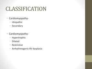
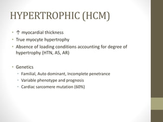
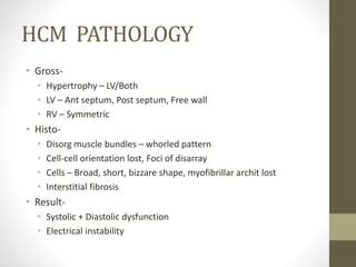
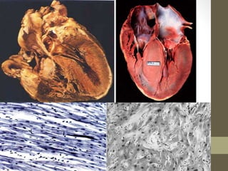
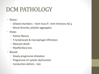
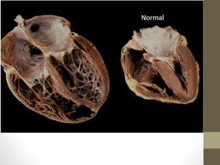
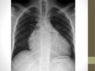
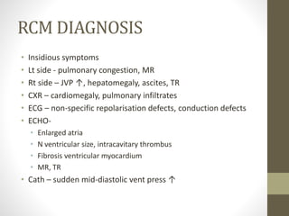
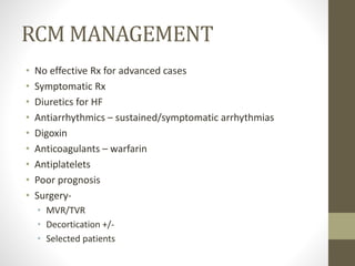
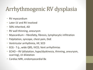
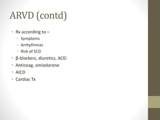
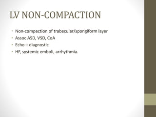
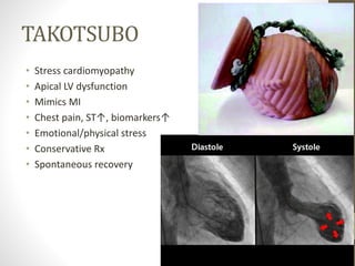
CARDIOMYOPATHY
What, How, Why, e.t.c ……………
CLASSIFICATION
• Cardiomyopathy-
• Idiopathic
• Secondary
• Cardiomyopathy-
• Hypertrophic
• Dilated
• Restrictive
• Arrhythmogenic RV dysplasia
HYPERTROPHIC (HCM)
• ↑ myocardial thickness
• True myocyte hypertrophy
• Absence of loading conditions accounting for degree of
hypertrophy (HTN, AS, AR)
• Genetics
• Familial, Auto dominant, Incomplete penetrance
• Variable phenotype and prognosis
• Cardiac sarcomere mutation (60%)
HCM PATHOLOGY
• Gross-
• Hypertrophy – LV/Both
• LV – Ant septum, Post septum, Free wall
• RV – Symmetric
• Histo-
• Disorg muscle bundles – whorled pattern
• Cell-cell orientation lost, Foci of disarray
• Cells – Broad, short, bizzare shape, myofibrillar archit lost
• Interstitial fibrosis
• Result-
• Systolic + Diastolic dysfunction
• Electrical instability
HCM PATHOPHYSIOLOGY
• Diast-
• LV compliance ↓, Filling abnormal, LVEDP
• Atrial press ↑
• Restrictive component +
• Syst-
• EF ↑
• LVOT obstruction – dynamic/static
• Ischaemic-
• ↓ Coronary flow reserve
• ↑ demand, supply N/↓
HCM DIAGNOSIS
• Unexplained/Disproportionate hypertrophy
• Screening of relatives
• History-
• Autopsy – SCD
• Any age
• Paroxysmal symptoms
• Exertional - dyspnoea, angina (typical/atypical), syncope
• Exam-
• JVP prominent a (RVH)
• Forceful apex, palpable atrial beat
• Pulse – rapid upstroke, low vol
• Ausc – Loud S4, ESM AA, PSM apex
HCM DIAGNOSIS 2
• CXR-
• LV enlargement
• LA RA enlargement
• PAH
• ECG-
• LVH, ST↓, T↓, deep S V1-V3
• P tall, M pattern
• AF, IVCD
• NSVT, VT, SVT
• ECHO-
• LVH, LV cavity↓, Diast dysfunc, LVOT gradient
• LA cavity↑
• MR, SAM
HCM DIAGNOSIS 3
• MRI- Myocardial fibrosis
• Cath-
• Chamber pressures
• Transvalvular & outlet gradients
• Systolic narrowing of coronary artery
HCM MANAGEMENT
• Asymptomatic-
• Genetic testing
• Screen relatives
• Annual follow-up
• Pharmacologic-
• β blocker, CCB, Verapamil, Amiodarone
• Anticoagulants
• IE suspicion high
• Surgical-
• Myotomy, myomectomy
• Papillary muscle remodelling
• MV repair/replacement
HCM MANAGEMENT 2
• Alcohol septal ablation-
• Septal artery
• Elderly, failed medical
• Pacing-
• AICD, RV pacing, CRT (AV sequential)
• Electrical cardioversion
• Stratify risk of SCD-
• Family history of sudden death (≥2 premature (<40 years) sudden
deaths)
• Unexplained syncope within previous year
• Abnormal exercise blood pressure
• Nonsustained ventricular tachycardia (≥3 beats at ≥120
beats/min)
• Severe left ventricular hypertrophy (>3 cm)
• Severe left ventricular outflow tract obstruction (>90 mmHg)
• Cardiac arrest (or sustained ventricular tachycardia)
DILATED (DCM)
• Chamber dilatation & Systolic dysfunction (LV, LV+RV)
• Absence of CAD, valve disease, pericardial disease
• Causes-
• Genetic-
• Autosomal dominant, incomplete penetrance
• X-linked – Duchenne, Becker’s, Emery Driefuss
• Acquired-
• Infectious myocarditis
• ChemoRx, RadioRx
• Alcohol, cocaine
• Nutritional deficiency – thiamine, calcium
• Iron overload
• Inflammatory, autoimmune disorders
• Endocrinopathy – hypothyroidism, DM
• Pregnancy, tachycardia
DCM PATHOLOGY
• Gross-
• Dilated chambers – Vent mass↑, Vent thickness N/↓
• Mural thrombi, platelet aggregates
• Histo-
• Patchy fibrosis
• T-lymphocyte & macrophage infiltration
• Myocyte death
• Myofibrillary loss
• Result-
• Slowly progressive dilatation
• Progressive LV systolic dysfunction
• Conduction defects - late
DCM DIAGNOSIS
• History-
• Progressive cardiac failure
• Insidious fatigue, dyspnoea, ↓exercise tolerance
• Arrhythmia – AF, VT, AV block
• Systemic embolism
• Incidental ECG, CXR, ECHO findings
• Exam-
• Normal
• CCF – Hepatomeg, ascites, pedal edema , JVP↑
• Cardiomegaly, apex - lateral, diffuse & dyskinetic
• Low pulse vol & press, Pulsus alternans, Low SBP
• Ausc – S2 spilt/paradox, loud P2, S4, S3, MR, TR
DCM DIAGNOSIS 2
• ECG – Sinus tachy, nonspecific ST (fibrosis), arrhythmia,
conduction defects
• CXR – Cardiomeg, ↑pulm markings, pleural effusion
• ECHO –
• LV dilatation, syst & diast dysfunc, intracavit thrombus
• Func MR TR
• Pulm art press↑
• Cardiac biomarkers - ↑ CKMB, Trop I&T, ANP
• MRI – Dimensions, fibrosis
• Exercise testing – Func class, progress, pre-Tx
• Cath – Pre-Tx
DCM MANAGEMENT
• Aims-
• Improve symptoms
• Attenuate disease progress
• Prevent complications (arrhythmia, stroke, sudden death)
• Pharmac-
• Diuretics
• ACEi, ARB, β blockers
• Digoxin, anticoagulants
• Antiarrhythmics – Caution (neg intotropic, proarrhythmic)
• Non-pharmac-
• PPI – AICD, CRT
• MVR
• Dor & Batista Sx
• LV assist device, artificial heart
• Cardiac Tx
RESTRICTIVE (RCM)
• ↑ stiffness of myocardium
• Vent press ↑↑ for small vol ↑
• Diast/syst volumes = N/↓
• N vent thickness
• Causes-
• Infiltrative – amyloidosis, sarcoidosis
• Storage – haemochromatosis, Fabry’s, glycogen storage
• Fibrotic – radiation, scleroderma, drugs (doxorubicin)
• Metabolic – carnitine def, fatty acid metab disorders
• Endomyocardial – endomyocardial fibrosis, hypereosinophilic syn
• Misc – carcinoid syndrome
RCM PATHOLOGY
• Heterogenous aetiology
• Gross-
• Biatrial dilatation
• No LVH, N heart wt
• Small vent cavity
• Histo-
• Idiopathic-
• Nonspecific, patchy interstitial fibrosis, myocyte disarray
• Endomyocardial fibrosis-
• Necrotic – eosinophilic abscess, necrotic arteritis, mural thrombus
• Thrombotic – endocardial + intracavitary thrombus
• Fibrotic – dense fibrosis, fill cavity, impaired valve function
• Pathophysiology-
• Impaired relaxation, ↓ volume, sudden mid-diastolic pressure ↑
RCM DIAGNOSIS
• Insidious symptoms
• Lt side - pulmonary congestion, MR
• Rt side – JVP ↑, hepatomegaly, ascites, TR
• CXR – cardiomegaly, pulmonary infiltrates
• ECG – non-specific repolarisation defects, conduction defects
• ECHO-
• Enlarged atria
• N ventricular size, intracavitary thrombus
• Fibrosis ventricular myocardium
• MR, TR
• Cath – sudden mid-diastolic vent press ↑
RCM MANAGEMENT
• No effective Rx for advanced cases
• Symptomatic Rx
• Diuretics for HF
• Antiarrhythmics – sustained/symptomatic arrhythmias
• Digoxin
• Anticoagulants – warfarin
• Antiplatelets
• Poor prognosis
• Surgery-
• MVR/TVR
• Decortication +/-
• Selected patients
OTHERS
Arrhythmogenic RV dysplasia
• RV myocardium
• Later LV and RV involved
• 50% inherited, AD
• RV wall thinning, aneurysm
• Myocardium – fibrofatty, fibrosis, lymphocytic infiltration
• Palpitation, syncope, chest pain, DoE
• Ventricular arrhythmia, HF, SCD
• ECG - T↓, wide QRS, IVCD, Vent arrhythmias
• ECHO – RV (dilatation, hypo/dyskinesia, thinning, aneurysm,
scarring), LV dilatation
• Cardiac MRI, endomyocardial Bx
ARVD (contd)
• Rx according to –
• Symptoms
• Arrhythmias
• Risk of SCD
• β-blockers, diuretics, ACEi
• Anticoag, amiodarone
• AICD
• Cardiac Tx
LV NON-COMPACTION
• Non-compaction of trabecular/spongiform layer
• Assoc ASD, VSD, CoA
• Echo – diagnostic
• HF, systemic emboli, arrhythmia.
TAKOTSUBO
• Stress cardiomyopathy
• Apical LV dysfunction
• Mimics MI
• Chest pain, ST↑, biomarkers↑
• Emotional/physical stress
• Conservative Rx
• Spontaneous recovery

Cardiomyopathy

  • 1.
    CARDIOMYOPATHY What, How, Why,e.t.c ……………
  • 2.
    CLASSIFICATION • Cardiomyopathy- • Idiopathic •Secondary • Cardiomyopathy- • Hypertrophic • Dilated • Restrictive • Arrhythmogenic RV dysplasia
  • 3.
    HYPERTROPHIC (HCM) • ↑myocardial thickness • True myocyte hypertrophy • Absence of loading conditions accounting for degree of hypertrophy (HTN, AS, AR) • Genetics • Familial, Auto dominant, Incomplete penetrance • Variable phenotype and prognosis • Cardiac sarcomere mutation (60%)
  • 4.
    HCM PATHOLOGY • Gross- •Hypertrophy – LV/Both • LV – Ant septum, Post septum, Free wall • RV – Symmetric • Histo- • Disorg muscle bundles – whorled pattern • Cell-cell orientation lost, Foci of disarray • Cells – Broad, short, bizzare shape, myofibrillar archit lost • Interstitial fibrosis • Result- • Systolic + Diastolic dysfunction • Electrical instability
  • 6.
    HCM PATHOPHYSIOLOGY • Diast- •LV compliance ↓, Filling abnormal, LVEDP • Atrial press ↑ • Restrictive component + • Syst- • EF ↑ • LVOT obstruction – dynamic/static • Ischaemic- • ↓ Coronary flow reserve • ↑ demand, supply N/↓
  • 7.
    HCM DIAGNOSIS • Unexplained/Disproportionatehypertrophy • Screening of relatives • History- • Autopsy – SCD • Any age • Paroxysmal symptoms • Exertional - dyspnoea, angina (typical/atypical), syncope • Exam- • JVP prominent a (RVH) • Forceful apex, palpable atrial beat • Pulse – rapid upstroke, low vol • Ausc – Loud S4, ESM AA, PSM apex
  • 8.
    HCM DIAGNOSIS 2 •CXR- • LV enlargement • LA RA enlargement • PAH • ECG- • LVH, ST↓, T↓, deep S V1-V3 • P tall, M pattern • AF, IVCD • NSVT, VT, SVT • ECHO- • LVH, LV cavity↓, Diast dysfunc, LVOT gradient • LA cavity↑ • MR, SAM
  • 9.
    HCM DIAGNOSIS 3 •MRI- Myocardial fibrosis • Cath- • Chamber pressures • Transvalvular & outlet gradients • Systolic narrowing of coronary artery
  • 10.
    HCM MANAGEMENT • Asymptomatic- •Genetic testing • Screen relatives • Annual follow-up • Pharmacologic- • β blocker, CCB, Verapamil, Amiodarone • Anticoagulants • IE suspicion high • Surgical- • Myotomy, myomectomy • Papillary muscle remodelling • MV repair/replacement
  • 11.
    HCM MANAGEMENT 2 •Alcohol septal ablation- • Septal artery • Elderly, failed medical • Pacing- • AICD, RV pacing, CRT (AV sequential) • Electrical cardioversion • Stratify risk of SCD- • Family history of sudden death (≥2 premature (<40 years) sudden deaths) • Unexplained syncope within previous year • Abnormal exercise blood pressure • Nonsustained ventricular tachycardia (≥3 beats at ≥120 beats/min) • Severe left ventricular hypertrophy (>3 cm) • Severe left ventricular outflow tract obstruction (>90 mmHg) • Cardiac arrest (or sustained ventricular tachycardia)
  • 12.
    DILATED (DCM) • Chamberdilatation & Systolic dysfunction (LV, LV+RV) • Absence of CAD, valve disease, pericardial disease • Causes- • Genetic- • Autosomal dominant, incomplete penetrance • X-linked – Duchenne, Becker’s, Emery Driefuss • Acquired- • Infectious myocarditis • ChemoRx, RadioRx • Alcohol, cocaine • Nutritional deficiency – thiamine, calcium • Iron overload • Inflammatory, autoimmune disorders • Endocrinopathy – hypothyroidism, DM • Pregnancy, tachycardia
  • 13.
    DCM PATHOLOGY • Gross- •Dilated chambers – Vent mass↑, Vent thickness N/↓ • Mural thrombi, platelet aggregates • Histo- • Patchy fibrosis • T-lymphocyte & macrophage infiltration • Myocyte death • Myofibrillary loss • Result- • Slowly progressive dilatation • Progressive LV systolic dysfunction • Conduction defects - late
  • 16.
    DCM DIAGNOSIS • History- •Progressive cardiac failure • Insidious fatigue, dyspnoea, ↓exercise tolerance • Arrhythmia – AF, VT, AV block • Systemic embolism • Incidental ECG, CXR, ECHO findings • Exam- • Normal • CCF – Hepatomeg, ascites, pedal edema , JVP↑ • Cardiomegaly, apex - lateral, diffuse & dyskinetic • Low pulse vol & press, Pulsus alternans, Low SBP • Ausc – S2 spilt/paradox, loud P2, S4, S3, MR, TR
  • 17.
    DCM DIAGNOSIS 2 •ECG – Sinus tachy, nonspecific ST (fibrosis), arrhythmia, conduction defects • CXR – Cardiomeg, ↑pulm markings, pleural effusion • ECHO – • LV dilatation, syst & diast dysfunc, intracavit thrombus • Func MR TR • Pulm art press↑ • Cardiac biomarkers - ↑ CKMB, Trop I&T, ANP • MRI – Dimensions, fibrosis • Exercise testing – Func class, progress, pre-Tx • Cath – Pre-Tx
  • 18.
    DCM MANAGEMENT • Aims- •Improve symptoms • Attenuate disease progress • Prevent complications (arrhythmia, stroke, sudden death) • Pharmac- • Diuretics • ACEi, ARB, β blockers • Digoxin, anticoagulants • Antiarrhythmics – Caution (neg intotropic, proarrhythmic) • Non-pharmac- • PPI – AICD, CRT • MVR • Dor & Batista Sx • LV assist device, artificial heart • Cardiac Tx
  • 19.
    RESTRICTIVE (RCM) • ↑stiffness of myocardium • Vent press ↑↑ for small vol ↑ • Diast/syst volumes = N/↓ • N vent thickness • Causes- • Infiltrative – amyloidosis, sarcoidosis • Storage – haemochromatosis, Fabry’s, glycogen storage • Fibrotic – radiation, scleroderma, drugs (doxorubicin) • Metabolic – carnitine def, fatty acid metab disorders • Endomyocardial – endomyocardial fibrosis, hypereosinophilic syn • Misc – carcinoid syndrome
  • 20.
    RCM PATHOLOGY • Heterogenousaetiology • Gross- • Biatrial dilatation • No LVH, N heart wt • Small vent cavity • Histo- • Idiopathic- • Nonspecific, patchy interstitial fibrosis, myocyte disarray • Endomyocardial fibrosis- • Necrotic – eosinophilic abscess, necrotic arteritis, mural thrombus • Thrombotic – endocardial + intracavitary thrombus • Fibrotic – dense fibrosis, fill cavity, impaired valve function • Pathophysiology- • Impaired relaxation, ↓ volume, sudden mid-diastolic pressure ↑
  • 21.
    RCM DIAGNOSIS • Insidioussymptoms • Lt side - pulmonary congestion, MR • Rt side – JVP ↑, hepatomegaly, ascites, TR • CXR – cardiomegaly, pulmonary infiltrates • ECG – non-specific repolarisation defects, conduction defects • ECHO- • Enlarged atria • N ventricular size, intracavitary thrombus • Fibrosis ventricular myocardium • MR, TR • Cath – sudden mid-diastolic vent press ↑
  • 22.
    RCM MANAGEMENT • Noeffective Rx for advanced cases • Symptomatic Rx • Diuretics for HF • Antiarrhythmics – sustained/symptomatic arrhythmias • Digoxin • Anticoagulants – warfarin • Antiplatelets • Poor prognosis • Surgery- • MVR/TVR • Decortication +/- • Selected patients
  • 23.
  • 24.
    Arrhythmogenic RV dysplasia •RV myocardium • Later LV and RV involved • 50% inherited, AD • RV wall thinning, aneurysm • Myocardium – fibrofatty, fibrosis, lymphocytic infiltration • Palpitation, syncope, chest pain, DoE • Ventricular arrhythmia, HF, SCD • ECG - T↓, wide QRS, IVCD, Vent arrhythmias • ECHO – RV (dilatation, hypo/dyskinesia, thinning, aneurysm, scarring), LV dilatation • Cardiac MRI, endomyocardial Bx
  • 25.
    ARVD (contd) • Rxaccording to – • Symptoms • Arrhythmias • Risk of SCD • β-blockers, diuretics, ACEi • Anticoag, amiodarone • AICD • Cardiac Tx
  • 26.
    LV NON-COMPACTION • Non-compactionof trabecular/spongiform layer • Assoc ASD, VSD, CoA • Echo – diagnostic • HF, systemic emboli, arrhythmia.
  • 27.
    TAKOTSUBO • Stress cardiomyopathy •Apical LV dysfunction • Mimics MI • Chest pain, ST↑, biomarkers↑ • Emotional/physical stress • Conservative Rx • Spontaneous recovery