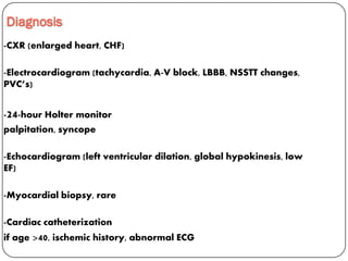
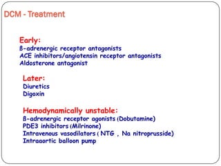
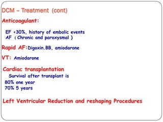
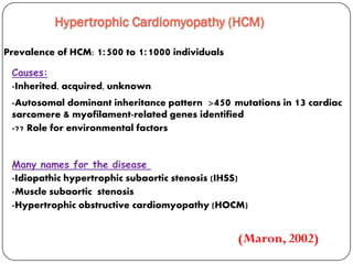
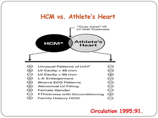
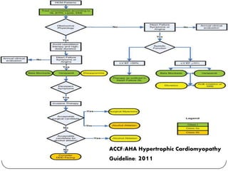
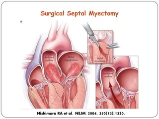
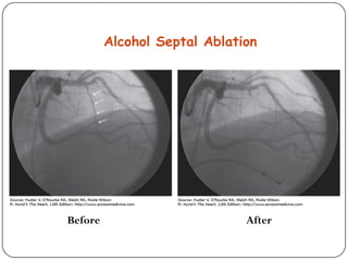
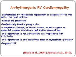
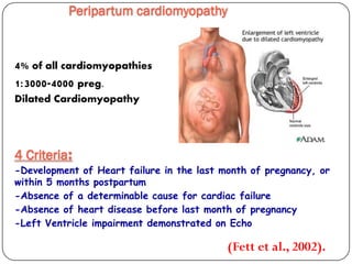
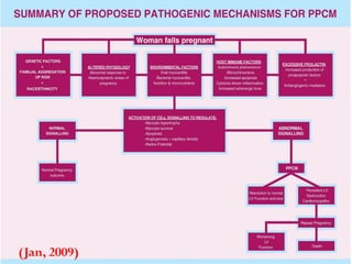
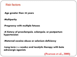
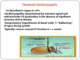
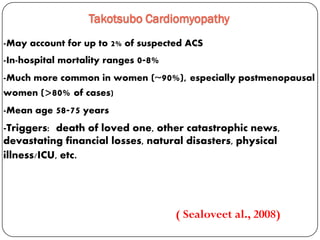
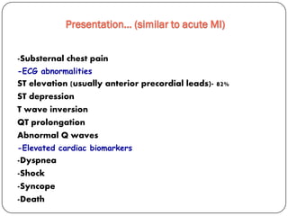
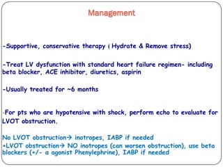
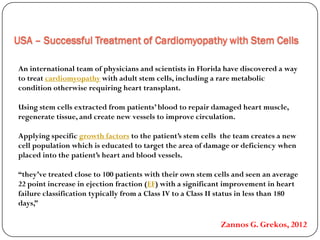
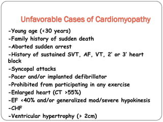
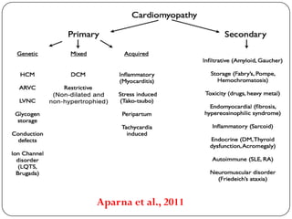
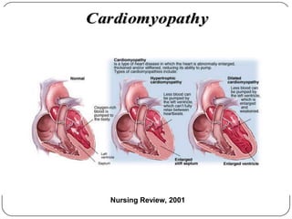
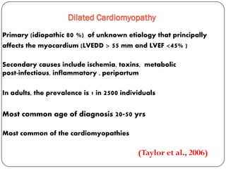
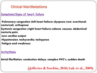
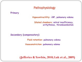
This document discusses various types of cardiomyopathy, including dilated cardiomyopathy, hypertrophic cardiomyopathy, restrictive cardiomyopathy, and Takotsubo cardiomyopathy. It covers the pathophysiology, clinical presentation, diagnosis, and management of each type. It also discusses prognosis and the use of stem cells in treating cardiomyopathy.
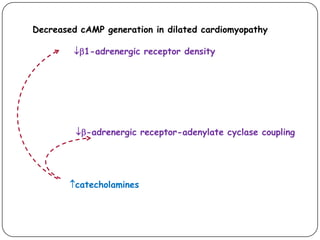
2. OBJECTIVES
-Discuss the pathophysiology and etiologies of various types
of cardiomyopathy
-Review current pharmacological and non pharmacological
managements of cardiomyopathy
-Discuss future trends in management
3. Cardiomyopathy is a disease of the heart muscle characterized by
abnormality in chamber size and wall thickness, or functional contractile
dysfunctions
2006 AHA defined cardiomyopathies as “a heterogeneous group of diseases
of the myocardium associated with mechanical &/or electrical dysfunction
which usually exhibit inappropriate ventricular hypertrophy or dilatation,
due to a variety of etiologies that frequently are genetic.
Definition
(Elliott et al., 2008).
6. Dilated Cardiomyopathy
Primary (idiopathic 80 %) of unknown etiology that principally
affects the myocardium (LVEDD > 55 mm and LVEF <45% )
Secondary causes include ischemia, toxins, metabolic
peripartumpost-infectious, inflammatory ,
In adults, the prevalence is 1 in 2500 individuals
Most common age of diagnosis 20-50 yrs
Most common of the cardiomyopathies
(Taylor et al., 2006)
17. DCM – Treatment (cont)
Anticoagulant:
EF <30%, history of embolic events
Chronic and paroxysmal ))AF
Rapid AF:Digoxin.BB, amiodarone
VT: Amiodarone
Cardiac transplantation
Survival after transplant is
80% one year
70% 5 years
Left Ventricular Reduction and reshaping Procedures
18. Hypertrophic Cardiomyopathy (HCM)
Prevalence of HCM: 1:500 to 1:1000 individuals
Many names for the disease
-Idiopathic hypertrophic subaortic stenosis (IHSS)
-Muscle subaortic stenosis
-Hypertrophic obstructive cardiomyopathy (HOCM)
Causes:
-Inherited, acquired, unknown
-Autosomal dominant inheritance pattern >450 mutations in 13 cardiac
sarcomere & myofilament-related genes identified
-?? Role for environmental factors
(Maron, 2002)
20. Pathophysiology of HCM
The pathophysiology of HCM involves 4 interrelated
processes:
-Left ventricular outflow obstruction
-Diastolic dysfunction
-Myocardial ischemia
-Mitral regurgitation
21. HCM Clinical Presentation
-Dyspnea on exertion (90%), orthopnea, PND
-Angina (70-80%)
-Syncope (20%), Presyncope (50%) outflow obstruction
worsens with increased contractility during exertional
activities
-Ejection systolic murmurs along the left sternal border
-Stroke
-HCM is most common cause of SCD in young people, including
athletes
26. Sudden Cardiac Death in HCM
-Most frequent in young adults
<30-35 years old
-Primary VF/VT
-Tend to die during or just
following vigorous physical
activity
-Often is 1st clinical
manifestation of disease
J Am Coll Cardiol. 2003;42(9):1693.
32. Restrictive Cardiomyopathies
-Hallmark: abnormal diastolic function
-Rigid ventricular wall with impaired ventricular filling
-Wall thickness may be normal or increased
-Systolic function usually remains normal
-Much less common then DCM or HCM
(Kushwaha et al., 1997).
34. Treatment Of RCM
-Treatment of the cause
-No satisfactory medical therapy
-Diuretics for extremely high filling pressures
-Vasodilators may decrease filling pressure
-(?) Calcium channel blockers to improve diastolic compliance
-Digitalis and other inotropic agents are not indicated
Heart transplantation-
35. Arrhythmogenic RV Cardiomyopathy
-Characterized by fibroadipose replacement of segments of the free
wall of the right ventricle
-Familial and progressive
-Predominately found in young adults
-Arrhythmias, syncope, or cardiac arrest, as well as global or
segmental chamber dilatation or wall motion abnormalities
-ICD implantation in ALL patients who are symptomatic with
arrhythmias
-ICD implantation vs anti-arrhythmic meds in asymptomatic patients?
-Prognosis????
(Basso et al., 2009) (Marcus et al., 2010).
36. Peripartum cardiomyopathy
(Fett et al., 2002).
4% of all cardiomyopathies
1:3000-4000 preg.
Dilated Cardiomyopathy
:Criteria4
-Development of Heart failure in the last month of pregnancy, or
within 5 months postpartum
-Absence of a determinable cause for cardiac failure
-Absence of heart disease before last month of pregnancy
-Left Ventricle impairment demonstrated on Echo
37. 37 of 48
Unfavorable Cases of Cardiomyopathy
Young age (<30 years)
Family history of sudden death
Aborted sudden death
History of sustained SVT,AF,VT, 2’ or 3’ heart block
Syncopal attacks
Pacer and/or implanted defibrillator
Prohibited from participating in any exercise
Enlarged heart (CT >55%)
EF <40% and/or generalized mod/severe hypokinesis
CHF
Substantial hypertrophy (> 2cm)
(Jan, 2009)
38. -Age greater than 30 years
-Multiparity
-Pregnancy with multiple fetuses
-A history of preeclampsia, eclampsia, or postpartum
hypertension
-Maternal cocaine abuse or selenium deficiency
-Long term (>4 weeks) oral tocolytic therapy with beta
adrenergic agonists
Risk factors
(Pearson et al., 2000)
39. DIAGNOSIS
ECG findings include
sinus tachycardia
nonspecific ST and T wave abnormalities
PR and QRS intervals may also be prolonged
(Elkayam et al., 2005)
FerritinCBC, BNP, TSH,Lab:
dimensional echocardiogram-Two
CXR
Cardiac catheterization
Myocardial biopsy?
/EF <55%Spherical LV /Left Atrial enlargement
41. Prognosis
-Mortality estimates range from 25-50%.
-Most deaths occur within 3 months postpartum
-Deaths usually caused by:
Progressive pump failure
Arrhythmias
Thromboembolic events
42. -Opinions widely vary
-Patients with PPCM and persistent left ventricular dysfunction > 6
ms are at extremely high risk of complications and death with
subsequent pregnancies
-These patients should be advised to avoid pregnancy unless,
successful cardiac transplantation is performed
Future pregnancy ??
-If future pregnancy desired:
Maternal Echocardiogram per trimester
Serial sonograms for growth
Consult, consult, consult
43. Takotsubo Cardiomyopathy
-Cardiomyopathy characterized by transient apical and
midventricular LV dysfunction in the absence of significant
coronary artery disease
-Compensatory hyperkinesis of basal walls “ballooning”
of apex during systole.
-Typically recover normal LV function in 1-4 weeks.
-1st described in Japan in 1991
44. -May account for up to 2% of suspected ACS
-In-hospital mortality ranges 0-8%
-Much more common in women (~90%), especially postmenopausal
women (>80% of cases)
-Mean age 58-75 years
Takotsubo Cardiomyopathy
( Sealoveet al., 2008)
-Triggers: death of loved one, other catastrophic news,
devastating financial losses, natural disasters, physical
illness/ICU, etc.
45. -Substernal chest pain
-ECG abnormalities
ST elevation (usually anterior precordial leads)- 82%
ST depression
T wave inversion
QT prolongation
Abnormal Q waves
-Elevated cardiac biomarkers
-Dyspnea
-Shock
-Syncope
-Death
Presentation… (similar to acute MI)
46. Hydrate & Remove stress))-Supportive, conservative therapy
-Treat LV dysfunction with standard heart failure regimen- including
beta blocker, ACE inhibitor, diuretics, aspirin
-Usually treated for ~6 months
-For pts who are hypotensive with shock, perform echo to evaluate for
LVOT obstruction.
No LVOT obstruction inotropes, IABP if needed
+LVOT obstruction NO inotropes (can worsen obstruction), use beta
blockers (+/- α agonist Phenylephrine), IABP if needed
Management
47. Prognosis
Overall, good prognosis. If
patient survives the acute phase,
long-term prognosis is excellent.
-0-8% in-hospital mortality, likely closer to 1-2%
-Recovery of LV function, typically in 1-4 weeks
-Late sudden death (rare) and recurrent disease (<10%) have
been reported
48. USA – Successful Treatment of Cardiomyopathy with Stem Cells
An international team of physicians and scientists in Florida have discovered a way
with adult stem cells, including a rare metaboliccardiomyopathyto treat
condition otherwise requiring heart transplant.
Using stem cells extracted from patients’ blood to repair damaged heart muscle,
regenerate tissue, and create new vessels to improve circulation.
the team creates a newto the patient’s stem cellsgrowth factorsApplying specific
cell population which is educated to target the area of damage or deficiency when
placed into the patient’s heart and blood vessels.
“they’ve treated close to 100 patients with their own stem cells and seen an average
) with a significant improvement in heartEFpoint increase in ejection fraction (22
failure classification typically from a Class IV to a Class II status in less than 180
days,”
Zannos G. Grekos, 2012
49. Unfavorable Cases of Cardiomyopathy
-Young age (<30 years)
-Family history of sudden death
-Aborted sudden arrest
-History of sustained SVT, AF, VT, 2’ or 3’ heart
block
-Syncopal attacks
-Pacer and/or implanted defibrillator
-Prohibited from participating in any exercise
-Enlarged heart (CT >55%)
-EF <40% and/or generalized mod/severe hypokinesis
-CHF
-Ventricular hypertrophy (> 2cm)