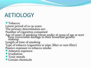
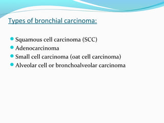
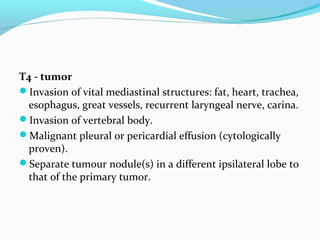
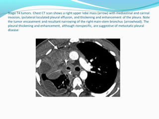
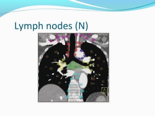
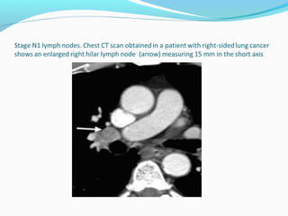
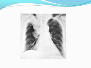
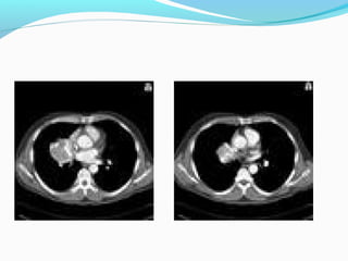
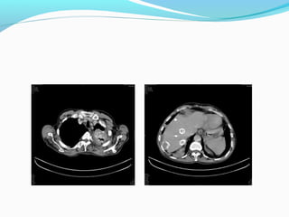
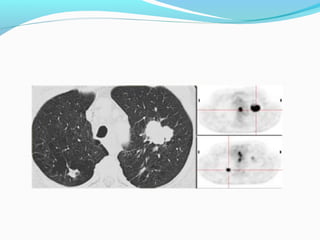
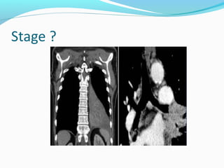
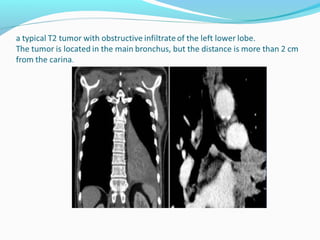
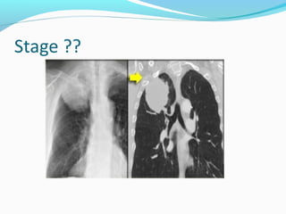
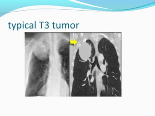
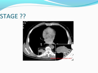
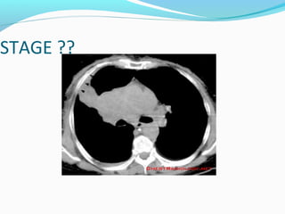
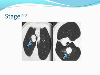
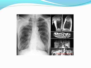
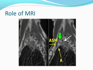
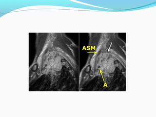
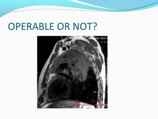
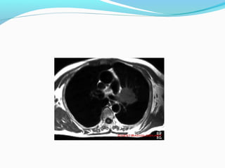
Dr shaista khan
AETIOLOGY
Tobacco:

Latent period of 10-30 years
The primary determinants are:
Number of cigarettes consumed
Age of onset of smoking (those under 16 years of age at start
have irreversible damage to their bronchial genetic
makeup
Length of time of smoking
Type of tobacco (cigarettes or pipe, filter or non-filter)
Passive exposure to tobacco smoke
Asbestos exposure
Irradiation
Toxic metals
Certain chemicals
Types of bronchial carcinoma:
Squamous cell carcinoma (SCC)
Adenocarcinoma
Small cell carcinoma (oat cell carcinoma)
Alveolar cell or bronchoalveolar carcinoma
Squamous cell carcinoma:
60% of all lung tumors
Associated with smoking and is rare in non-smokers
Squamous metaplasia -> carcinoma in situ -> invasive
carcinoma
Adenocarcinoma:
15% of lung tumours
Has a tendency to be more peripheral, arising in the
small bronchial glands
Most common in women
Is the type seen in non-smokers
Small cell (oat cell) carcinoma:
20% of lung tumours
Arises from the chromaffin cells
Highly malignant
Hormone production by the tumour is common
A benign form of a small cell carcinoma is a carcinoid
tumour
Alveolar cell carcinoma:
5% of lung tumours
Arises in the distal airways
Often diffuse, multifocal and bilateral
Resistant to radio-/chemotherapy
Very poor prognosis
TNM STAGING
(T)
T1 - tumor
Diameter of 3 cm or smaller and surrounded by lung
or visceral pleura or
endobronchial tumor distal to the lobar bronchus
T2 - tumor
Greater than 3 and smaller than 7 cm
Invasion of the visceral pleura
Atelectasis or obstructive pneumopathy involving less
than the whole lung
Tumor involving the main bronchus 2 cm or more
distal to the carina.
T3 - tumor
Tumor with atelectasis or obstructive pneumonitis of the
entire lung
Tumor in the main bronchus within 2 cm of the carina but
not invading it
Tumor of any size with invasion of non-vital structures
such as the chest wall, mediastinal pleura, diaphragm,
pericardium.
Separate tumour nodules in the same lobe as the primary
tumor.
T4 - tumor
Invasion of vital mediastinal structures: fat, heart, trachea,
esophagus, great vessels, recurrent laryngeal nerve, carina.
Invasion of vertebral body.
Malignant pleural or pericardial effusion (cytologically
proven).
Separate tumour nodule(s) in a different ipsilateral lobe to
that of the primary tumor.
Lymph nodes (N)
N1 - Nodes
N1-nodes are ipsilateral nodes within the lung up to

hilar nodes.
N1 alters the prognosis but not the management.
N 2 NODES.
Nodes in the ipsilateral mediastinum
N3 - Nodes
N3-nodes are clearly unresectable.

These are contralateral mediastinal or contralateral
hilar nodes or any scalene or supraclavicular nodes.
CALCIFICATION
FDG UPTAKE
STAGE ??
STAGE ??
PANCOAST TUMOR
OPERABLE OR NOT?
THANKS

Carcinoma bronchus

  • 1.
  • 2.
    AETIOLOGY Tobacco: Latent period of10-30 years The primary determinants are: Number of cigarettes consumed Age of onset of smoking (those under 16 years of age at start have irreversible damage to their bronchial genetic makeup Length of time of smoking Type of tobacco (cigarettes or pipe, filter or non-filter) Passive exposure to tobacco smoke Asbestos exposure Irradiation Toxic metals Certain chemicals
  • 3.
    Types of bronchialcarcinoma: Squamous cell carcinoma (SCC) Adenocarcinoma Small cell carcinoma (oat cell carcinoma) Alveolar cell or bronchoalveolar carcinoma
  • 4.
    Squamous cell carcinoma: 60%of all lung tumors Associated with smoking and is rare in non-smokers Squamous metaplasia -> carcinoma in situ -> invasive carcinoma
  • 5.
    Adenocarcinoma: 15% of lungtumours Has a tendency to be more peripheral, arising in the small bronchial glands Most common in women Is the type seen in non-smokers
  • 6.
    Small cell (oatcell) carcinoma: 20% of lung tumours Arises from the chromaffin cells Highly malignant Hormone production by the tumour is common A benign form of a small cell carcinoma is a carcinoid tumour
  • 7.
    Alveolar cell carcinoma: 5%of lung tumours Arises in the distal airways Often diffuse, multifocal and bilateral Resistant to radio-/chemotherapy Very poor prognosis
  • 10.
    TNM STAGING (T) T1 -tumor Diameter of 3 cm or smaller and surrounded by lung or visceral pleura or endobronchial tumor distal to the lobar bronchus
  • 13.
    T2 - tumor Greaterthan 3 and smaller than 7 cm Invasion of the visceral pleura Atelectasis or obstructive pneumopathy involving less than the whole lung Tumor involving the main bronchus 2 cm or more distal to the carina.
  • 16.
    T3 - tumor Tumorwith atelectasis or obstructive pneumonitis of the entire lung Tumor in the main bronchus within 2 cm of the carina but not invading it Tumor of any size with invasion of non-vital structures such as the chest wall, mediastinal pleura, diaphragm, pericardium. Separate tumour nodules in the same lobe as the primary tumor.
  • 19.
    T4 - tumor Invasionof vital mediastinal structures: fat, heart, trachea, esophagus, great vessels, recurrent laryngeal nerve, carina. Invasion of vertebral body. Malignant pleural or pericardial effusion (cytologically proven). Separate tumour nodule(s) in a different ipsilateral lobe to that of the primary tumor.
  • 23.
  • 24.
    N1 - Nodes N1-nodesare ipsilateral nodes within the lung up to hilar nodes. N1 alters the prognosis but not the management.
  • 26.
    N 2 NODES. Nodesin the ipsilateral mediastinum
  • 28.
    N3 - Nodes N3-nodesare clearly unresectable. These are contralateral mediastinal or contralateral hilar nodes or any scalene or supraclavicular nodes.
  • 39.
  • 45.
  • 50.
  • 51.
  • 54.
  • 57.
  • 59.