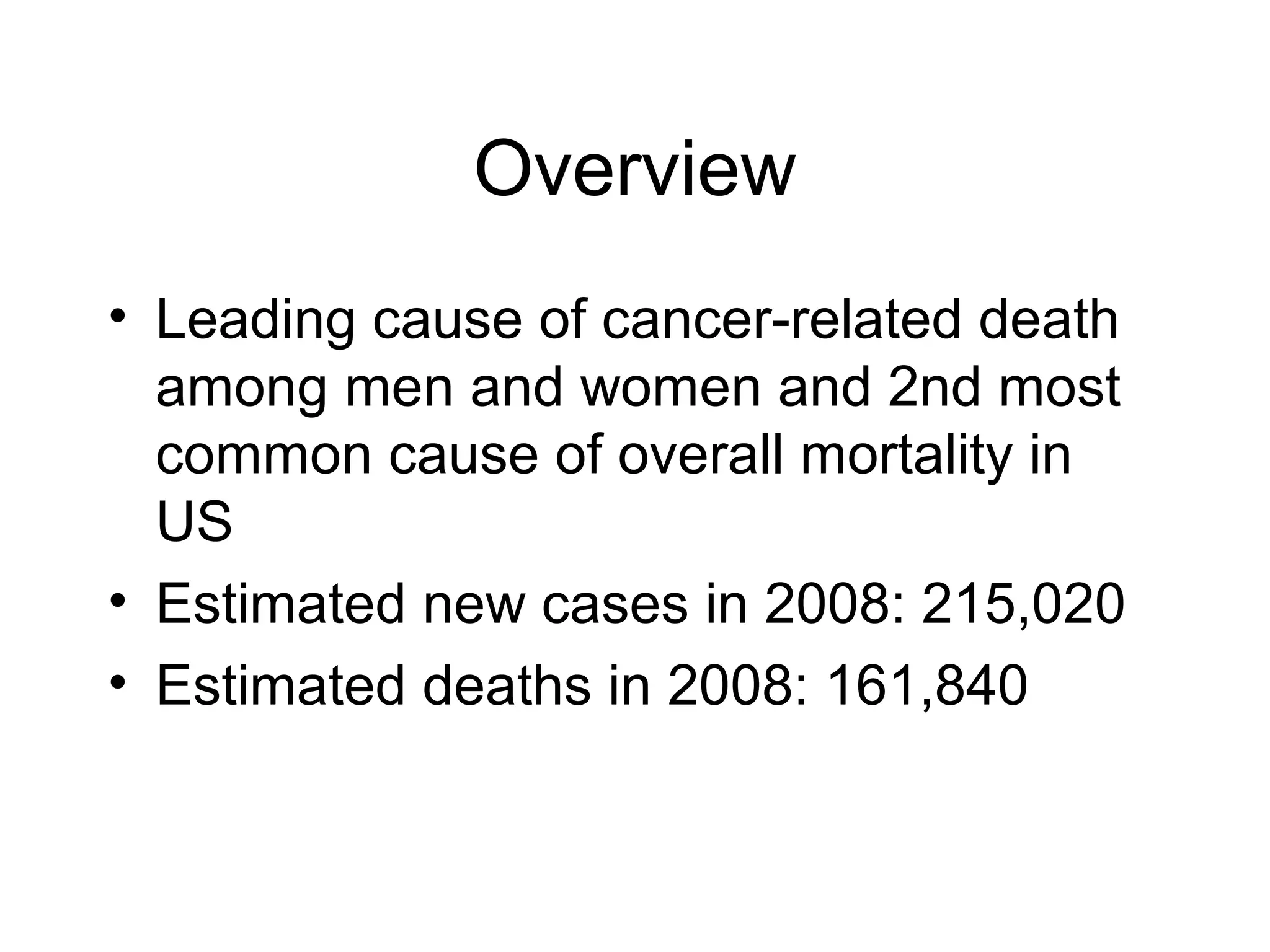
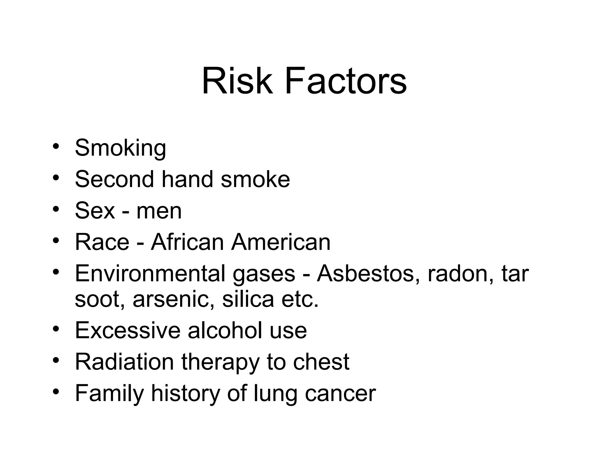
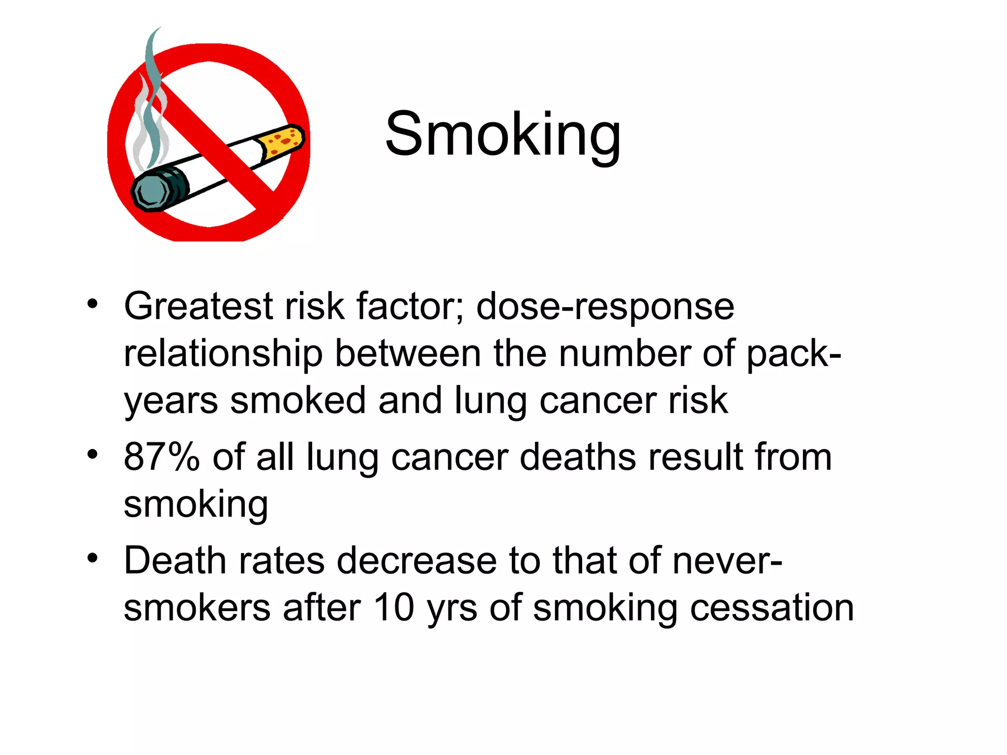
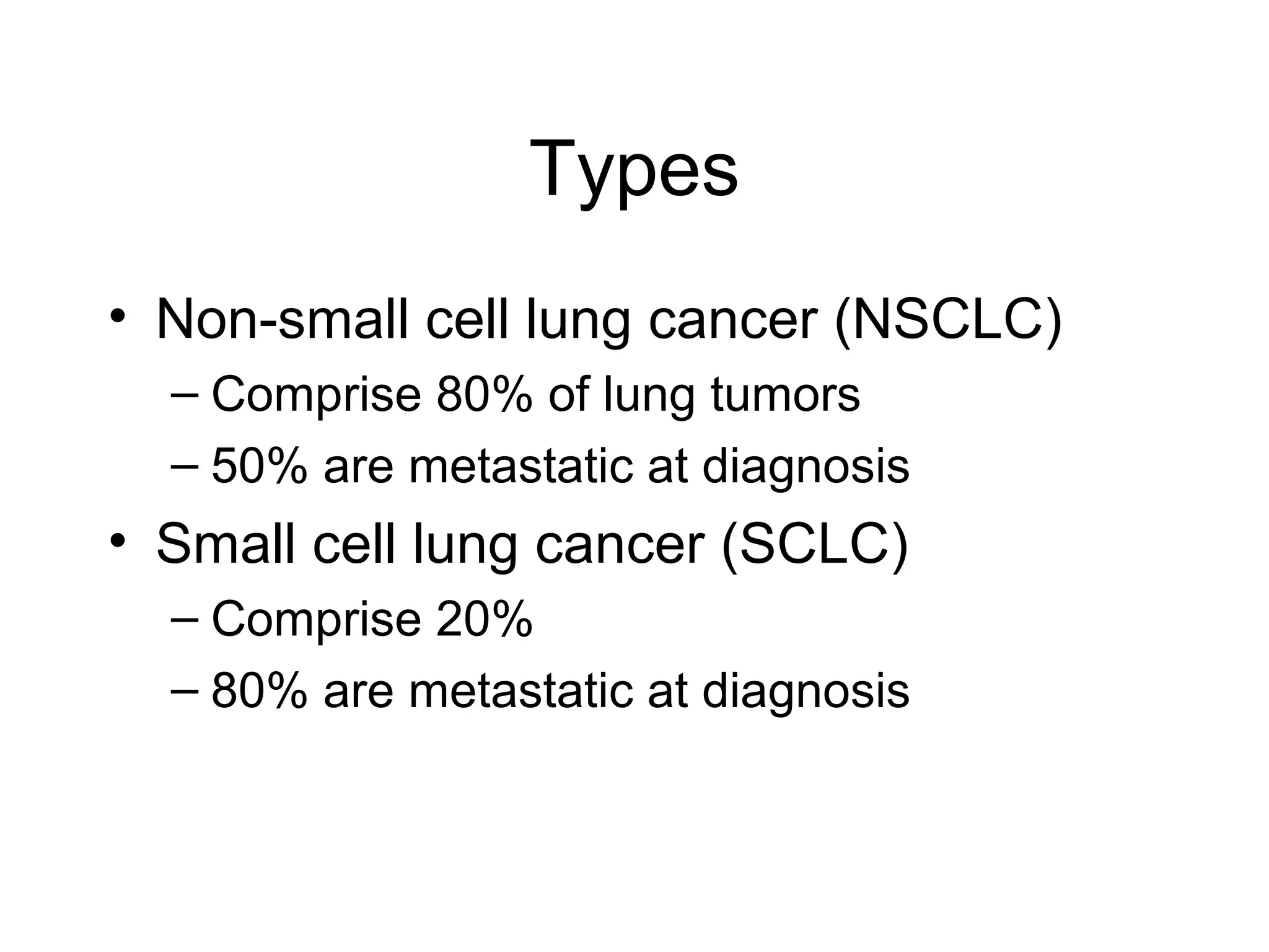
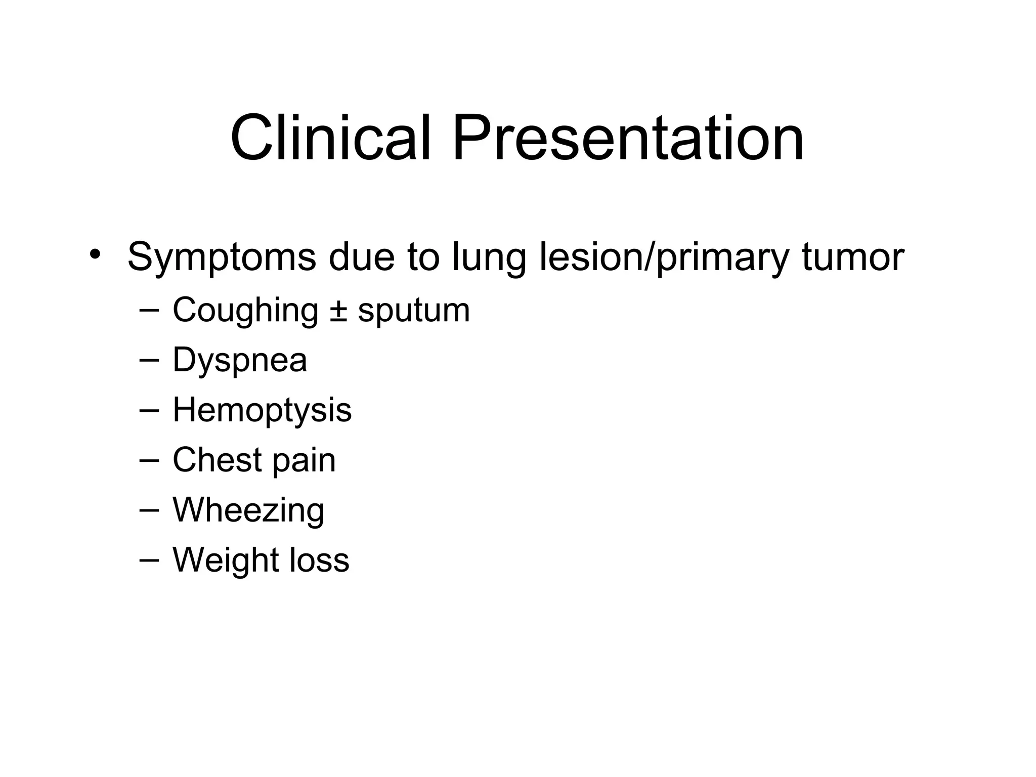
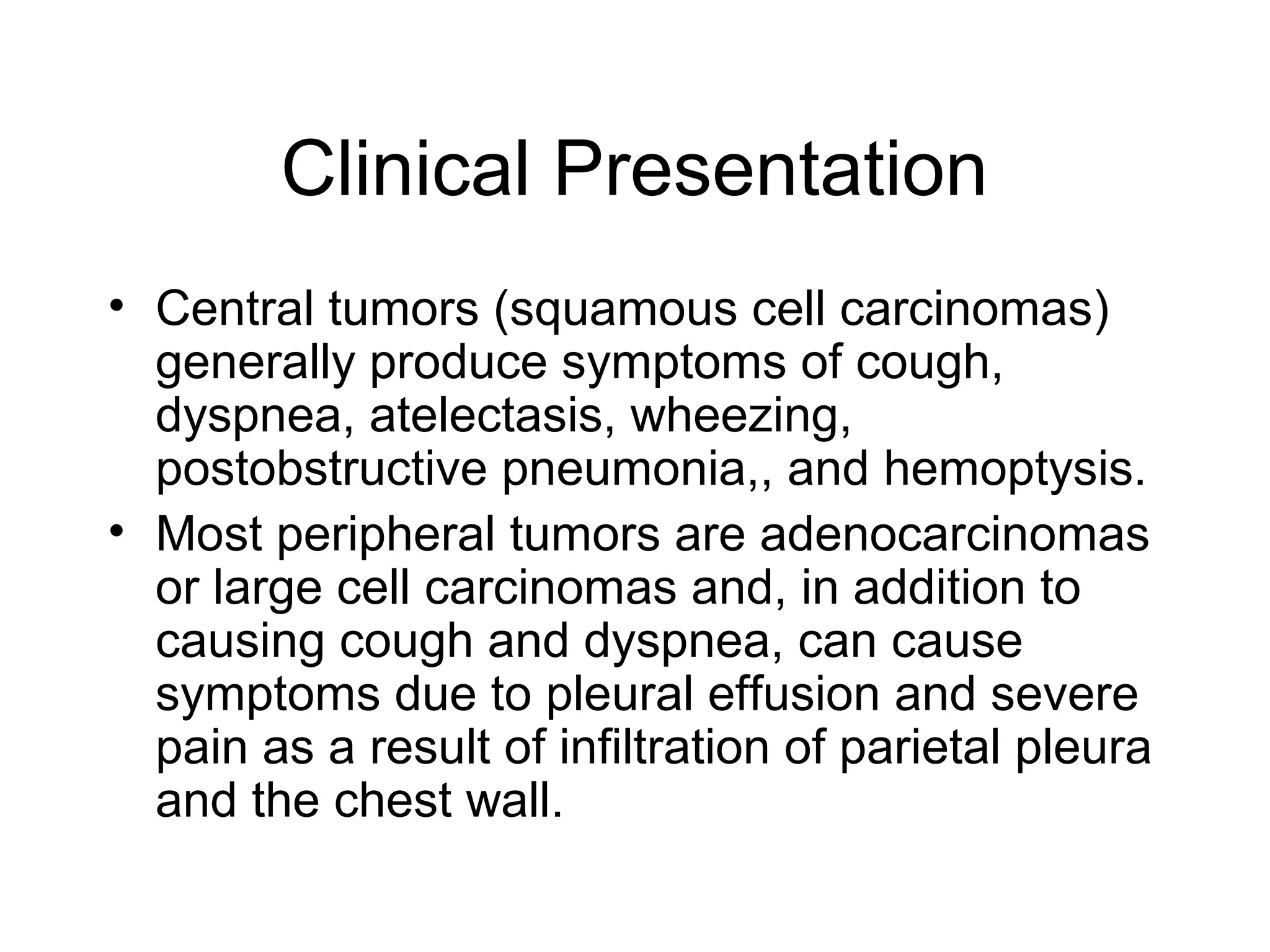
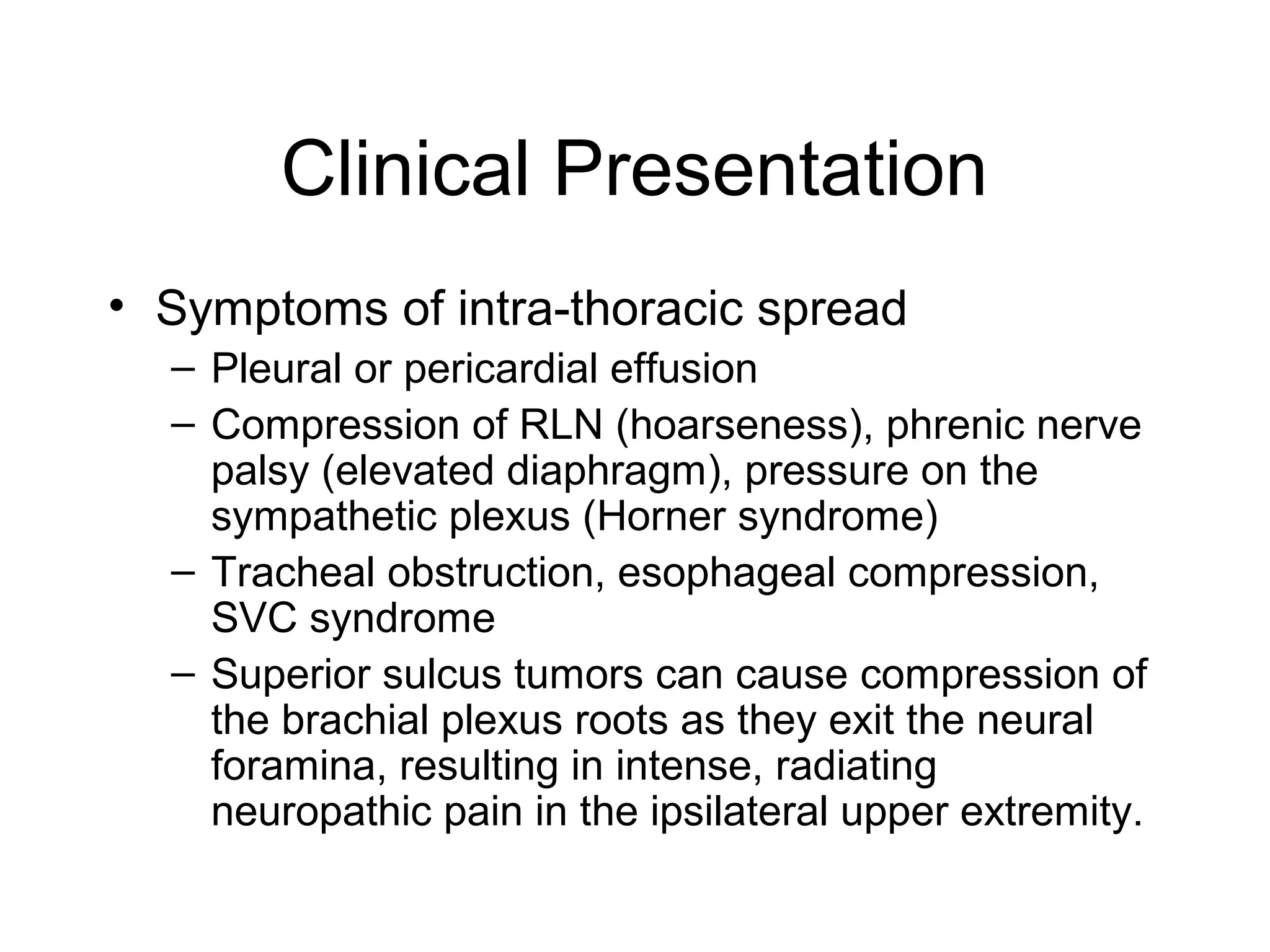
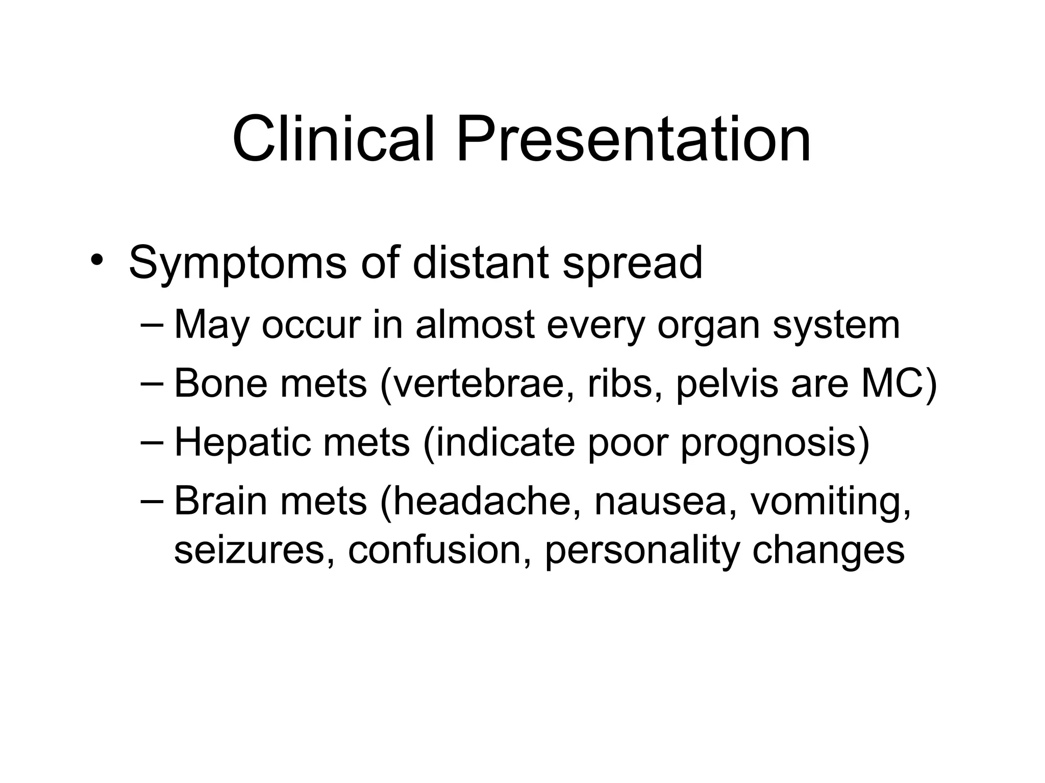
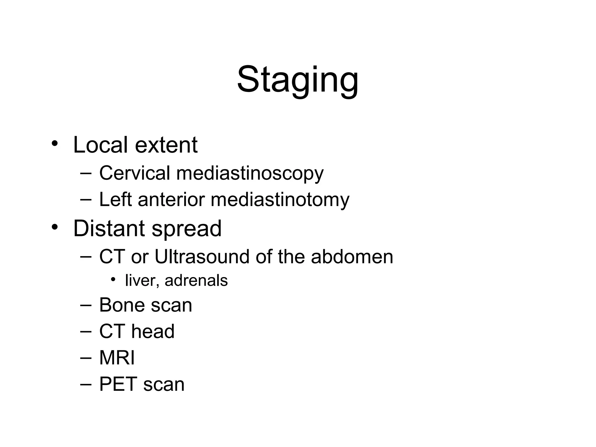
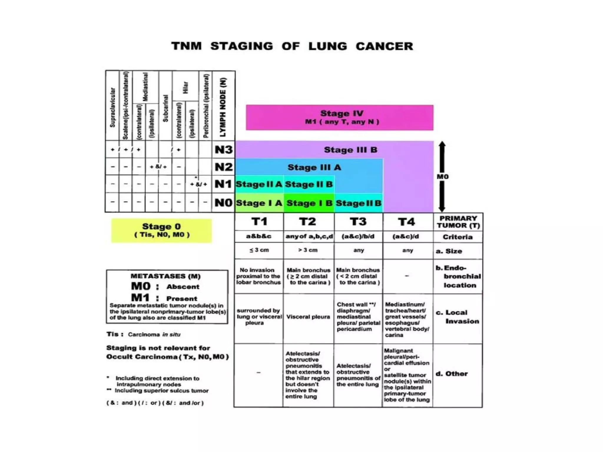
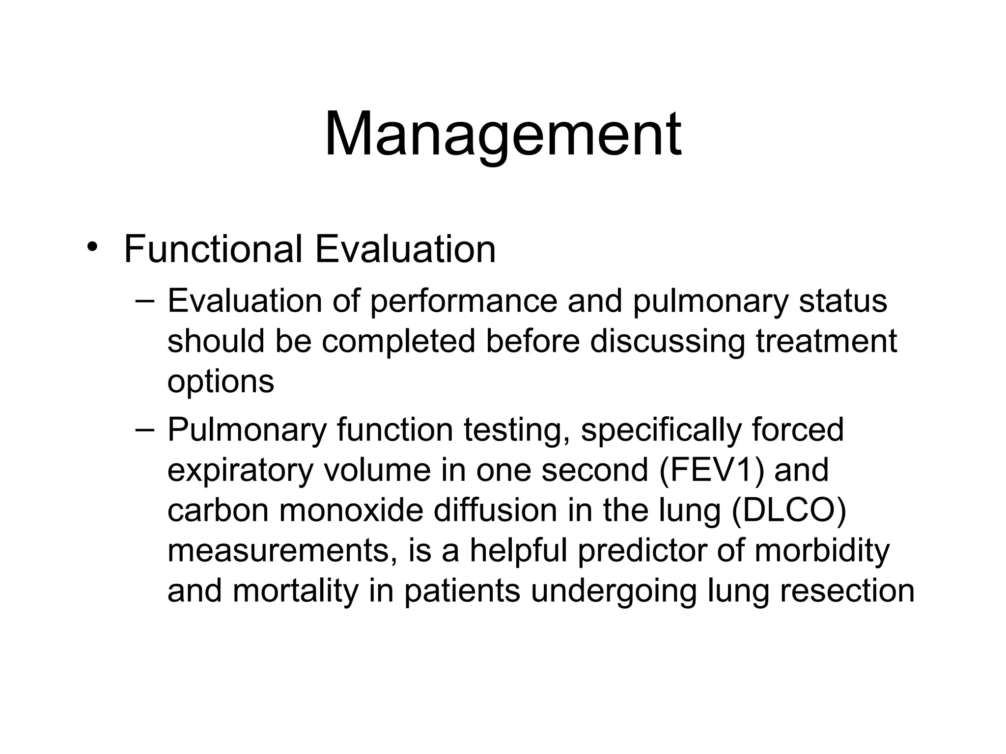
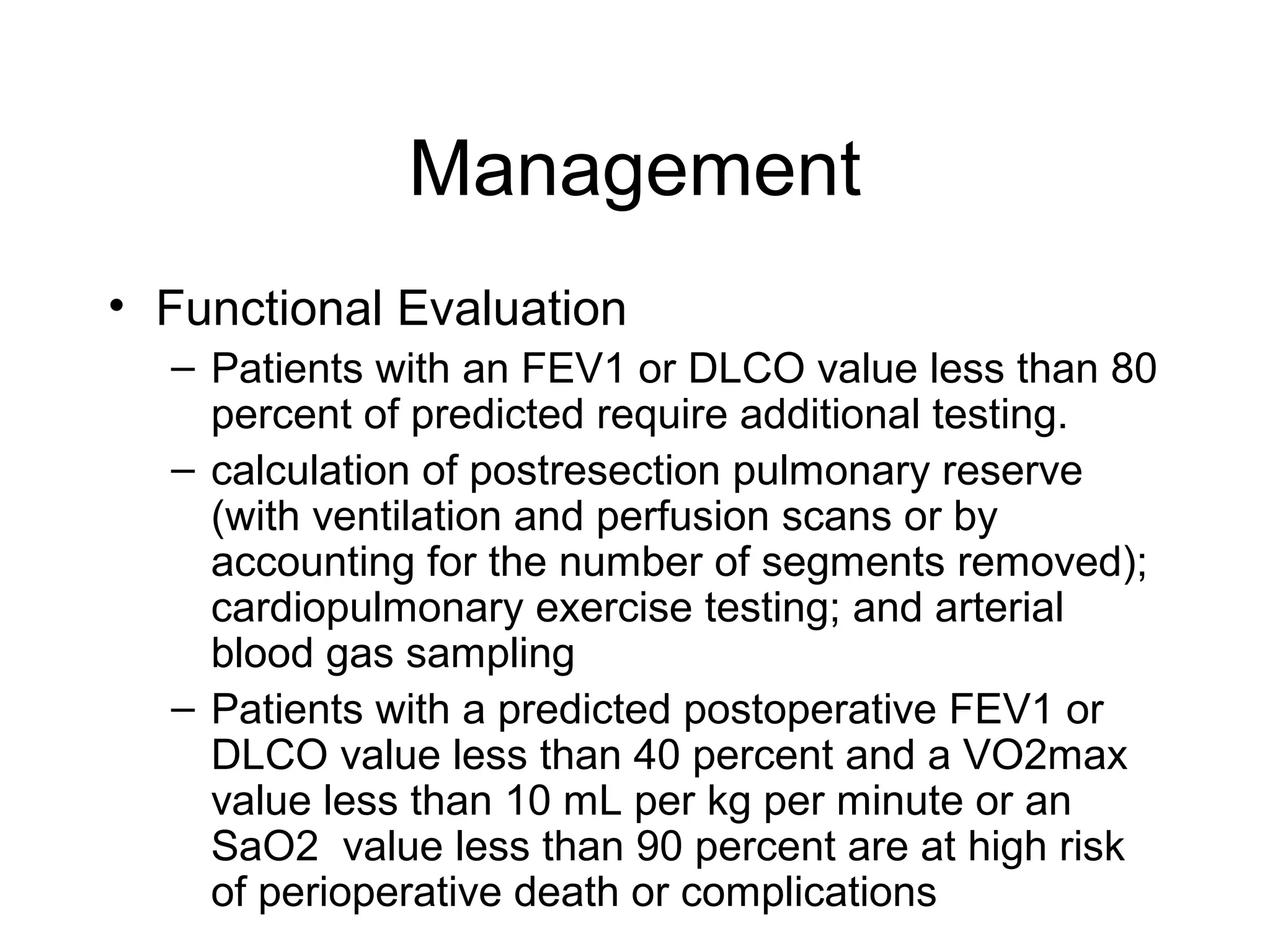
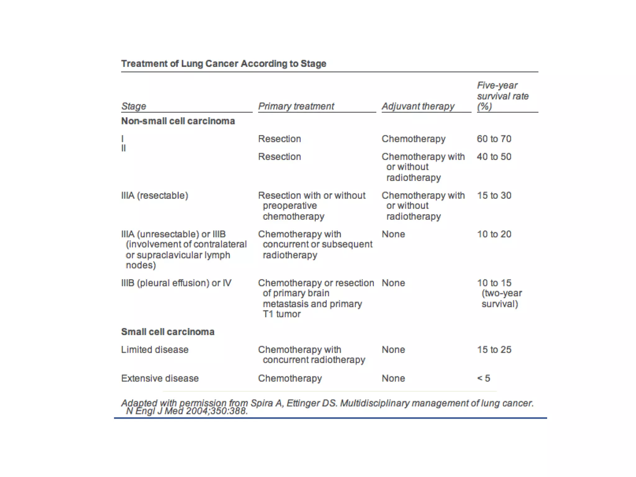
Lung Neoplasms
Sanjay Munireddy
Dept of Surgery
Sinai Hospital of Baltimore
June17, 2008
Overview
• Leading cause of cancer-related death
among men and women and 2nd most
common cause of overall mortality in
US
• Estimated new cases in 2008: 215,020
• Estimated deaths in 2008: 161,840
Epidemiology
• Recent continued decline in incidence
among men (79.4 cases per 100,00)
• Stabilization of incidence in women
(52.6 cases per 100,00)
• Greatest incidence in AA men (107.6
cases per 100,000)
NCI SEER Cancer Data
Risk Factors
•
•
•
•
•

Smoking
Second hand smoke
Sex - men
Race - African American
Environmental gases - Asbestos, radon, tar
soot, arsenic, silica etc.
• Excessive alcohol use
• Radiation therapy to chest
• Family history of lung cancer
Smoking
• Greatest risk factor; dose-response
relationship between the number of packyears smoked and lung cancer risk
• 87% of all lung cancer deaths result from
smoking
• Death rates decrease to that of neversmokers after 10 yrs of smoking cessation
1999 WHO Classification of
Lung Tumors
• Epithelial
– Malignant
•
•
•
•
•
•

Squamous cell carcinoma
Small cell carcinoma
Adenocarcinoma
Large cell carcinoma
Adenosquamous cell carcinoma
Carcinomas with pleomorphic, sarcomatoid or
sarcomatous elements
• Carcinoid tumor
Types
• Non-small cell lung cancer (NSCLC)
– Comprise 80% of lung tumors
– 50% are metastatic at diagnosis

• Small cell lung cancer (SCLC)
– Comprise 20%
– 80% are metastatic at diagnosis
Adenocarcinoma of Lung
• Most common type of lung cancer
• Comprises 30-40% in smokers and 60-80% in
non-smokers
• Arises from terminal bronchioles
• Usually develops in the peripheral portions of
the lung
• Slow growing than squamous cell ca.
• Often is associated with a peripheral scar or
honeycombing due to response to tumor
Squamous Cell Carcinoma of
Lung
• Comprise 25-40% of lung cancers; rates are
declining due to reduction in smoking
• Dose-response relationship of smoking is
strongest with this type of cancer
• Usually occurs in the lung’s central portions
or in one of the main airway branches.
• Can form cavities in the lung if they grow to a
large size
• Slow growing
Large Cell Carcinoma of Lung
• Accounts for 10-15% of lung tumors
• Diagnosis of exclusion; cannot diagnose on
small biopsies or in lymph node metastases
• Usually large peripheral mass with necrosis
• Often associated with peripheral eosinophilia
and leukocytosis, due to tumor production of
colony stimulating factor
Small Cell Carcinoma of Lung
• Also called undifferentiated or oat cell
carcinoma
• Accounts for 10-15% of lung tumors
• Almost always caused by smoking
• Fast growing compared to NSCLC
• Usually metastatic in about 70% of cases at
the time of diagnosis
• Without treatment, has the most aggressive
clinical course of any type of pulmonary tumor
• Median survival from diagnosis of only 2 to 4
months.
Clinical Presentation
• Majority are symptomatic at
presentation (>85%)
• Symptoms are broadly classified as
– Due to lung lesion
– Due to intra-thoracic spread
– Due to distant mets
– Due to paraneoplastic syndrome
Clinical Presentation
• Symptoms due to lung lesion/primary tumor
–
–
–
–
–
–

Coughing ± sputum
Dyspnea
Hemoptysis
Chest pain
Wheezing
Weight loss
Clinical Presentation
• Central tumors (squamous cell carcinomas)
generally produce symptoms of cough,
dyspnea, atelectasis, wheezing,
postobstructive pneumonia,, and hemoptysis.
• Most peripheral tumors are adenocarcinomas
or large cell carcinomas and, in addition to
causing cough and dyspnea, can cause
symptoms due to pleural effusion and severe
pain as a result of infiltration of parietal pleura
and the chest wall.
Clinical Presentation
• Symptoms of intra-thoracic spread
– Pleural or pericardial effusion
– Compression of RLN (hoarseness), phrenic nerve
palsy (elevated diaphragm), pressure on the
sympathetic plexus (Horner syndrome)
– Tracheal obstruction, esophageal compression,
SVC syndrome
– Superior sulcus tumors can cause compression of
the brachial plexus roots as they exit the neural
foramina, resulting in intense, radiating
neuropathic pain in the ipsilateral upper extremity.
Clinical Presentation
• Symptoms of distant spread
– May occur in almost every organ system
– Bone mets (vertebrae, ribs, pelvis are MC)
– Hepatic mets (indicate poor prognosis)
– Brain mets (headache, nausea, vomiting,
seizures, confusion, personality changes
Clinical Presentation
• Paraneoplastic syndromes (10%)
– Squamous cell carcinoma: hypercalcemia
due to parathyroidlike hormone production.
– Adenocarcinomas: Clubbing, hypertrophic
pulmonary osteoarthropathy and the
Trousseau syndrome of hypercoagulability
– Small cell carcinomas: SIADH, Ectopic
ACTH production, Lambert-Eaton
myasthenic syndrome
Diagnosis
• History & physical
–
–
–
–
–

Wt. loss, respiratory distress
Lymphadenopathy
Horner syndrome
SVC syndrome (usually SCLC)
Absence of breath sounds, dullness, pleural
effusions
– Bone pain
– Neurological deficits
Diagnosis
• CXR
• Sputum cytologic studies: centrally located
endobronchial tumors exfoliate malignant
cells into sputum
• Thoracentesis
• FNAB
• Bronchoscopy with BAL, brushings, biopsies
• Staging work-up
– Local extent
– Distant spread
Staging
• In the United States, the standard staging
workup includes at least the following:
– Complete history and physical examination
– CT scan of the chest and upper abdomen
(including liver and adrenals)
– Complete blood cell counts
– Liver and kidney functions tests
– Serum electrolytes
Staging
• Local extent
– Cervical mediastinoscopy
– Left anterior mediastinotomy

• Distant spread
– CT or Ultrasound of the abdomen
• liver, adrenals

–
–
–
–

Bone scan
CT head
MRI
PET scan
Management
• Functional Evaluation
– Evaluation of performance and pulmonary status
should be completed before discussing treatment
options
– Pulmonary function testing, specifically forced
expiratory volume in one second (FEV1) and
carbon monoxide diffusion in the lung (DLCO)
measurements, is a helpful predictor of morbidity
and mortality in patients undergoing lung resection
Management
• Functional Evaluation
– Patients with an FEV1 or DLCO value less than 80
percent of predicted require additional testing.
– calculation of postresection pulmonary reserve
(with ventilation and perfusion scans or by
accounting for the number of segments removed);
cardiopulmonary exercise testing; and arterial
blood gas sampling
– Patients with a predicted postoperative FEV1 or
DLCO value less than 40 percent and a VO2max
value less than 10 mL per kg per minute or an
SaO2 value less than 90 percent are at high risk
of perioperative death or complications
Lung cancer-overview-munireddy-20092810

Lung cancer-overview-munireddy-20092810

  • 1.
    Lung Neoplasms Sanjay Munireddy Deptof Surgery Sinai Hospital of Baltimore June17, 2008
  • 2.
    Overview • Leading causeof cancer-related death among men and women and 2nd most common cause of overall mortality in US • Estimated new cases in 2008: 215,020 • Estimated deaths in 2008: 161,840
  • 3.
    Epidemiology • Recent continueddecline in incidence among men (79.4 cases per 100,00) • Stabilization of incidence in women (52.6 cases per 100,00) • Greatest incidence in AA men (107.6 cases per 100,000) NCI SEER Cancer Data
  • 4.
    Risk Factors • • • • • Smoking Second handsmoke Sex - men Race - African American Environmental gases - Asbestos, radon, tar soot, arsenic, silica etc. • Excessive alcohol use • Radiation therapy to chest • Family history of lung cancer
  • 5.
    Smoking • Greatest riskfactor; dose-response relationship between the number of packyears smoked and lung cancer risk • 87% of all lung cancer deaths result from smoking • Death rates decrease to that of neversmokers after 10 yrs of smoking cessation
  • 6.
    1999 WHO Classificationof Lung Tumors • Epithelial – Malignant • • • • • • Squamous cell carcinoma Small cell carcinoma Adenocarcinoma Large cell carcinoma Adenosquamous cell carcinoma Carcinomas with pleomorphic, sarcomatoid or sarcomatous elements • Carcinoid tumor
  • 7.
    Types • Non-small celllung cancer (NSCLC) – Comprise 80% of lung tumors – 50% are metastatic at diagnosis • Small cell lung cancer (SCLC) – Comprise 20% – 80% are metastatic at diagnosis
  • 8.
    Adenocarcinoma of Lung •Most common type of lung cancer • Comprises 30-40% in smokers and 60-80% in non-smokers • Arises from terminal bronchioles • Usually develops in the peripheral portions of the lung • Slow growing than squamous cell ca. • Often is associated with a peripheral scar or honeycombing due to response to tumor
  • 9.
    Squamous Cell Carcinomaof Lung • Comprise 25-40% of lung cancers; rates are declining due to reduction in smoking • Dose-response relationship of smoking is strongest with this type of cancer • Usually occurs in the lung’s central portions or in one of the main airway branches. • Can form cavities in the lung if they grow to a large size • Slow growing
  • 10.
    Large Cell Carcinomaof Lung • Accounts for 10-15% of lung tumors • Diagnosis of exclusion; cannot diagnose on small biopsies or in lymph node metastases • Usually large peripheral mass with necrosis • Often associated with peripheral eosinophilia and leukocytosis, due to tumor production of colony stimulating factor
  • 11.
    Small Cell Carcinomaof Lung • Also called undifferentiated or oat cell carcinoma • Accounts for 10-15% of lung tumors • Almost always caused by smoking • Fast growing compared to NSCLC • Usually metastatic in about 70% of cases at the time of diagnosis • Without treatment, has the most aggressive clinical course of any type of pulmonary tumor • Median survival from diagnosis of only 2 to 4 months.
  • 12.
    Clinical Presentation • Majorityare symptomatic at presentation (>85%) • Symptoms are broadly classified as – Due to lung lesion – Due to intra-thoracic spread – Due to distant mets – Due to paraneoplastic syndrome
  • 13.
    Clinical Presentation • Symptomsdue to lung lesion/primary tumor – – – – – – Coughing ± sputum Dyspnea Hemoptysis Chest pain Wheezing Weight loss
  • 14.
    Clinical Presentation • Centraltumors (squamous cell carcinomas) generally produce symptoms of cough, dyspnea, atelectasis, wheezing, postobstructive pneumonia,, and hemoptysis. • Most peripheral tumors are adenocarcinomas or large cell carcinomas and, in addition to causing cough and dyspnea, can cause symptoms due to pleural effusion and severe pain as a result of infiltration of parietal pleura and the chest wall.
  • 15.
    Clinical Presentation • Symptomsof intra-thoracic spread – Pleural or pericardial effusion – Compression of RLN (hoarseness), phrenic nerve palsy (elevated diaphragm), pressure on the sympathetic plexus (Horner syndrome) – Tracheal obstruction, esophageal compression, SVC syndrome – Superior sulcus tumors can cause compression of the brachial plexus roots as they exit the neural foramina, resulting in intense, radiating neuropathic pain in the ipsilateral upper extremity.
  • 16.
    Clinical Presentation • Symptomsof distant spread – May occur in almost every organ system – Bone mets (vertebrae, ribs, pelvis are MC) – Hepatic mets (indicate poor prognosis) – Brain mets (headache, nausea, vomiting, seizures, confusion, personality changes
  • 17.
    Clinical Presentation • Paraneoplasticsyndromes (10%) – Squamous cell carcinoma: hypercalcemia due to parathyroidlike hormone production. – Adenocarcinomas: Clubbing, hypertrophic pulmonary osteoarthropathy and the Trousseau syndrome of hypercoagulability – Small cell carcinomas: SIADH, Ectopic ACTH production, Lambert-Eaton myasthenic syndrome
  • 18.
    Diagnosis • History &physical – – – – – Wt. loss, respiratory distress Lymphadenopathy Horner syndrome SVC syndrome (usually SCLC) Absence of breath sounds, dullness, pleural effusions – Bone pain – Neurological deficits
  • 19.
    Diagnosis • CXR • Sputumcytologic studies: centrally located endobronchial tumors exfoliate malignant cells into sputum • Thoracentesis • FNAB • Bronchoscopy with BAL, brushings, biopsies • Staging work-up – Local extent – Distant spread
  • 20.
    Staging • In theUnited States, the standard staging workup includes at least the following: – Complete history and physical examination – CT scan of the chest and upper abdomen (including liver and adrenals) – Complete blood cell counts – Liver and kidney functions tests – Serum electrolytes
  • 21.
    Staging • Local extent –Cervical mediastinoscopy – Left anterior mediastinotomy • Distant spread – CT or Ultrasound of the abdomen • liver, adrenals – – – – Bone scan CT head MRI PET scan
  • 23.
    Management • Functional Evaluation –Evaluation of performance and pulmonary status should be completed before discussing treatment options – Pulmonary function testing, specifically forced expiratory volume in one second (FEV1) and carbon monoxide diffusion in the lung (DLCO) measurements, is a helpful predictor of morbidity and mortality in patients undergoing lung resection
  • 24.
    Management • Functional Evaluation –Patients with an FEV1 or DLCO value less than 80 percent of predicted require additional testing. – calculation of postresection pulmonary reserve (with ventilation and perfusion scans or by accounting for the number of segments removed); cardiopulmonary exercise testing; and arterial blood gas sampling – Patients with a predicted postoperative FEV1 or DLCO value less than 40 percent and a VO2max value less than 10 mL per kg per minute or an SaO2 value less than 90 percent are at high risk of perioperative death or complications