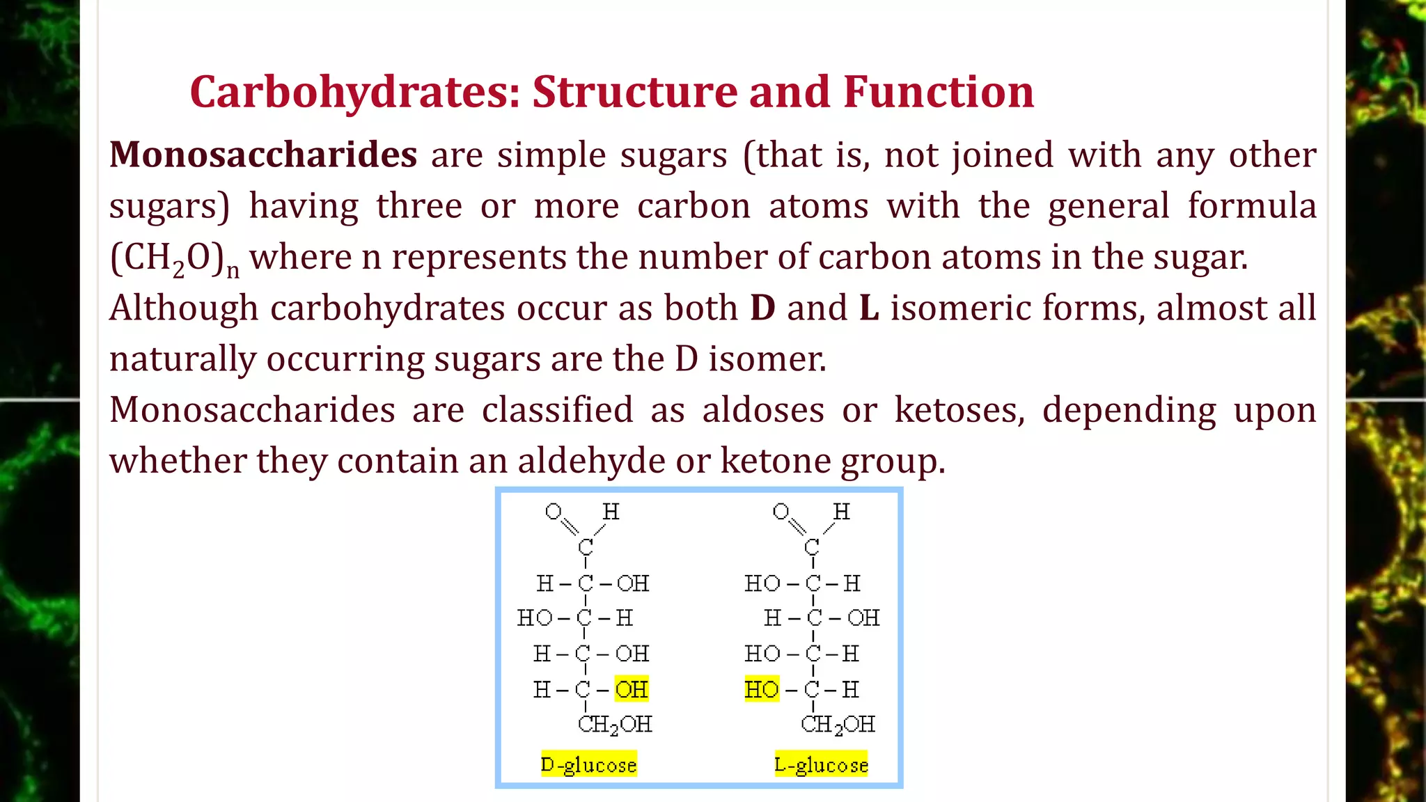
This tackles the topic on Lipids, generally. This include its uses, structures, metabolism and Chemical reactions involved with it.
This is a great help for learners in Junior High School, senior high school and those who are majoring in Science.
This tackles the topic on Lipids, generally. This include its uses, structures, metabolism and Chemical reactions involved with it.
This is a great help for learners in Junior High School, senior high school and those who are majoring in Science.
Austin Biomolecules: open access is a peer reviewed, scholarly journal dedicated to publish articles covering all areas of Biomolecules.
The journal aims to promote latest information and provide a forum for doctors, researchers, physicians, and healthcare professionals to find most recent advances in the areas of Biomolecules. Austin Biomolecules: open access accepts research articles, reviews, mini reviews, case reports and rapid communications covering all aspects of Biomolecules.
Austin Biomolecules: open access strongly supports the scientific up gradation and fortification in related scientific research community by enhancing access to peer reviewed scientific literary works. Austin Publishing Group also brings universally peer reviewed journals under one roof thereby promoting knowledge sharing, mutual promotion of multidisciplinary science.
bio chemistry
كيمياء حيوية جامعة الملك سعود
chemistry
كيمياء جامعية
0503964728
محمد منير كيمياء
ابو يوسف
all branched of chemistry bio chemistry - organic chemistry - inorganic chemistry - analytically - spectra - d-block
nucleic acid, glucose, fructose, preparation of sucrose, monosaccahrides, disaccharides, pedptide bond, glycosidic linkage, gluconic acid, DNA, RNA, Structure of amines, zwitter ion of amino acids, fibrous and globular protein,denaturation of proteins, Chemical properties of glucose, alpha helix and beta folded structure, ring structure of glucose and fructose, biomolecules, polyhydroxy aldose, poly hydroxy ketose
Carbohydrates are the sugars, starches and fibers found in fruits, grains, vegetables and milk products. Though often maligned in trendy diets, carbohydrates — one of the basic food groups — are important to a healthy diet.
Introduction
Classification of carbohydrate
Monosaccharide
The Common Monosaccharide Have Cyclic Structure
Organism Contain a Variety of Hexose Derivatives
Monosaccharide Are Reducing Agents
Disaccharide
Polysaccharide
Type of polysaccharide
Some Homopolysaccharides Are Stored Forms of Fuel
Some Homopolysaccharides Serve Structural Roles
Homopolysaccharied Folding
Reaction of Sugar
Conclusion
Reference
Lec 1 Carbohydrates-1.pptx the signs and symptoms of Kwashiorkor and Marasmus...phatimamohamett054
For security reasons why you think some students do the signs and symptoms of Kwashiorkor disease ppt biochemistry and Marasmus and Marasmus and Marasmus and Marasmus and Marasmus and Marasmus and the children are the signs of all that would have small mouth
Carbohydrate is an organic compound that consists only of carbon (C), hydrogen & oxygen. The primary function of carbohydrates is to provide energy for the body.
Simple carbohydrates have one or two sugar molecules.
Complex carbohydrates have three or more sugar molecules, such as legumes, bread, rice, pasta.
Biochemistry or biological chemistry is the study of chemical processes within and relating to living organisms. Biochemistry is the application of chemistry to the study of biological processes at the cellular and molecular level. It emerged as a distinct discipline around the beginning of the 20th century when scientists combined chemistry, physiology, and biology to investigate the chemistry of living systems.
This features the types of chemical reactions: Combustion, Neutralization, Precipitation and RedOx Reactions.
There are sample in each of the type of reaction that can help the learners understand more about each type.
This tackles the basics and the easiest concept of Chemical reactions. This features only the four basic types of chemical reactions: synthesis, decomposition, metathesis, and ion - exchange reaction.
This is a basic concept because there is a pattern to be followed in each type of reaction.
More types of chemical reactions will be given on my next set of presentation entitled, "Everything You Want to Know About Chemical Reactions."
Reproduction of plants and simple animalsRAJEEVBAYAN1
This is a concept that tackles about the different methods of reproduction, asexual and sexual.
This features how organisms reproduce to offspring other than man and higher animals.
This covers the topic on Proteins, in general. This discusses the different amino acids, bonds formed, structure of proteins and also the different chemical reactions involved with it.
This material is a great help for high school students and students taking up medical and science courses.
Basics of Chemistry: Chemical stoichiometryRAJEEVBAYAN1
This material presents quantitative method of numerical measurements involved in a chemical reaction.
this involves quantities such as the measures of mass in grams and the amount of substance in moles.
I am hoping that this material will help to make the concept easier.
This showcases the basics of the laws governing behavior of gases which includes:
1. Boyle's Law
2. Charles's Law
3. Gay - Lussac's Law
4. Combined Gas Law
5. Avogadro's Law
6. Ideal Gas Law
7. Dalton's Law on Partial Pressures
8. Graham's Law of Diffusion
Observation of Io’s Resurfacing via Plume Deposition Using Ground-based Adapt...Sérgio Sacani
Since volcanic activity was first discovered on Io from Voyager images in 1979, changes
on Io’s surface have been monitored from both spacecraft and ground-based telescopes.
Here, we present the highest spatial resolution images of Io ever obtained from a groundbased telescope. These images, acquired by the SHARK-VIS instrument on the Large
Binocular Telescope, show evidence of a major resurfacing event on Io’s trailing hemisphere. When compared to the most recent spacecraft images, the SHARK-VIS images
show that a plume deposit from a powerful eruption at Pillan Patera has covered part
of the long-lived Pele plume deposit. Although this type of resurfacing event may be common on Io, few have been detected due to the rarity of spacecraft visits and the previously low spatial resolution available from Earth-based telescopes. The SHARK-VIS instrument ushers in a new era of high resolution imaging of Io’s surface using adaptive
optics at visible wavelengths.
Earliest Galaxies in the JADES Origins Field: Luminosity Function and Cosmic ...Sérgio Sacani
We characterize the earliest galaxy population in the JADES Origins Field (JOF), the deepest
imaging field observed with JWST. We make use of the ancillary Hubble optical images (5 filters
spanning 0.4−0.9µm) and novel JWST images with 14 filters spanning 0.8−5µm, including 7 mediumband filters, and reaching total exposure times of up to 46 hours per filter. We combine all our data
at > 2.3µm to construct an ultradeep image, reaching as deep as ≈ 31.4 AB mag in the stack and
30.3-31.0 AB mag (5σ, r = 0.1” circular aperture) in individual filters. We measure photometric
redshifts and use robust selection criteria to identify a sample of eight galaxy candidates at redshifts
z = 11.5 − 15. These objects show compact half-light radii of R1/2 ∼ 50 − 200pc, stellar masses of
M⋆ ∼ 107−108M⊙, and star-formation rates of SFR ∼ 0.1−1 M⊙ yr−1
. Our search finds no candidates
at 15 < z < 20, placing upper limits at these redshifts. We develop a forward modeling approach to
infer the properties of the evolving luminosity function without binning in redshift or luminosity that
marginalizes over the photometric redshift uncertainty of our candidate galaxies and incorporates the
impact of non-detections. We find a z = 12 luminosity function in good agreement with prior results,
and that the luminosity function normalization and UV luminosity density decline by a factor of ∼ 2.5
from z = 12 to z = 14. We discuss the possible implications of our results in the context of theoretical
models for evolution of the dark matter halo mass function.
Richard's entangled aventures in wonderlandRichard Gill
Since the loophole-free Bell experiments of 2020 and the Nobel prizes in physics of 2022, critics of Bell's work have retreated to the fortress of super-determinism. Now, super-determinism is a derogatory word - it just means "determinism". Palmer, Hance and Hossenfelder argue that quantum mechanics and determinism are not incompatible, using a sophisticated mathematical construction based on a subtle thinning of allowed states and measurements in quantum mechanics, such that what is left appears to make Bell's argument fail, without altering the empirical predictions of quantum mechanics. I think however that it is a smoke screen, and the slogan "lost in math" comes to my mind. I will discuss some other recent disproofs of Bell's theorem using the language of causality based on causal graphs. Causal thinking is also central to law and justice. I will mention surprising connections to my work on serial killer nurse cases, in particular the Dutch case of Lucia de Berk and the current UK case of Lucy Letby.
This pdf is about the Schizophrenia.
For more details visit on YouTube; @SELF-EXPLANATORY;
https://www.youtube.com/channel/UCAiarMZDNhe1A3Rnpr_WkzA/videos
Thanks...!
A brief information about the SCOP protein database used in bioinformatics.
The Structural Classification of Proteins (SCOP) database is a comprehensive and authoritative resource for the structural and evolutionary relationships of proteins. It provides a detailed and curated classification of protein structures, grouping them into families, superfamilies, and folds based on their structural and sequence similarities.
2. OVERVIEW
Living things are composed of lifeless molecules.
These molecules, when isolated and examined individually, conform to all to all the physical
and chemical laws that describes the behavior of inanimate matter.
Living things possess extraordinary attributes not shown by collections of inanimate matter.
If we examine some of these special properties, we can approach the study of biochemistry
with a better understanding of the fundamental questions it seeks to answer.
BIOCHEMISTRY is the study of life at the molecular level.
It focuses on the compounds found within the living organisms, such as enzymes and
deoxyribonucleic acids (DNA), and the processes in which they function, such as catalysis
and replication.
Carbon, with four valence electrons, can bond to as many as four of the same or different
atoms or groups.
Structures containing carbon may be in a linear, branched, cyclic, or tetrahedral (four – faced
pyramid – shaped) form.
For biochemical reactions to occur, there usually must be an optimal spatial arrangement
between the reacting molecules.
3. Carbohydrates: Structure and Function
Carbohydrates (commonly called sugars) are aldehydes and
ketones containing multiple hydroxyl groups; the word
“carbohydrates” simply indicates a hydrate of a carbon –
containing compound.
Carbohydrates exist as individual molecules, called
monosaccharides, or as a number of individual
carbohydrate molecules joined by glycosidic bonds;
carbohydrates joined through covalent bonds are called
oligo- (a few) or poly- (more than three) saccharides,
depending on how many carbohydrates are joined as one
molecule.
4. Carbohydrates: Structure and Function
Starch is the storage form of carbohydrates in plants;
glycogen is the storage form of carbohydrates in animals;
both are polysaccharides.
The monosaccharides ribose and deoxyribose comprise part
of RNA and DNA, respectively; these molecules are
responsible for transmitting genetic information.
Other biochemically important carbohydrates include glucose
(a monosaccharide that is the principal source of energy for
most cells), fructose (a monosaccharide found in fruit and
honey), and lactose (a disaccharide found in milk).
Polysaccharides such as cellulose provide structure in the cell
walls of bacteria and plants.
5. Carbohydrates: Structure and Function
Monosaccharides are simple sugars (that is, not joined with any other
sugars) having three or more carbon atoms with the general formula
(CH2O)n where n represents the number of carbon atoms in the sugar.
Although carbohydrates occur as both D and L isomeric forms, almost all
naturally occurring sugars are the D isomer.
Monosaccharides are classified as aldoses or ketoses, depending upon
whether they contain an aldehyde or ketone group.
6. Carbohydrates: Structure and Function
The simplest monosaccharides are trioses (three carbon
sugars); glyceraldehyde is the simplest 3 – carbon aldose and
dihydroxyacetone is the simplest 3 – carbon ketone.
7. Carbohydrates: Structure and Function
Ribose, an important pentose (5 – carbon sugar), is a
constituent of RNA and also an intermediate in a metabolic
pathway called the pentose phosphate pathway.
Two important metabolic hexoses (6 – carbon
sugars) are glucose, an aldose, and fructose, a ketose.
In solution, pentoses and hexoses are found almost
in ring forms: Haworth projections are
representations of monosaccharide ring structures.
8. Carbohydrates: Structure and Function
The designations α and β indicate whether the hydroxyl group of
the anomeric carbon (the new asymmetric center of a
carbohydrate in a ring form) lies below (α) or above (β) the plane
of the ring when the anomeric carbon is to the right, and the ring
is numbered in clockwise direction in Haworth projection.
9. Carbohydrates: Structure and Function
In ribose, the carbonyl carbon (C-1) can react with the C-4 hydroxyl
group, with rotation around C-3 to C-4 bond, to form a five – membered
ring called a furanose; the C-1 of ribose also can react with the C-5
hydroxyl group, to form a six – membered ring called a pyranose.
10. Carbohydrates: Structure and Function
In glucose solutions,
the carbonyl carbon
(C-1) reacts with the
C-5 hydroxyl group
to form a pyranose
ring.
In fructose solutions,
the ketone carbon at
C-2 reacts with the
C-5 hydroxyl group
to form a furanose
ring.
11. Carbohydrates: Structure and Function
Monosaccharides are typically phosphorylated, a modification
that imparts certain characteristics to a sugar.
Phosphorylated carbohydrates are sources of energy because the
phosphate bond releases energy when it is hydrolyzed.
Phosphorylated carbohydrates are anionic; their negative charge
renders them to participate in electrostatic interactions.
Phosphorylated carbohydrates are reactive intermediates in
many metabolic pathways and in the synthesis of purines and
pyrimidines.
12. Carbohydrates: Structure and Function
A disaccharide is
composed of two
monosaccharides joined by
O – glycosidic bond
between the hydroxyl
group at the anomeric
carbon of the first
carbohydrate and a carbon
of the second
carbohydrate.
13. Carbohydrates: Structure and Function
Sucrose (glucose-α,(1⟶2)-β-fructose) contains glucose and
fructose linked between C-1 of α-D-glucose and C-2 of β-D-
fructose, with the bond from glucose projecting below (α) and
the bond from fructose projecting above (β) the plane of the
rings.
Lactose is galactose-β(1⟶4)-glucose.
14. Carbohydrates: Structure and Function
An oligosaccharide is a general form
used for a carbohydrate containing
between two and eight
monosaccharides; alternatively, a prefix
indicating the number of carbon atoms
may be used to denote the specific
number of monosaccharides.
Polysaccharides, also called complex
carbohydrates, contain more than
eight of the same or different
monosaccharides.
15. Carbohydrates: Structure and Function
Glycogen, the main storage form of glucose in animal cells, is a
large branched polymer of glucose units linked by α(1⟶4)
and α(1⟶6) glycosidic bonds.
a. Branching, which occurs approximately once per 10
glucose units, increases the volubility of glycogen and
creates additional terminal units, points at which glycogen
synthesis and degradation occurs.
b. Glycogen is stored as granules in
the cytosol, primarily in liver and
skeletal muscle cells.
16. Carbohydrates: Structure and Function
c. Glycogen catabolism and synthesis (a primary function of
the liver) help regulate the concentration of glucose in the
blood,
1. When the blood glucose concentration is low, glycogen is
catabolized to glucose in a process called glycogenolysis
and then released into the blood.
2. When the blood glucose concentration is high (implying
an adequate glucose supply), glucose is removed from the
blood for glycogen synthesis; this process, called
glycogenesis, permits excess glucose to be stored.
17. Carbohydrates: Structure and Function
Starch and cellulose are the main storage forms of glucose in plant cells.
a. Starch, as a glucose reservoir for metabolic reactions, occurs in the
forms of amylose and amylopectin.
(1) Amylose is composed of glucose units linked by α(1⟶4) glycosidic
bonds, forming only straight chain structures.
(2) Amylopectin is composed of glucose units linked by α(1⟶4) and
α(1⟶6) glycosidic bonds, forming straight chain and branched
structures, respectively; branching occurs less frequently than in
glycogen, approximately once per 30 glucose units.
18. Carbohydrates: Structure and Function
Cellulose serves a structural role in plants; it is a straight
chain of glucose units linked by β(1⟶4) glycosidic bonds.
(1) The β(1⟶4) linkages of cellulose impart the high tensile
strength characteristic of woody and fibrous plants.
(2) Humans lack the enzymes (cellulases) necessary to digest
cellulose, resulting in their inability to digest certain parts
of plants.
19. Carbohydrate Metabolism
Carbohydrates serve as sources of energy, storage forms of energy,
intermediates in metabolic processes, and structural compounds.
Complex carbohydrates are catabolized to glucose, which in turn is used to
generate energy as adenosine triphosphate (ATP) and reducing power in the
form of reduced nicotinamide adenine dinucleotide phosphate (NADPH).
Glucose is stored as glycogen when glucose supply is plentiful; conversely,
glycogen is used to produce when the glucose supply is inadequate.
When the need for glucose exceeds its supply, certain specialized tissues can
convert noncarbohydrates to glucose
(a) In mammals, the liver and kidneys are the principle organs for
gluconeogenesis, synthesis of new glucose molecules from
noncarbohydrate precursors.
(b) Humans can synthesize new glucose from pyruvate, lactate, tricarboxylic
acid (TCA) cycle intermediates, glycerol, and most, but not all, amino acids.
20. Carbohydrate Metabolism: Glycogenolysis
When the blood glucose concentration is low, glycogen is degraded and
transported by the blood to organs that must have a constant glucose
supply, such as the brain (which lacks energy stores) and skeletal
muscles (which have large stores of glycogen but utilizes large quantities
of glucose).
Recall that glycogen is a large branched
polymer of glucose units, linked by
α(1⟶4) and α(1⟶6) glycosidic bonds
that form straight chains and branches,
respectively; the highly branched
structure makes glycogen a starch with
many nonreducing ends (final glucose
residues without a free anomeric
carbon).
21. Carbohydrate Metabolism: Glycogenolysis
Glycogen phosphorylase cleaves glucose from glycogen only as a result of
a cascade of events that convert the enzyme from its inactive to its active
form.
(1) Glycogen phosphorylase exists in a b form
(dephosphorylated) and an a form
(phosphorylated); only the a form is the active
enzyme.
(2) Glycogen phosphorylase b form
(dephosphorylated) is converted to glycogen
phosphorylase a through the action of another
enzyme, phosphorylase kinase.
(3) To convert inactive glycogen phosphorylase b to active glycogen
phosphorylase a, phosphorylase kinase simply adds inorganic phosphate
to the enzyme.
22. Carbohydrate Metabolism: Glycogenolysis
(4) Glycogen phosphorylase a is converted to glycogen phosphorylase b
through he action of the enzyme phosphoprotein phosphatase.
(5) Phosphorylase kinase, like glycogen phosphorylase, also exists in an
active a and an inactive b form; it is activated by the cyclic adenosine
monophosphate (cAMP) – dependent protein kinase; it is also partially
activated by calcium ions; one of its subunits is the calcium binding
protein calmodulin.
The glycogen phosphorylase reaction removes
glucose units one at a time until fur units remain
on a branch; the limit branch is a four – unit
branch.
23. Carbohydrate Metabolism: Glycogenolysis
When four glucose units remain on a branch, a debranching enzyme
moves the three outer units of the limit branch to the nonreducing end of
another branch to be available for removal by glycogen phosphorylase.
The last glucose unit of the original branch is attached to glycogen
through an α(1⟶6) glycosidic bond; the debranching enzyme hydrolyzes
this final glycosidic bond.
24. Carbohydrate Metabolism: Glycogenolysis
The enzyme phosphoglucomutase catalyzes the conversion of glucose 1 –
P to glucose 6 – P.
a. There is no glucose 6 – P transport system across cell membranes;
glucose 6 – P remains in the cell where it was created.
b. In muscle cells, glucose 6 – P can enter glycolysis.
25. Carbohydrate Metabolism: Glycogenolysis
c. Liver, intestine and kidney cells contain the enzyme glucose 6 –
phosphatase, which removes the phosphate group from glucose 6 – P to
form glucose; glucose then enter the blood for delivery to cells that need
it.
(1) Brain and skeletal muscle lack glucose 6 – P in these cells that need
it quickly.
(2) Muscles store glycogen to have a ready supply of glucose; brain cells
cannot live for more than five minutes without glucose; lack of
glucose 6 – phosphatase in these cells ensures that their intracellular
glucose supply is not sent to less glucose – dependent cells.
26. Carbohydrate Metabolism: Glycogenesis
Glycogen synthesis occurs
when blood glucose levels are
high, allowing excess glucose
to be stored.
a. The liver is a major
glycogen storage site; liver
glycogen regulates glucose
levels in the blood.
b. Muscle is a major glycogen
storage site; muscle
glycogen is a fuel reservoir
for ATP synthesis, required
by active muscle.
27. Carbohydrate Metabolism: Glycogenesis
In the first step, the enzyme
hexokinase catalyzes the
conversion of glucose to
glucose 6 – P, just as in
glycolysis.
The enzyme
phosphoglucomutase catalyzes
the conversion of glucose 6 – P
to glucose 1 – P.
Glucose 1 – P is activated by
complexing with uridine
triphosphate (UTP) to form
uridine diphosphate (UDP)-
glucose.
28. Carbohydrate Metabolism: Glycogenesis
Glucose 1-P reacts with UTP,
catalyzed by glucose 1-
phosphate uridylytransferase
(also called UDP-glucose
pyrophosphorylase), to form
UDP – glucose (UDPG) and
pyrophosphate.
Pyrophosphate is rapidly
hydrolyzed to two molecules of
inorganic phosphate in a
reaction catalyzed by
pyrophosphatase.
29. Carbohydrate Metabolism: Glycogenesis
Hydrolysis of pyrophosphate
has a large negative free
energy, but the formation of
UDPG has a free energy change
of nearly zero.
The free energy release that
accompanies the hydrolysis of
pyrophosphate drives the
reaction that forms UDPG.
The formation of UDPG is an
essentially irreversible
reaction.
30. Carbohydrate Metabolism: Glycogenesis
UDPG is added at the C – 4 hydroxyl
group of a nonreducing end of the
glycogen molecule.
a. Glycogen synthase, which
catalyzes this reaction, adds
glucose units only if the glycogen
molecule already contains more
than four glucose units.
b. Glycogen synthase has an active
(dephosphorylated) form and an
inactive (dephosphorylated)
form; each of these forms is
regulated by a separate enzyme.
31. Carbohydrate Metabolism: Glycogenesis
(1) Phosphoprotein phosphatase
dephosphorylates the glycogen
synthase to its active form.
(2) The enzyme cAMP-dependent
protein kinase phosphorylates
the glycogen synthase to its
inactive form
Branches-α(1⟶6) linkages – are added to the growing glycogen chain
through the action of α(1⟶4)-glucan-branching enzyme, which
transfers several glucose units to an interior glucose unit.
32. Carbohydrate Metabolism:
Regulation of Glycogen Synthesis and Degradation
Covalent modification of individual
enzymes controls their activities.
a. Phosphorylation of glycogen synthase,
the enzyme catalyzing glycogen synthesis,
has the opposite effect of
phosphorylation of glycogen
phosphorylase, the enzyme catalyzing
glycogen degradation.
b. When phosphorylated, glycogen
phosphorylase is active and glycogen
synthase is inactive; dephosphorylated
glycogen phosphorylase is inactive while
dephosphorylated glycogen synthase is
active and vice versa.
33. Carbohydrate Metabolism:
Regulation of Glycogen Synthesis and Degradation
The hormones insulin, glucagon, and
epinephrine (adrenalin) control whether
glycogen synthesis or degradation occurs.
a. Insulin lowers the blood glucose
concentration by increasing the
uptake of glucose by the liver and
other cells, thereby promoting
glycogen synthesis from the excess
blood glucose; glucose inhibits
glycogen phosphorylase a (the active
enzyme), to prevent the degradation
of glycogen.
34. Carbohydrate Metabolism:
Regulation of Glycogen Synthesis and Degradation
b. The effect of glucagon and epinephrine
is the reverse of insulin; glucagon and
epinephrine increase the blood glucose
level by stimulating glycogen breakdown.
(1) Epinephrine causes glycogen
degradation, primarily in skeletal
muscles; glucagon causes glycogen
degradation, primarily in the liver.
(2) The effect of both hormones are
mediated by the cAMP.
35. Carbohydrate Metabolism:
Regulation of Glycogen Synthesis and Degradation
(3) Epinephrine and glucagon bind to receptors in the
plasma membrane of skeletal muscle and liver cells,
respectively; their binding causes a signal coupling protein to
activate adenylate cyclase, the enzyme that catalyzes the
formation of cAMP.
(4) The cAMP activates a protein kinase that phosphorylates
both glycogen phosphorylase (activating it) and glycogen
synthase (inactivating it).
(5) The molecule cAMP is a second messenger in signal
transduction, while the hormones binding to their receptors
are the first messengers.
36. Carbohydrate Metabolism:
The Pentose Phosphate Pathway
The pentose phosphate pathway, also called
the hexose monophosphate shunt or the
phosphogluconate pathway, has two
functions: to generate reducing equivalents
and produce ribose 5 – phosphate.
Many biochemical reactions consume energy;
biochemical systems supply this energy
through molecules that carry reducing
equivalents (for example NADH and NADPH)
and through molecules that carry high-
energy phosphate bonds ( for example, ATP,
guanosine triphosphate [GTP], and UTP).
37. Carbohydrate Metabolism:
The Pentose Phosphate Pathway
The pentose phosphate pathway generates
reducing equivalents (conserved electrons and
hydrogen atoms) in the reduced form of
nicotinamide adenine dinucleotide phosphate
(NADPH).
a. NADPH is the phosphorylated form of reduced
nicotinamide adenine dinucleotide (NADH).
b. NADPH is generated in a series of reactions
comprising the oxidation – reduction phase of
the pentose phosphate pathway
c. The pentose phosphate pathway occurs in the
cytosol of the cells and is particularly
important in anabolic tissues because NADPH
is needed for biosynthesis of certain
important molecules such as fatty acids and
steroids.
38. Carbohydrate Metabolism:
The Pentose Phosphate Pathway
Ribose 5 – phosphate, another important
product of the pentose phosphate pathway, is
used in the synthesis of nucleotides (DNA and
RNA).
a. Ribose 5-P can be interconverted into
other carbohydrates containing three,
four, five, six or seven carbon atoms
b. A cell often uses the pentose phosphate
pathway because it needs NADPH; the
easy interconversion of ribose 5-P is
practical because it provides other sugars
that can enter glycolysis directly.
39. Carbohydrate Metabolism:
The Pentose Phosphate Pathway
Whether glucose proceeds through glycolysis
or the pentose phosphate pathway is
determined by the cell’s requirement for ATP
(produced in glycolysis) versus its
requirement for NADPH and ribose 5-
phosphate (produced in the pentose
phosphate pathway).
The reactions of the pentose phosphate
pathway occur in three main recognizable
stages: the oxidation – reduction stage, the
isomerization – epimerization stage, and the
carbon bond cleavage – rearrangement stage.
40. Carbohydrate Metabolism:
The Pentose Phosphate Pathway
Glucose 6 – phosphate (generated in the first
step of glycolysis) is oxidatively
dehydrogenated at C – 1, producing 6 –
phosphogluconolactone.
(1) Glucose 6 – P dehydrogenase catalyzes this
reaction and requires the cofactor
oxidized nicotinamide adenine
dinucleotide phosphate (NADP+), which is
reduced to NADPH + H+.
(2) This reaction is the rate – limiting step of
the pentose phosphate pathway and is
regulated by the level of NADP+ in the cell.
41. Carbohydrate Metabolism:
The Pentose Phosphate Pathway
In the next step, which is the commitment step of
the pathway, 6 – phosphoglucolactone is
hydrolyzed to 6 – phosphogluconate;
gluconolactonase catalyzes this reaction.
The compound 6 – phosphogluconate is
oxidatively decarboxylated to ribulose 5 –
phosphate (ribulose 5-P) by 6 – phosphogluconate
dehydrogenase; the cofactor NADP+ is reduced to
NADPH + H+.
The formation of ribulose 5-P completes the
oxidation-reduction stage of the pentose
phosphate pathway; all of the NADPHthat will be
generated by the entire pathway has been
generated at this point; for each molecule of
glucose entering the pathway, two molecules of
NADPH are generated.
42. Carbohydrate Metabolism:
The Pentose Phosphate Pathway
In the isomerization-epimerization stage,
ribulose 5-P is isomerized to ribose 5-
phosphate (ribose 5-P) in a reaction
catalyzed by ribulose 5-P isomerase; ribulose
5-P is also epimerized to xylulose 5-
phosphate in a reaction catalyzed by ribulose
5-P epimerase.
The remaining steps in this pathway (the
carbon-carbon bond cleavage –
rearrangement stage) are simply a
rearrangement of carbon atoms in which
three five-carbon sugars are converted to two
six-carbon sugars plus a three-carbon sugar.
43. Carbohydrate Metabolism:
The Pentose Phosphate Pathway
(1) Ribose 5-P and its epimer xylulose 5-phosphate are
converted, via a two – carbon transfer, to
glyceraldehyde 3 – phosphate (a 3 – carbon sugar)
and sedoheptulose 7 – phosphate (a 7-carbon
sugar); transketolase catalyzes this reaction.
(2) Glyceraldehyde 3-phosphate and sedoheptulose 7-
phosphate are converted, via three-carbon transfer,
to erythrose 4-pjosphate (a 4 – carbon sugar) and
fructose 6-phosphate (a 6-carbon sugar);
transaldolase catalyzes this reaction.
(3) Erythrose 4 – phosphate reacts with xylulose 5 –
phosphate, via a two-carbon transfer to produce
glyceraldehyde 3-phosphate and fructose 6-
phosphate; transketolase catalyzes this reaction.
44. Carbohydrate Metabolism:
Gluconeogenesis
Gluconeogenesis is the synthesis of glucose
from noncarbohydrate precursors.
Lactate (an end product of anaerobic
glycolysis), most amino acids (from the
breakdown of dietary protein), and glycerol
(an end product of triacylglycerol catabolism)
are the usual noncarbohydrate precursors.
Lactate and amino acids are converted to
either pyruvate or oxaloacetate, both of which
are starting molecules for gluconeogenesis.
(1) Lactate forms pyruvate in the reverse of
the reaction catalyzed by lactate
dehydrogenase in glycolysis.
45. Carbohydrate Metabolism:
Gluconeogenesis
2) All amino acids except leucine and lysine
can be converted to either pyruvate or
oxaloacetate and are glucogenic.
Glycerol is converted to dihydroxyacetone
phosphate, which can enter the
gluconeogenesis pathway.
(1) Glycerol is first phosphorylated to glycerol
3-phosphate (glycerol 3-P), catalyzed by
glycerol kinase.
(2) Glycerol 3-P is converted to
dihydroxyacetone phosphate, catalyzed by
glycerol 3-P dehydrogenase.
46. Carbohydrate Metabolism:
Gluconeogenesis
Some of the reactions of gluconeogenesis are
simply a reversal of glycolysis and use the
same enzymes as glycolysis, but other
reactions use enzymes unique to
gluconeogenesis; the reciprocal regulation of
the separate enzyme systems enables an
organism to meet its needs for both glycolysis
and gluconeogenesis.
Gluconeogenesis is necessary to maintain
adequate glucose levels in the blood,
especially when the diet is deficient in glucose
or when the need for glucose exceeds its
supply, such as during fasting.
47. Carbohydrate Metabolism:
Gluconeogenesis
The liver is the major organ that performs
gluconeogenesis; the kidney is the second.
The intracellular ATP concentration
influences gluconeogenesis activity.
a. Low ATP levels inhibit gluconeogenesis;
when ATP levels are low, acetyl coenzyme
A (CoA) reacts with oxaloacetate to from
citrate, and the TCA cycle proceeds to
produce more ATP.
b. High ATP levels promote gluconeogenesis;
a high ATP concentration decreases the
activity of the TCA activity allows
oxaloacetate, the starting point for both
TCA and gluconeogenesis, to be available
for gluconeogenesis.
48. Carbohydrate Metabolism:
Gluconeogenesis
All reactions of gluconeogenesis except the first
reaction, which is catalyzed by mitochondrial
pyruvate carboxylase, occur in the cytosol of
cells.
Four unique enzymes characterize
gluconeogenesis; pyruvate carboxylase,
phosphoenolpyruvate carboxykinase, fructose
1,6-bisphosphatase, and glucose 6-
phosphatase; the remainder of the enzymes on
this pathway are the enzymes of glycolysis.
The first reaction unique to gluconeogenesis,
the conversion of pyruvate to oxaloacetate,
takes place in the mitochondria; the remainder
of the reactions take place in the cytosol.
49. Carbohydrate Metabolism:
Gluconeogenesis
a. CO2 is added to pyruvate, forming
oxaloacetate; this reaction is catalyzed by
pyruvate carboxylase and is the rate – limiting
step of gluconeogenesis.
(1) Biotin, a vitamin, is a prosthetic group of
pyruvate carboxylase and carries the activated
CO2 molecule.
(2) One molecule of ATP is hydrolyzed to
adenosine diphosphate (ADP) and inorganic
phosphate (Pi).
(3) Acetyl CoA increases the activity of pyruvate
carboxylase; an increase in acetyl CoA signals
a need for moreoxaloacetate, the molecule that
brings acetyl CoA into the TCA cycle.
50. Carbohydrate Metabolism:
Gluconeogenesis
(4) Mitochondrial membranes do not possess a
transport system for oxaloacetate, yet oxaloacetate
must be transported to the cytosol for the
remaining steps of gluconeogenesis.
(5) In a simple reversal of the normal TCA cycle
reaction, oxaloacetate is reduced to malate
(catalyzed by malate dehydrogenase); malate can
cross the mitochondrial membrane; once in
cytosol, malate is reoxidized to oxaloacetate and
gluconeogenesis proceeds.
b. In the second reactionunique to
gluconeogenesis, oxaloacetate is
decarboxylated and phosphorylated to
phosphoenolpypyruvate;
phosphoenolpypyruvate carboxykinase catalyzes
this reaction and GTP provides the high-energy
phosphate group necessary for the reaction to
proceed; GDP and CO2 are also produced.
51. Carbohydrate Metabolism:
Gluconeogenesis
c. Phosphoenolpypyruvate follows the reverse
reactions of glycolysis until fructose 1,6-
bisphosphate (fructose 1,6-bis-P) is formed.
d. In the third reaction unique to gluconeogenesis,
fructose 1,6-bis-P is converted to fructose 6-P by
the enzyme fructose 1,6-bisphosphatase.
(1) This reaction, which is the reverse of the
phosphofructokinase (PFK) reaction in glycolysis,
is highly regulated by the same molecules that
regulate PFK.
(2) In glycolysis, AMP activates PFK; in
gluconeogenesis, AMP inhibits fructose 1,6-
bisphosphatase; AMP indicates a low cellular
energy charge and a need for ATP production
through glycolysis.
52. Carbohydrate Metabolism:
Gluconeogenesis
(3) In glycolysis, an increase in citrate inhibits PFK;
in gluconeogenesis an increase in citrate activates
fructose 1,6-bisphosphatase; citrate is a product of
the first TCA cycle reaction; accumulation of citrate
indicates a low need for glycolysis and high
availability of oxaloacetate for gluconeogenesis.
(4) In glycolysis, fructose 2,6-bisphosphate (fructose
2,6-bisP) activates PFK; in gluconeogenesis, fructose
2,6-bisP inhibits fructose 1,6-bisphosphatase;
abundant glucose triggers a hormone cascade that
increases fructose 2,6-bisP, leading to an increase in
glycolysis and a decrease ingluconeogenesis; low
glucose levels cause low fructose 2,6-bisP levels and
a decrease in PFK activity.
53. Carbohydrate Metabolism:
Gluconeogenesis
e. Fructose 6-P is converted to glucose 6-P by the
reversal of the glycolystic phosphoglucose
isomerase reaction.
f. In the fourth and last reaction unique to
gluconeogenesis, glucose 6-P is converted to
glucose by hydrolysis of the phosphate group;
glucose 6-phosphatase, which catalyzes the
reaction, is present only in the liver and
kidneys; thus only in the liver and kidneys are
capable for gluconeogenesis.
Glycolysis and gluconeogenesis are coordinately
regulated so that both processes do not operate
simultaneously (futile cycling) and hence waste
energy.
54. Carbohydrate Metabolism:
Gluconeogenesis
a. Glycolysis is controlled by regulation of its
enzymes and also by the concentration of its
substrate, glucose, in the blood
b. Gluconeogenesis is controlled by regulation
of its enzymes and also by the concentration
of its substrates (lactate, amino acids,
pyruvate, oxaloacetate, and glycerol) in the
blood.
In the liver, the conversion of lactate (produced
by anaerobic glycolysis in active skeletal muscle)
into glucose via gluconeogenesis is called the
Cori cycle.
55. Carbohydrate Metabolism:
Gluconeogenesis
a. Lactate is transported via the blood form
active skeletal muscle to the liver, where it is
converted to pyruvate.
b. Pyruvate is converted to glucose by
gluconeogenesis
c. The glucose synthesized in the liver is then
transported in the blood to active skeletal
muscle, where it is oxidized to produce energy
for muscle contraction.
56.
57. 1. Draw the structures (open chain and ring forms, where
appropriate) of glucose, fructose, galactose, ribose ,
glyceraldehyde and dihydroxyacetone.
2. Show the dehydration of two monosaccharides producing
sucrose, lactose and maltose structurally.
3. Explain how glucose is stored in animals and in plants.
4. List reactions with products of glycolysis, glycogenolysis,
glycogenesis, pentose phosphate pathway and gluconeogenesis.