SlideShare a Scribd company logo
1 of 110
Download to read offline
STRUCTURE AND CHEMICAL
REACTIONS OF PROTEINS
PREPARED BY RGB
PROTEIN COMPOSITION
Proteins are precisely defined sequences of amino acids
(residues) linked through peptide bonds to form polymers
(hundreds or thousands of molecules combined to produce a
single macromolecule.
A peptide is a molecule formed by linking together a small
number of amino acids (from two to a dozen) through
peptide bonds; both peptides and proteins usually have a
free amine group on one end of a molecule (the N – terminal
residue) and a free carboxyl group on the other end of the
molecule (the C – terminal residue).
The structure, function, and activity of a protein is
determined to a large extent by the number, sequence, and
chemical properties of its constituent amino acids.
PROTEIN COMPOSITION
All proteins are composed of varying sequence of 20 amino
acids called common amino acids.
a. Other amino acids that occur in proteins are
modifications of the common amino acids
b. Modifications include addition of functional groups and
bond formation between certain groups within the amino
acid.
c. Amino acids containing modifications are labeled derived
or modified amino acids; modifications occur after the
amino acids are incorporated into the protein.
The names of 20 common amino acids are usually written as
three-letter abbreviation with the first letter capitalized or
as a single capital letter designation.
STRUCTURE OF AMINO ACIDS
Amino acids contain a central carbon (C), called the α
carbon, to which four substituents are bound: an amine
group (-NH2), a carboxylic acid group (-COOH), a hydrogen
atom (H), and a group unique to each amino acid called the
side chain or R group.
a. Three of the four substituents on the α carbon (-NH2, -
COOH, and –H) are common to all amino acids.
b. The fourth substituent, the side chain (indicated by R),
varies and confers distinctive chemical properties to each
amino acid.
c. In standard nomenclature for amino acids, the carbons of
the R group are labeled with Greek letters, starting with β
for the first carbon attached to the α carbon.
STRUCTURE OF AMINO ACIDS
For example, in glutamic acid (three linear carbons in the R
group), the carbons are labeled β, γ, and δ, with the δ carbon
being he final carbonyl carbon in this R group.
All amino acids found in proteins are called α amino acids
because the amine group is bonded to the α carbon; however,
amino acids with an amine group attached to other carbon
atoms have important biochemical functions.
a. β-alanine, β-amino acid, is a building block of pantothenic
acid, which is one of the structural units of coenzyme A.
b. γ-aminobutyric acid (GABA), a γ-amino acid, is involved in
the transmission of nerve impulses.
STRUCTURE OF AMINO ACIDS
Because four different substituents are bonded to the α
carbon of an amino acid, the α carbon is the chiral center and
the amino acids (except glycine) are optically active.
a. Amino acids can be either D- or L-stereoisomers,
depending on the orientation of substituents around the
α carbon.
b. All amino acids found in proteins are L-amino acids; D-
amino acids are found in certain bacterial cell walls and
in some peptide antibiotics.
The structure and function of an amino acid is determined to
a large extent by the characteristics of its R group.
STRUCTURE OF AMINO ACIDS
One simple way to remember amino acids is to classify them
as having one of three different types of side chains (R
groups); amino acids with nonpolar side chains, amino acids
with charged polar side chains, and amino acids with
uncharged polar side chains.
a. The nine amino acids described below have a nonpolar
side chain and various structures and sizes.
1. Glycine (Gly, G), the simplest amino acid, has two
identical substituents (two hydrogen atoms) bonded to
the α carbon; because the α carbon is achiral, glycine is
the only amino acid that is not optically active.
2. Alanine (Ala, A) has a methyl group; valine (Val, V) has an
isopropyl group; leucine (LEU, L) and isoleucine (Ile, I)
contain isomers of butane.
STRUCTURE OF AMINO ACIDS
3. Phenylalanine (Phe, F) is so named because of its R group
can be described as a phenyl group attached to the methyl
group of alanine.
4. Proline (Pro, P) is uniquely in that its α-amino group, as
well as its α carbon, are incorporated into a single cyclic
structure; proline is more correctly classified as an imino
acid rather than an amino acid, because its α-NH2 group
contains a secondary nitrogen (one that is bonded to two
other alkyl (R) groups).
5. The R group of tryptophan (Trp, W) contains indole (a five-
member, nitrogen-containing ring fused to a benzene ring).
6. Methionine (Met, M) contains sulfur in a thioether (R-S-R)
linkage.
STRUCTURE OF AMINO ACIDS
b. The five amino acids described below here have charged
polar side chains.
(1) Aspartate and glutamate are the only two common
amino acids that bear a negative charge on the R group at
neutral pH.
- Aspartate (Asp, D) contains a carboxyl group (-COOH)
separated from the α carbon by a methylene (-CH2-) group.
- Glutamate (Glu, E) contains a carboxyl group (-COOH)
separated from the α carbon by two methylene groups.
(2) Lysine, arginine, and histidine are the only three
common amino acids that bear a positive charge on the R
group at neutral pH.
STRUCTURE OF AMINO ACIDS
- Lysine (Lys, K) contains a butylamine side chain.
- Arginine (Arg, R) contains a guanidinium group
separated from the α carbon by three methylene
groups.
- Histidine (His, H) contains a five –member
heterocyclic ring, known as a imidazole group.
c. The seven amino acids described below have
uncharged polar side chains.
(1) Serine (Ser, S) contains a hydroxymethyl (-
CH2OH) group attached to the α carbon; threonine
(Thr, T) contains ethanol attached to the α carbon;
tyrosine (Tyr, Y) contains phenol (a benzene ring
bearing a hydroxyl group); cysteine (Cys, C) contains
a sulfhydryl (-SH) group.
STRUCTURE OF AMINO ACIDS
(2) Asparagine (Asn, N) and glutamine (Gln, Q) are structurally similar to aspartate
and glutamate, respectively, except that an amide group replaces the carboxyl group
in the side chain of each; the designation Asx and Glx represent the sum of aspartate
and asparagine, or glutamate and glutamine, in a protein respectively.
CHEMICAL PROPERTIES
OF AMINO ACIDS
The chemical properties of the R group determine the chemical properties of each
amino acid.
The number of ionizable groups in an amino acid determine the extent to which it will
be ionized at a given pH in the body.
Amino acids may be categorized according to the functional groups and chemical
properties of their side chains; some amino acids groups fit more than one
classification.
a. The amino acids with aliphatic (non-ring) side chains are glycine, alanine, valine,
leucine and isoleucine.
- Glycine and alanine have the smallest aliphatic side chains; they play an important
structural role in proteins by fitting into spaces too small for a larger amino acid.
- Valine, leucine, and isoleucine have longer aliphatic side chains that are
hydrophobic; they tend to congregate in structures that enable them to avoid water.
CHEMICAL PROPERTIES
OF AMINO ACIDS
b. The aromatic (benzene-related) amino acids, phenylalanine, tyrosine, and
tryptophan, all have the aromatic group attached to the α carbon via a methylene (-
CH2-) group.
- Phenylalanine and tryptophan are hydrophobic and tend to form structures that
enable them to avoid water.
- The hydroxyl group on tyrosine makes it more hydrophilic and a more chemically
reactive acid than phenylalanine.
- Although histidine and proline contain ring structures, they are usually classified
differently.
c. Cysteine and methionine, the two sulfur – containing amino acids, are hydrophobic.
- Two cysteine residues can bond via their thiol (-SH) groups to form the amino acid
cystine; the thiol group of cysteine is chemically reactive and easily forms a disulfide
(-S-S-) through oxidation.
CHEMICAL PROPERTIES
OF AMINO ACIDS
- The formation of disulfide bonds is important in protein structures; disulfide bonds
commonly join two different peptide chains together or link two different cysteine
residues within the same peptide.
d. Serine and threonine have a side chain hydroxyl group that makes them both
hydrophilic and chemically reactive.
e. In proline, the incorporation of nitrogen from the α-amine group into the side
chain constrains the rotation of the molecule around the C – N bond; this bond
constraint affect the structure of a protein causing it to bend at the position of
proline residue.
f. The acidic amino acids aspartate and glutamate are dicarboxylic, monobasic amino
acids because they have two carboxyl groups (one bound to the α carbon and one
in the side chain) but only one amine group (bound to the α carbon); the carboxyl
groups of both amino acids are negatively charged at physiologic pH (7.4).
CHEMICAL PROPERTIES
OF AMINO ACIDS
g. Amino acids with basic side chains are lysine,
arginine, and histidine.
- The side chain amine group makes these amino acids
extremely polar and hydrophilic; at physiologic pH,
both lysine and arginine have positively charged
amine group.
- At physiologic pH, the imidazole ring in histidine
exists primarily in the unchanged form because the
imidazole ring has pK’a of approximately 6.
h. Asparagine and glutamine, the two amino acids that
contain an amide group in the side chain, are not
charged at physiologic pH.
IONIZATION OF AMINO ACIDS
All free amino acids contain at least two ionizable groups, the
α-carboxyl group and the α-amine group; the charge on the
amino acid R groups within a protein affects the three-
dimensional structure and reactivity of the protein.
Except for the first and last amino acids in a protein, all of the
α-carboxyl and the α-amine groups within a protein
participate in peptide bonds and thus do not contribute to
the protein’s overall charge.
The overall charge of a protein is determined by the charge
on the amino acid side chains (the charge on the R groups)
and the charge on the protein’s N- terminal and C-terminal
residues.
IONIZATION OF AMINO ACIDS
a. The pK’a of the α COOH is 2.35; at physiologic pH, the
proton is removed and the carboxyl group is in the
ionized form with a-1 charge.
b. The pK’a of the α NH2 is 9.69; at physiologic pH, the NH2 is
protonated to the ionized form (-NH3
—1) with a +1
charge.
All amino acids contain both an acidic group (the α-carboxyl
group) and a basic group (the α-amine group) in the same
molecule.
a. An amino acid without an ionizable side chain will have
one +1 charge (from the α-amine group) and one –1
charge (from the α-carboxyl group) at physiologic pH; its
net charge is zero.
b. A charged compound with a net charge of zero is called a
zwitterion.
IONIZATION OF AMINO ACIDS
The pH at which a compound exists as zwitterion is the
isoelectric point (pl).
The pH of the environment determines the degree of
ionization of an amino acid; this is illustrated in the titration
of the two ionizable groups of alanine.
a. At pH <2.35, the two ionizable groups (the α COOH and
the α NH2) are completely protonated; the carboxyl group
is un-ionized and esists as – COOH; the amine group has a
+1 charge and exists as – NH3
+1.
b. At pH = 2.35, which is the pK’a of the carboxyl group,
equal amounts of the ionized (-COO—1) and unionized (-
COOH) carboxyl group are present.
IONIZATION OF AMINO ACIDS
c. As the pH increases beyond 2.35, the carboxyl group on
most of the molecules remains unprotonated and thus is
negatively charged; the amine group is still protonated
because the pH has not reached the amine pK’a of 9.69,
and hence the amine group on most molecules is still
charged.
d. At the pH midway between the two pK’a values, the
negative charge from the carboxyl group equals the
positive charge from the amine group; this pH
([2.35+9.69]2 = 6.02) is the isoelectric point of alanine,
the pH at which alanine exists as zwitterion.
e. At pH=9.69, which is the pK’a of the amine group, equal
amounts of un-ionized (-NH2) and ionized (-NH3
+1) amine
groups are present; the carboxyl group, well beyond its
pK’a, remains ionized (-COO—1)
IONIZATION OF AMINO ACIDS
f. At pH>9.69, none of the ionizable groups of alanine are
protonated; the carboxyl group has a negative charge (-COO—
1) and the amine group is un-ionized (-NH2).
An ionizable group in the side chain of an amino acid has its
own pK’a; in these amino acids, the side chain pK’a also
affects the isoelectric point.
LEVELS OF PROTEIN STRUCTURE
All proteins have at least three levels of structures:
primary, secondary, and tertiary.
Proteins composed of more than one polypeptide chain
also have a quaternary structure.
Monomers are proteins composed of only one
polypeptide chain; proteins composed of two or more
polypeptide chains are called oligomers and are
classified by number of polypeptide chains; dimers
have two chains; trimers, three chains, tetramers, four
chains, and so on.
The primary, secondary, tertiary, and quaternary
structure of a protein controls its three-dimensional
shape, which partly determines its activity.
PRIMARY STRUCTURE OF PROTEIN
Amino acids are linked by peptide bonds.
a. A peptide bond forms as result of a condensation
reaction between the carbonyl carbon from the α COOH
of one amino acid and the amine nitrogen from the α
NH2 of the next amino acid; the reaction eliminates a
molecule of water.
b. The peptide bond is rigid and planar; it has a bond
length intermediate between that of a single bond and a
double bond.
c. The partial double-bond character of the peptide bond
restricts its ability to rotate, making it the backbone of
the protein.
d. Other bonds in amino acids are single bonds and
therefore have more freedom of rotation than peptide
bonds.
Insulin
PRIMARY STRUCTURE OF PROTEIN
The primary structure of a protein is the sequence and
number of amino acids linked by peptide bonds.
a. The convention for writing the primary structure of an
amino acid is to name its sequence of amino acid from
left to right.
b. The first amino acid (the N-terminal amino acid) has a
free α-amine group; the carboxyl group of this first
amino acid participates in the formation of the first
peptide bond.
c. Subsequent amino acids are written to the right of the
N-terminal amino acid; all of these residues are linked
by peptide bonds at their α-amine and α-carboxyl
groups.
d. The last amino acid (the C –terminal amino acid) has a
free α-carboxyl group.
Insulin
SECONDARY STRUCTURE OF PROTEIN
Intramolecular and intermolecular hydrogen bonding in
the primary structure give the protein its secondary
structure.
The α helix, the β pleated sheet, and the collagen triple
helix are three different types of secondary structure.
The α helix occurs in many different fibrous and globular
proteins; it is formed by intramolecular hydrogen bonds
between the carboxyl group oxygen of one amino acid and
the amine hydrogen of the fourth amino acid in front of it.
a. Every carboxyl oxygen and amine hydrogen participates
in the intramolecular bonding; the combined forces of
all these hydrogen bonds give the protein an overall
rodlike structure.
SECONDARY STRUCTURE OF PROTEIN
b. Formation of the α helix occurs spontaneously in a pattern of 3.6
amino acids per turn of the α helix.
c. Amino acids that are three to four residues apart in the primary
structure are spatially close to each other in the α helix; R groups that
are adjacent to each other in the primary structure are spatially
distant in the α helix.
d. The extent of α-helical structure in a protein depends on its
constituent amino acids; some amino acids (Leu, Ala, Glu, Met) tend to
favor α helix formation, whereas others (Val, Ser, Asp, Asn) tend to
destabilize it.
The β pleated sheet is the secondary structure of silk, a fibrous protein
produced by spiders and insects; it is formed by intermolecular hydrogen
bonds between a carboxyl group oxygen in one amino acid and an amine
group hydrogen in an adjacent polypeptide.
SECONDARY STRUCTURE OF PROTEIN
a. A parallel β pleated sheet is formed when adjacent
polypeptids are oriented in the same direction (from N-
terminal to C-terminal or vice versa).
b. An antiparallel β pleated sheet is formed when adjacent
polypeptides are oriented opposite directions (one
chain oriented from the N- to C- terminal and adjacent
chain oriented from C- to N- terminal).
c. Proteins that have β pleated sheets assume an
extended, rather than a rod-shaped or coiled, structure.
Collagen, the most abundant structural protein found in
vertebrates, is a major component of connective tissues,
such as bone, teeth, cartilage, and tendon; the secondary
structure of collagen is a triple helix composed of three
entwined polypeptide chains and bound by intermolecular
hydrogen bonds.
SECONDARY STRUCTURE OF PROTEIN
a. The collagen triple helix has a high
concentration of proline and glycine; often
every third residue in each of the three
chains is a glycine molecule.
b. Two derived amino acids, hydroxyproline
and hydroxylysine, occur frequently; the
sequence – Gly – Pro – hydroxyproline –
occurs frequently.
c. There are 3.3 amino acids per turn of the
triple helix, with approximately every third
residue projecting into the helix interior.
d. The high concentration of glycine (the
smallest amino acid) prevents crowding in
the interior of the helix.
TERTIARY STRUCTURE OF PROTEIN
A protein’s tertiary structure results from interactions between
amino acid side chains of each of the residues; these interactions
between R groups cause a polypeptide to assume its normal
three-dimensional configuration.
Many types of noncovalent interactions are involved, such as van
der Waals forces, ionic bonding, hydrophobic bonding, and
hydrogen bonding.
a. Residues with hydrophilic side groups usually are located on
the exterior of the molecule, oriented to interact with the
aqueous environment.
b. Residues with hydrophobic side groups usually are located
on the interior o the molecule, oriented so the hydrophobic
groups can interact with each other and exclude water.
One type of covalent bond that influences tertiary structures is
the disulfide bond of the derived amino acid.
QUATERNARY STRUCTURE OF PROTEIN
Most proteins are composed of more than one polypeptide
chain; these polypeptide subunits associate with each
other in a defined geometric arrangement called the
quaternary structure.
Formation of the quaternary structure involves the same
noncovalent interactions as in the formation of a tertiary
structure, but the interactions occur between polypeptide
chains (interchain) rather than within a polypeptide chain
(intrachain).
Oligomeric proteins composed of identical subunits are
called homogeneous; those composed of different subunits
are called heterogeneous.
IDENTIFICATION OF PROTEIN
The amino acid composition of protein can be discerned by
cleaving all the peptide bonds, then separating and identifying
the constituent amino acids.
The amino acid sequence of a protein can be discerned by using
various methods to cleave only selected peptide bonds, then
assimilating all the information to deduce the amino acid
sequence; this process is similar to putting together the pieces of
the puzzle.
Proteins are also identified and characterized by spectroscopy,
ultracentrifugation, chromatography, and electrophoresis.
More sophisticated aspects of protein characterization are
accomplished by X-ray diffraction, circular dichroism
spectroscopy, nuclear magnetic resonance spectroscopy, and
mass spectroscopy.
DETERMINING AMINO ACID
COMPOSITION AND SEQUENCE
In the first stage of determining the amino acid composition of a
protein, all the peptide bonds are hydrolyzed by an acid or a base.
a. In acid hydrolysis, the protein solution is usually treated with 6 N
hydrochloric acid at 110OC for 24 hours.
b. In base hydrolysis, the protein solution is usually treated with 2 to 4
N sodium hydroxide at 100 OC for 4 to 8 hours.
c. Under acid hydrolysis, aspartate cannot be distinguished from
asparagine and glutamate cannot be distinguished from glutamine;
the totals for both types of molecules are identified as Asx and Glx,
respectively.
d. Tryptophan is acid-labile and requires base hydrolysis for
identification.
DETERMINING AMINO ACID
COMPOSITION AND SEQUENCE
In the second stage of determining the amino acid
composition of a protein, the hydrolyzed amino acids are
separated, identified, and quantitated.
a. Amino acids can be separated by thin layer
chromatography or liquid chromatography, which is the
separation of molecules on a solid support according to
their physical or chemical properties.
b. Amino acids can be identified and quantitated by
reacting them with reagents that produce a colored or
fluorescent product, the intensity of which is
proportional to the concentration of the amino acid
present.
DETERMINING AMINO ACID
COMPOSITION AND SEQUENCE
(1)Ninhydrin, a reagent that turns blue when reacted with
amino acids and yellow when reacted with imino acids
(proline), is sensitive to microgram (10—6 g) quantities
of amino acids.
(2)Fluorescamine, a reagent that reacts with the α-amine
group to produce a fluorescent derivative, is more
sensitive than ninhydrin; fluorescamine can detect
nanogram (10—9 g) quantities of amino acids.
After determining the amino acid composition of the
protein, the determination of the structure includes
identifying the N-terminal residue and C-terminal residue.
DETERMINING AMINO ACID
COMPOSITION AND SEQUENCE
a. The N-terminal amino acid is identified by the reaction
of its free amine group in the intact protein.
(1)The reagent fluoronitrobenzene (FDNB) reacts with the
free amine group of a protein to form a covalent
dinitrophenyl derivative.
(2)The protein is subjected to acid hydrolysis and
chromatographic separation as previously described.
(3)The dinitrophenyl derivative has different
chromatographic properties than the other amino acids
and can thus be identified.
b. The free amine group in the side chain of lysine also
reacts with FDNB and must be considered when
interpreting results.
DETERMINING AMINO ACID
COMPOSITION AND SEQUENCE
c. An N-terminal amino acid that is modified on the
amine group will not have a free amine group to
react with FDNB.
d. Other reagents, such as dabsyl chloride and dansyl
chloride, frequently replace FDNB; they permit
detection of smaller quantities of the amino acid.
e. The Edman degradation is a method of identifying an
entire sequence of amino acids in a protein by
successively releasing individual N-terminal
residues that can be separated from the intact
protein and identified.
DETERMINING AMINO ACID
COMPOSITION AND SEQUENCE
(1)The reagent phenyl isothiocyanate (PITC, also called Edman’s reagent) reacts
with the N-terminal amino acid to form a thiazolinone derivative, which is then
extracted with organic solvent and converted to a more stable molecule called a
phenylthiohydantoin (PTH) amino acid.
(2)The technique used in the Edman degradation separates the N-terminal residue
only and leaves the rest of the polypeptide intact.
(3)The separated PTH amino acid is then identified using standard identification
techniques, such as chromatography and electrophoresis.
(4)The next amino acid is the new N-terminal amino acid and is sequentially labeled
and removed without altering the remaining protein.
(5)The cycles are repeated, with a new N-terminal amino acid generated during
each cycle, until all amino acids have been separated and identified in sequence.
DETERMINING AMINO ACID
COMPOSITION AND SEQUENCE
f. The identification of the C-terminal residue in a protein
is accomplished by treating the intact protein with a
carboxypeptidase, a class of enzymes that selectively
catalyze the removal of the C-terminal amino acid from
the peptide.
(1)Other enzymes that cleave specific peptide bonds on
their carboxyl side include trypsin, which cleaves the
residues that contain positively charged R groups (Lys
and Arg), and chymotrypsin, which cleaves at residues
with aromatic R groups (Phe, Tyr, and Trp) and bulky
aliphatic R groups (Ile and Val).
(2)Cyanogen bromide is a chemical that cleaves at the
carboxyl side of the peptide bond of methionine.
DISSOCIATION OF PROTEIN SUBUNITS
Proteins with multiple subunits must be broken down to
individual subunits to characterize their amino acid composition
and sequence.
Denaturation is the process of treating a protein with heat or
chemical reagents to break it down into its constituent subunits.
a. Denaturation causes loss of a protein’s secondary, tertiary, and
quaternary structure, but its primary structure may be
maintained.
b. In vivo, changes in pH, ionic strength, and temperature
denature proteins; in vitro, detergents, urea, organic solvents,
acids or bases denature proteins.
c. Denatured proteins lose their normal biochemical activity and
are nonfunctional.
d. Disulfide binds in proteins are disrupted when reducing
agents, such as β-mercaptoethanol or dithiothreitol, are added.
IDENTIFICATION, SEPARATION AND
CHARACTERIZATION OF PROTEINS
The presence of a protein and its integrity can be
detected by spectroscopy, a technique for
identifying and quantitating unknown molecules
by virtue of their interaction with light.
a. Absorption spectroscopy identifies peptide
bonds and certain functional groups by
measuring the absorbance of different
wavelengths UV and Visible light.
1. Peptide bonds absorb UV light
in the range of 180 to 230
nanometers (nm)
2. The aromatic rings in
phenylalanine, tryptophan
and tyrosine absorb UV in 260
to 300 nm.
3. Denaturation changes in secondary and
tertiary structure appear as a difference in
light absorbance between 180 to 230 nm.
IDENTIFICATION, SEPARATION AND
CHARACTERIZATION OF PROTEINS
Fluorescence spectroscopy, a more sensitive
measurement than UV or visible spectroscopy, detects the
presence and quantity of a protein.
1. Fluorescence occurs when a molecule in an excited
state, produced from absorption of short wavelength
(high energy) light, re-emits some of its excess energy
not emitted as light dissipates as heat.
2. Only proteins containing amino acids with aromatic
side chains (Phe, Tyr, Trp) will naturally fluoresce.
3. A protein that does not naturally fluoresce can be
made to fluoresce by covalently bonding a fluorescent
group to the protein.
IDENTIFICATION, SEPARATION AND
CHARACTERIZATION OF PROTEINS
Ultracentrifugation separates proteins according to
size.
a. The sedimentation coefficient of a protein,
subjected to a centrifugal (outward) force, depends
on he protein’s mass, density, and shape and on the
density of the suspending solution.
b. The Svedberg (S) unit, a measure of the
sedimentation coefficient of a particle, is equal to
10—13 second.
c. The magnitude of the sedimentation coefficient
gives a relative value that can characterize the
molecular weight of protein.
IDENTIFICATION, SEPARATION AND
CHARACTERIZATION OF PROTEINS
Gel filtration chromatography (also called size exclusion
chromatography) separates proteins according to size.
a. Inert porous beads are packed into a column; the pore
size is selected to correspond to the molecular weight
of molecules small enough to enter the pores.
b. A buffered protein solution is then applied to the top of
the packed column.
c. High molecular weight proteins are too large to enter
the pores in the beads and thus go directly to the
column.
d. Low molecular weight proteins enter the pores and
thus have a longer path to traverse on their way
through the column; some midsize proteins can enter
the pores while others cannot; they travel through the
column at rates between high and low molecular
weight proteins.
e. The elution time of each
protein is related to its
molecular size; large
proteins elute first,
midsize protein next and
small proteins elute last.
IDENTIFICATION, SEPARATION AND
CHARACTERIZATION OF PROTEINS
Ion exchange chromatography separates
proteins according to their charge.
a. Inert beads coated with either negatively
or positively charged groups are packed
into a column, and a buffered protein
solution is then applied to the top of the
packed column.
b. Proteins with a net positive charge will
bind, via ionic bonds, to beads coated with
negatively charged groups (cation
exchange); conversely, proteins with net
negative charge will bind to beads coated
with positively charged groups (anion
exchange).
c. The addition of buffer solution containing
a higher concentration of the same type of
ions as in the original protein solution
displaces, or exchanges, the proteins ionic
bonding to the charged groups on the
beads; this allows the researcher to
control when the protein of interest elutes
from the column.
IDENTIFICATION, SEPARATION AND
CHARACTERIZATION OF PROTEINS
Affinity chromatography separates proteins that bind
specifically with certain chemical groups.
a. Inert beads coated with specific chemical groups are
packed into a column; a buffered protein solution is
then applied to the top of the packed column.
b. A protein with an affinity for the specific chemical
group will bond to that group and consequently will
be retained on the column; proteins that lack affinity
will not bind and will flow directly through the
column.
c. Addition of higher concentration of a soluble form of
the specific chemical group displaces the protein from
the binding sites on the beads; the protein then elutes
separately.
IDENTIFICATION, SEPARATION AND
CHARACTERIZATION OF PROTEINS
Electrophoresis, the separation of charged
molecules by their movement on a solid support
while exposed to an electric field, also aids in the
separation of different proteins.
a. Electrophoresis separates proteins on the
basis of their mass if they can be made to
have the same charge.
b. The anionic detergent Sodium dodecyl
sulfate (SDS) is added to a protein solution;
SDS binds strongly to most proteins, causing
denaturation and imparting a large net
negative charge to each protein.
IDENTIFICATION, SEPARATION AND
CHARACTERIZATION OF PROTEINS
c. The large negative charge assumed by each
protein masks its intrinsic charge; each
protein in the SDS solution tends to have the
same charge-to-mass ratio and a similar
shape.
d. The SDS-treated proteins are placed on the
solid support, along with several marker
proteins of known molecular weight, and are
exposed to an electric field; the proteins vary
in the distance they move on the support in
direct relation to their molecular weight.
IDENTIFICATION, SEPARATION AND
CHARACTERIZATION OF PROTEINS
Isoelectric focusing separates proteins on the
basis of their isoelectric points.
a. The support medium is a gel in which a pH
gradient has been established.
b. The protein mixture is applied to the gel;
proteins migrate through the gel until they
reach the pH at which they have no net
charge (their pl); with no net charge,
proteins cease to migrate.
c. Because each protein has its own pl,
separation of a mixture of proteins can be
achieved.
DETERMINATION OF HIGHER
LEVELS OF PROTEIN STRUCTURE
The three-dimensional structure of a protein (its
secondary, tertiary and quaternary structure)
may be determined by X-ray diffraction.
The amount and type of secondary structure in a
protein is determined by circular dichroism
(CD) spectroscopy.
Nuclear magnetic resonance spectroscopy
(NMR) provides structural and functional
information about the environment of a
particular atom of a protein.
Mass Spectroscopy (MS) can determine the
structure of very small quantities of protein.
DIVERSITY OF PROTEIN FUNCTIONS
The different functions of PROTEINS include…
▪ …are involved in some way in all the biochemical reactions that make life possible; each
species possesses proteins that are distinct from those of all other species.
▪ …transport substances; for example, the protein hemoglobin transports both oxygen and
carbon dioxide in the blood.
▪ …catalyze biochemical reactions; most enzymes are proteins.
▪ …help maintain structure; for example, the protein collagen constitutes part of fibrous
connective tissues in skin, bone, tendon, cartilage, blood vessels, and teeth.
▪ …facilitate movement; for example, the proteins actin and myosin mediate muscle
contraction.
▪ …serve as storage compounds; for example, the protein ferritin stores iron in the body.
▪ … protect organisms against foreign invaders, such as viruses, bacteria and other toxins
they produce; for example, immunoglobulins (antibodies) are proteins that react with
foreign compounds (antigen).
▪ Serve as hormones, the messengers and regulators of metabolism; for example, the protein
hormone insulin regulates glucose levels in the blood.
TRANSPORT ROLE OF PROTEINS: HEMOGLOBIN
Hemoglobin (Hb), an oligomeric protein found in
erythrocytes (red blood cells), is composed of four
polypeptide chains (subunits), two each of two identical
subunits.
Hemoglobin is a globular protein with considerable α-
helical secondary structure; different hemoglobin types
vary in subunit composition.
a. Hb A, the major type of adult hemoglobin of two α and
two β polypeptide chains.
b. Hb A2, composing approximately 3% of adult
hemoglobin, is composed of two α and two δ
polypeptide chains.
c. Hb F (fetal hemoglobin), found in fetuses and newborns,
is composed of two α and two γ polypeptide chains.
TRANSPORT ROLE OF PROTEINS: HEMOGLOBIN
Each of the four polypeptide chains in hemoglobin is tightly
bound to its own molecule of heme, which functions as a
prosthetic group (a nonprotein portion of a protein
molecule that is necessary for its activity).
a. Heme is a heterocyclic molecule that contains an iron
atom in its center; the heme in the hemoglobin is called
Fe (II) heme, iron protoporphyrin IX, or
ferroprotoporphyrin IX.
b. Protophyrin IX , the nonmetal portion of the heme
molecule, consists of four pyrrole rings linked by
methane bridges to form a tetrapyrrole structure.
TRANSPORT ROLE OF PROTEINS: HEMOGLOBIN
c. The iron atom bound in the center of the
protoporphyrin ring is the actual binding site of O2 in
the hemoglobin molecule; each iron atom binds one O2
molecule.
d. Hemoglobin binds O2 when iron is in the reduced state
(abbr. Fe+2, Iron (II), ferrous); hemoglobin does not bind
when iron is in the oxidized – Fe+3, Iron (III), ferric –
state.
e. Because each of the four polypeptide chains in
hemoglobin has its own heme group, one hemoglobin
binds four O2 molecules (the iron in each heme binds
one O2 molecule).
f. The four heme groups are located in nonpolar crevices
near the surface of the hemoglobin molecule, but far
apart from each other.
TRANSPORT ROLE OF PROTEINS: HEMOGLOBIN
Hemoglobin binds O2 for transport to cells that require oxygen
and then releases O2 at those cells; oxyhemoglobin is hemoglobin
bound to O2; deoxyhemoglobin is hemoglobin without bound O2.
a. O2 binding to hemoglobin is cooperative; binding of the first
O2 molecule to the first heme group facilitates binding of
subsequent O2 molecules to the other three heme groups.
b. The cooperative binding of O2 to hemoglobin demonstrates an
allosteric effect, an effect in which the binding of one
molecule at a specific site of protein facilitates the binding of
subsequent molecules at a distant sites on a protein; in
hemoglobin, the binding of the first O2 molecule facilitates the
binding of subsequent O2 molecules, even though the binding
sites are distant from each other.
TRANSPORT ROLE OF PROTEINS: HEMOGLOBIN
c. O2 binding to deoxyhemoglobin breaks 8 ionic bonds and
causes partial disruption of its normal quaternary structure as
it becomes oxyhemoglobin.
d. The molecule 2,3-bisphosphoglycerate (BPG) causes
hemoglobin to release O2 molecules at body cells needing
oxygen.
(1)One molecule of BPG binds in the central cavity of
deoxyhemoglobin molecule, stabilizing its quaternary
structure and thereby favoring the deoxygenated form of
hemoglobin.
(2)Upon reoxygenation, the conformation of hemoglobin
changes, making the central activity of the protein too small
for BPG; without BPG, hemoglobin can take up O2 and form
oxyhemoglobin.
TRANSPORT ROLE OF PROTEINS: HEMOGLOBIN
e. An oxygen dissociation curve is a graphic representation of the
saturation of binding sites (heme groups) as a function of the
amount of O2 present; O2 is measured as the partial pressures
of gaseous O2 (PO2) in units of torr.
1. The PO2 at which 50% of binding sites are saturated with O2 is
termed the P50.
2. The P50 for different O2 binding proteins is unique and
predicts the affinity of the protein for O2.
3. A partial pressure of 26 torr is required to saturate half the
binding sites in hemoglobin, but only 1 torr saturates half the
binding sites in myoglobin, indicating that myoglobin has a
higher affinity for O2 than hemoglobin.
4. The sigmoidal (S-shaped) oxygen dissociation curve for
hemoglobin reflects the cooperative binding of O2 to
hemoglobin; in contrast, the hyperbolic oxygen dissociation
curve for myoglobin suggests no cooperative binding.
TRANSPORT ROLE OF PROTEINS: HEMOGLOBIN
The Bohr effect describes the relationships between the binding of O2, CO2, and H+1 to
hemoglobin.
a. Metabolically active tissues require O2 and produce CO2 and H+1 as by – products;
hemoglobin releases O2 to these tissues and picks up CO2 and H+1.
b. Accumulation of CO2 and H+1 in the hemoglobin molecule decreases its affinity for O2.
c. CO2 generates H+1 in erythrocytes by the reaction CO2 + H2O⇄ H+1 + HCO3
+1, catalyzed by
carbonic anhydrase.
d. Hemoglobin transports CO2 to the lungs, which eliminate CO2 in expired air; H+1 is handled
in two ways:
1. hemoglobin’s four N-terminal amine groups and the imidazole groups of its histidine
residues take up H+1; these groups are responsible for hemoglobin’s blood-buffering
capacity.
2. Deoxyhemoglobin can take up H+1 without a resultant change in pH, if the extent to which
H+1 is generated from CO2 is just sufficient to meet the ability of deoxyhemoglobin to
accept H+1; this is referred to as the isohydric carriage of CO2 and is independent of
hemoglobin’s buffering ability.
CATALYTIC ROLE OF PROTEINS: ENZYMES
Many proteins function as biological catalysts, catalysts in living
organisms are called enzymes.
a. Enzymes accelerate biochemical reaction rates by reducing the
energy of activation needed to reach the transition state
between reactant and product.
b. In enzyme-catalyzed biochemical reactions, the reactant
molecule that binds first to the enzyme is called the substrate.
c. The active site of an enzyme are those amino acids that come
into direct contact with the substrate and bind it.
d. Two different models describe how substrates bind to enzymes;
the lock-and-key model assumes that the substrate and enzyme
have complementary shapes that fit perfectly; the induced fit
model assumes that enzyme’s active site changes its shape to fit
the substrate’s molecular configuration as the substrate binds.
CATALYTIC ROLE OF PROTEINS: ENZYMES
e. Like all true catalysts, enzymes are not consumed by reactions
they catalyze; after participating in a reaction, they recycle and
catalyze additional reactions.
f. Enzymes are selective in the reactions they catalyze; many
enzymes catalyze only one specific biological reaction.
g. Some enzymes require nonprotein groups called cofactors to
function optimally; a cofactor may be a metal ion such as Zn+2, or
an organic molecule, such as NAD+1 (nicotinamide adenine
dinucleotide); organic molecules that functions as cofactors are
called coenzymes; a cofactor bound to an enzyme is known as a
prosthetic group.
h. An enzyme without its cofactor is called an apoenzyme; an
enzyme associated with its cofactor is called a holoenzyme.
i. Enzymes requiring metal ions as cofactors are known as
metalloenzymes.
CATALYTIC ROLE OF PROTEINS: ENZYMES
j. Many organisms are unable to synthesize certain essential
coenzymes and thus must consume these coenzyme in the diet;
in humans, most the water-soluble vitamins are dietary
precursors of coenzymes.
k. Isoenzymes are either structurally different forms of the same
enzyme or oligomeric proteins with various combinations of
different subunits; isoenzymes usually occur in different tissues
of the same organism, use different substrates, but catalyze
similar types of reactions.
l. Enzymes are classified as allosteric or nonallosteric; the activity
of an allosteric enzyme is controlled by the binding of a second
molecule, called an effector or modulator; the binding of the
modulator may either decrease or increase the enzyme’s
activity; nonallosteric enzymes show no modulation through
binding of other molecules.
CATALYTIC ROLE OF PROTEINS: ENZYMES
Enzymes require optimal conditions for optimal catalytic activity.
a. Because enzymes must bind precisely with their substrates, anything
that changes the enzyme’s three – dimensional shape changes its
ability to catalyze reactions; enzymes that have been altered through
physical or chemical means so they are no longer active are called
denatured.
b. Enzymes require an optimal temperature for optimal activity;
enzymes are heat-labile and denature at temperatures exceeding
normal physiologic temperature (37OC).
c. Enzymes require optimal pH and ionic strength of the surrounding
medium affect the changes on specific amino acid residues; the
charges on the amino acid residues affect the molecule’s configuration
and therefore its activity.
d. Enzyme-catalyzed reactions require optimal concentrations of
enzyme, substrate, and cofactors for the maximal reaction rate to be
attained.
CATALYTIC ROLE OF PROTEINS: ENZYMES
Enzymes are named by attaching the suffix “ase” to the type of
reaction they catalyze.
a. Oxidoreductases are class 1 enzymes; they catalyze oxidation-
reduction reactions; for example, alcohol dehydrogenase
catalyzes the oxidation of an alcohol to an aldehyde.
b. Transferases are class 2 enzymes; they catalyze the transfer of a
group from one molecule to another; for example,
phosphotransferase catalyzes the transfer of phosphoryl group
from one molecule to another.
c. Hydrolases are class 3 enzymes; they catalyze the breaking of
covalent bonds using water; for example, peptidase hydrolyzes a
peptide bond.
CATALYTIC ROLE OF PROTEINS: ENZYMES
d. Lyases are class 4 enzymes; they either remove a group by
splitting a bond and forming a double bond or add a group to a
double bond and form a single bond; for example, decarboxylase
removes a carboxyl group to form carbon dioxide.
e. Isomerases are class 5 enzymes; they catalyze internal atom
rearrangements in a molecule; for example, racemase catalyzes
the rearrangement (isomerization) of substituents on an alpha
carbon atom.
f. Ligases are class 6 enzymes; they catalyze the formation of
covalent bond and therefore require energy, usually supplied by
the hydrolysis of high-energy phosphate bonds; for example,
pyruvate carboxylase catalyzes the formation of carbon-to-
carbon bond from pyruvate and carbon dioxide to form the
compound oxaloacetate.
CATALYTIC ROLE OF PROTEINS: ENZYMES
Kinetics, the study of the rate (velocity) of reactions, uses Michaelis-
Menten equation to describe the rate of enzyme-catalyzed reactions; the
Michaelis-Menten equation generally predicts the reaction rate for single
substrate reactions involving nonallosteric enzymes only.
a. In first-order reactions involving a single substrate, the initial reaction
rate (Vi) depends only on the concentration of the substrate [S]; in
zero-order reactions involving a single substrate, the rate of reaction
remains the same regardless of the concentration of the substrate
because all the active sites on the enzyme are already saturated with
substrate.
b. For single-substrate reactions at a constant enzyme concentration,
nonallosteric enzymes display first-order kinetics as the
concentration of substrate increases; when the concentration of the
substrate is high enough to saturate all available enzyme active sites,
the reaction displays zero-order kinetics.
CATALYTIC ROLE OF PROTEINS: ENZYMES
c. The Michaelis-Menten equation describes enzyme-
catalyzed reactions that follow the hyperbolic curve
shown.
d. The concentration of a substrate required to achieve one-
half of an enzyme’s maximal velocity is a constant termed
Km; each enzyme has a unique, characteristic Km; the lower
the Km, the higher the affinity of the enzyme for its
substrate.
e. The Lineweaver-Burk plot is a plot of a reciprocal of the
Michaelis-Menten equation; the reciprocal of the
Michaelis-Menten equation is equivalent to a straight-line
equation of the form y=ax+b, where a is the slope of the
line and b is the y-intercept of the line.
CATALYTIC ROLE OF PROTEINS: ENZYMES
f. A Lineweaver-Burk provides a way to find the Km
and Vmax for a particular reaction because its x-
axis (1/[S]) intercept is – 1/Km and its y-axis
(1/Vi) intercept is 1/Vmax.
g. The Lineweaver-Burk plot also is called a double-
reciprocal plot because 1/[S] and 1/Vi are plotted
on the x-axis and y-axis respectively.
h. The slope of a Lineweaver-Burke plot is equal to
Km/Vmax.
CATALYTIC ROLE OF PROTEINS: ENZYMES
The rate of enzyme-catalyzed reactions may be
affected by the presence of inhibitors or by allosteric
effects.
a. Inhibitors are substances that decrease the rate
of an enzyme-catalyzed reaction.
b. Competitive inhibitors are substances with a
molecular structure similar to the substrate; they
compete with the substrate for binding at the
enzyme’s active site, but do not change the
enzymes affinity for the substrate; competitive
inhibition may be overcome by increasing the
substrate concentration.
CATALYTIC ROLE OF PROTEINS: ENZYMES
c. Noncompetitive inhibitors are substances bearing
no structural similarity to the substrate; they
bind to the free enzyme or enzyme-substrate
complex at a site other than the active site, thus
reducing the enzyme’s affinity for the substrate;
noncompetitive inhibition cannot be overcome by
increasing the substrate concentration.
d. Irreversible inhibitors form a covalent bond at the
enzyme’s active site, thus rendering the enzyme
inactive.
e. A Lineweaver-Burk plot of reaction rates in the
presence of an inhibitor can distinguish the type
of the inhibitor.
CATALYTIC ROLE OF PROTEINS: ENZYMES
(1)A competitive inhibitor alters the Km but not the
Vmax; because the competitive inhibitor resembles
and competes with the substrate for binding at
the active site, more than the usual concentration
of substrate (Km) is needed to achieve one-half
maximal velocity.
(2)A noncompetitive inhibitor decreases the Vmax,
does not alter the Km; a noncompetitive inhibitor
decreases the overall maximum reaction velocity
(decreased Vmax), but it does not change the
concentration of the substrate required to
achieve one-half the lowered maximal velocity
(Km) because it does not bind at the active site.
CATALYTIC ROLE OF PROTEINS: ENZYMES
f. Allosteric effects are the changes in an enzyme’s
activity caused by conformational changes in the
protein at sites distant from the active site.
g. Allosteric activators increase the reaction rate;
allosteric inhibitors decrease the reaction rate.
h. Allosteric effects are important in ensuring that
the body’s metabolic processes are coordinated
to meet the body’s metabolic needs under
different circumstances.
The turnover number (kcat) of an enzyme is a
measure of an enzyme’s activity; it is defined as the
number of moles of substrate transformed per
minute per mole of enzyme under optimal
conditions.
STRUCTURAL ROLE OF PROTEINS: COLLAGEN
Collagen is an extracellular fibrous protein capable
of providing great structural support; it is water –
insoluble and the most abundant protein in
vertebrates.
The secondary structure of collagen is a triple helix,
a rigid structure that gives the protein a high tensile
strength.
Collagen is composed of three individual polypeptide
chains joined by interchain cross-links into a triple-
helix; collagen’s secondary and tertiary structures
include many intramolecular and intermolecular
covalent cross-links.
Humans have at least 5 types of collagen, each with a
different amino acid composition.
STRUCTURAL ROLE OF PROTEINS: COLLAGEN
The body makes mature collagen from simpler, less mature forms of the
protein called procollagen and tropocollagen.
a. Procollagen, an immature and inactive form of collagen, is a
propeptide (a peptide that must be modified before it is functional).
(1) In the rough endoplasmic reticulum, procollagen is modified by
hydroxylation of certain amino acid residues and addition of
carbohydrate units; procollagen polypeptides containing these
modifications form a triple-helical structure.
(2) The procollagen still contains extra amino acids, which must be
removed.
b. Hydrolysis of the peptide bonds linking the extra amino acids to
procollagen yields tropocollagen, the next level of collagen maturity.
c. Collagen, the mature protein, is produced when cross-linking occurs
within and among the three helical chains to produce a triple helix.
(1) Intramolecular cross-links form between lysine residues.
(2) Intermolecular cross-links form between hydroxylysine and lysine
residues.
MOVEMENT ROLE OF PROTEINS: ACTIN AND MYOSIN
Actin and myosin, two proteins found in muscle cells,
mediate muscle contraction.
Muscle cell cytoplasm (called sarcoplasm) contains
fibers called myofibrils, which are composed of
repeating functional units called sarcomeres.
Two types of protein filaments are found in the
sarcomere: thick filaments composed primarily of
myosin, and thin filaments composed primarily of
actin but also containing the proteins tropomyosin
and troponin.
Electron microscopy preparations of skeletal muscle
reveal that a sarcomere contains alternating regions
of dark A band and a light I band.
MOVEMENT ROLE OF PROTEINS: ACTIN AND MYOSIN
a. The A band contains both thick and thin
filaments.
b. The central, less dense, region of the A band,
called the H zone, contains only thick filaments
(myosin filaments).
c. The H zone is bisected by a dark M line, which
results from the overlap of thick filaments in this
region.
d. The I band, which contains only thin filaments
(actin filaments), is bisected by a dense Z line, an
area rich in proteins other than actin.
Sequential binding and release between the proteins
of the thick and thin filaments is the biochemical
event that brings about muscle movement.
MOVEMENT ROLE OF PROTEINS: ACTIN AND MYOSIN
a. Myosin, the major structural protein of the thick
filaments, is a large, oligomeric, fibrous protein with
globular portions that appear as projections or heads
from the thick filament.
b. Myosin is also an enzyme that catalyzes the hydrolysis
of adenosine triphosphate (ATP) to adenosine
diphosphate (ADP); ATP hydrolysis provides the
energy for muscle contraction.
c. Myosin is a binding protein; the binding and
subsequent release of myosin and actin causes
muscle movement.
d. The small, monomeric protein, actin, is the primary
component of thin filaments; the proteins
tropomyosin and troponin are bound to actin;
tropomyosin is a double- stranded α helix; troponin is
composed of three subunits, one of which binds
calcium ions (Ca+2).
MOVEMENT ROLE OF PROTEINS: ACTIN AND MYOSIN
e. Calcium ions are the physiologic regulators of muscle contraction; Ca+2 is sequestered in
the sarcoplasmic reticulum (analogous to the endoplasmic reticulum of nonmuscle cells)
in resting skeletal muscle.
(1) A nerve impulse causes release of Ca+2from the sarcoplasmic reticulum into the
sarcoplasm.
(2) Released Ca+2 binds to troponin and induces a conformational change in both troponin
and tropomyosin.
f. The sliding filament model explains how contraction occurs after muscles have been
stimulated by Ca+2 movement into the sarcoplasm.
(1) The length of thick and thin filaments does not change; rather, the sarcomere shortens on
contraction because the thick and thin filaments slide past from each other.
(2) As calcium binds to troponin, the myosin globular head (called a cross bridge) binds to
actin.
(3) The subsequent calcium-mediated conformational change in tropomyosin induces a
tilting and sliding of the myosin globular head; sliding continues until the myosin globule
detaches from actin and becomes available to bind to actin again at a new site.
STORAGE ROLE OF PROTEINS: FERRITIN
Ferritin, a major protein involved in the storage of iron
in body tissues, is an apoprotein (a protein without its
prosthetic group) to which iron can reversibly bind.
The structure of ferritin is well suited to its role.
a. Ferritin is a very large protein, containing 24
subunits, with a large internal cavity for iron storage.
b. Each molecule of ferritin can store as many as 4, 500
iron ions, although in most individuals less than 3,
000 are stored.
c. Channels from ferritin’s surface to its cavity allow the
uptake and release of iron.
Ferritin facilitates the conversion of Iron (II) or Fe+2 – to
the Iron (III) or Fe+3 – form of iron; iron is stored in its
ferric form.
PROTECTIVE ROLE OF PROTEINS: IMMUNOGLOBULINS
Immunoglobulins (Ig), also called antibodies (Ab),
are specific proteins produced in response to an
antigen (Ag) (a specific foreign compound in the
body).
a. Immunoglobulins play a key role in a part of the
body’s immune system called the humoral
immune system.
b. The humoral immune system relies on
immunoglobulins to mark antigens for
destruction by phagocytes (leukocytes, or
specialized white blood cells that ingest and
break down antigens as well as worn-out,
diseased, or foreign cells).
PROTECTIVE ROLE OF PROTEINS: IMMUNOGLOBULINS
c. Immunoglobulins occur on the cell membrane of B-
lymphocytes, where they act as binding sites for antigens.
d. Antigens also induce clonal selection, a proliferation of B
cells and helper T cells (leukocytes that recognize and
bind to the antigen).
e. Binding of antigen to immunoglobulin at the surface of B-
lymphocytes induces helper T-lymphocytes to bind to B-
lymphocytes, which in turn causes B-lymphocytes to
rapidly divide and multiply.
f. Immature B-lymphocytes (called plasma cells) resulting
from this rapid cell proliferation secrete large amounts of
free immunoglobulin, which circulates the blood.
g. This free immunoglobulin binds to free antigen and
“marks” them for destruction by phagocytes.
PROTECTIVE ROLE OF PROTEINS: IMMUNOGLOBULINS
Immunoglobulins are glycoproteins (compounds in which protein is linked to
carbohydrate), with protein constituting the major portion of the molecule.
a. The protein portion of a basic immunoglobulin molecule is a four-chain
polypeptide, with each chain having both intra- and inter- chain disulfide bonds.
b. There are two identical light (L) chains and two identical heavy (H) chains, each
with a variable and constant region.
(1)The N-terminal portion of both heavy and light chains is the variable region
because its amino acid sequence differs in each immunoglobulin.
(2)The C-terminal portions of the light chains and the remainders of the heavy
chains are called constant regions because their amino acid sequence is the
same in each immunoglobulin.
c. The N-terminal region of each chain binds to antigen; the C-terminal region
binds the chain to its host cell.
PROTECTIVE ROLE OF PROTEINS: IMMUNOGLOBULINS
Humans have five classes of immunoglobulins, each with a
characteristic type of heavy chain (labeled α, δ, ϵ, γ, or μ);
there are two different types of light chains, but these occur
in all five classes of hemoglobin.
a. Immunoglobulin G (IgG), the most abundant
immunoglobulin, accounts for approximately 70% of
immunoglobulins produced; IgG is a monomer and is so
named because it contains γ heavy chains.
b. IgM, usually the first immunoglobulin produced in
response to an antigen, is a pentamer containing μ heavy
chains.
c. IgA, the primary immunoglobulin found in such secretions
as saliva, accounts for 15% to 20% of immunoglobulins; it
is a monomer in humans and is so named because it
contains α heavy chains.
PROTECTIVE ROLE OF PROTEINS: IMMUNOGLOBULINS
d. IgD, a monomer present in only trace amounts, is of
unknown function and is named for its δ heavy chains.
e. IgE is a monomer named for its ϵ heavy chains; it is
associated with allergic reactions, such as hay fever,
and is found most frequently in histamine-containing
cells, such as mast cells and basophils.
An immunoglobulin binds an antigen at the variable
regions of its heavy and light chains.
a. The specific antigen-binding site is called the
hypervariable region because its amino acid sequence
differs in every immunoglobulin; the hypervariable
region forms a cleft in which the antigen binds via
many noncovalent bonds and interactions.
PROTECTIVE ROLE OF PROTEINS: IMMUNOGLOBULINS
b. An antigen may be multivalent (contains more than
one group that the body will recognize as foreign)
and hence may elicit the production of more than
one immunoglobulin.
c. Because each antigen induces production of a unique
immunoglobulin, the immune system requires a very
large number of immunoglobulins (1 million to 1
billion)
d. The production of this quantity of proteins is
accomplished by rearrangements of DNA that
encodes immunoglobulins; this biochemical feat
allows great diversity in immunoglobulin production
with minimal amounts of cell-stored DNA.
PROTECTIVE ROLE OF PROTEINS: IMMUNOGLOBULINS
Organisms exhibit two main classes of immune responses to
invasion by foreign compounds.
a. In the primary response to antigen exposure,
immunoglobulins (mostly IgM and IgG) appear in the blood
approximately 7 days after invasion of an antigen; the blood
concentration of immunoglobulins rises, reaches a plateau,
then declines in approximately another 7 to 10 days.
b. Special B-lymphocytes called memory cells are already
“primed” to react quickly against the antigen after the
primary response occurs; they are part of secondary
response to an antigen.
c. Exposure to the same antigen for a second time provokes
the memory cells to produce immunoglobulins more
quickly, at higher concentrations, and for a longer duration
than in the primary response.
ROLE OF PROTEINS AS HORMONES: INSULIN
A hormone is a potent compound produced at one site in the
body that travels to another target site to exert its effects.
Hormones are chemically classified as either polypeptides,
steroids, or amines; polypeptide hormones include growth
hormone, adrenocorticotrophic hormone (ACTH), thyroid-
stimulating hormone, oxytocin and insulin.
The hormone, insulin, is synthesized by the β cells of the
pancreas and travels in the blood to its target sites (for
example, to liver cells or adipose cells).
Insulin causes its target cells to take up glucose from the
blood; the net effect is that the target cells have glucose to
oxidize for energy and the blood glucose concentration drops.
ROLE OF PROTEINS AS HORMONES: INSULIN
Two processing steps are required to produce insulin:
a. A compound called preproinsulin is synthesized on
ribosomes in the pancreas; this compound contains an N-
terminal signal sequence that targets preproinsulin to
the membrane of the endoplasmic reticulum.
b. The signal sequence is removed is removed to yield
proinsulin, which is targeted to the Golgi apparatus; in the
Golgi apparatus, additional internal residues are removed
to yield functional insulin.
Mature insulin consists of two polypeptide chains covalently
linked by the disulfide bonds of cystine residues.
FUNCTIONS OF CONJUGATED PROTEINS
Many important biochemical proteins,
called conjugated proteins, consists partly
of protein and partly of some other
molecule class.
Proteins containing one or more covalently
bound carbohydrate residue as prosthetic
groups are called glycoproteins.
Proteins complexed with lipids in
noncovalent interactions are lipoproteins.
Proteins tightly bound to nucleic acids are
nucleoproteins.
FUNCTIONS OF GLYCOPROTEINS
In glycoproteins, the carbohydrate, typically N-
acetylglucosamine or N-acetylgalactosamine, is bonded
to a serine, threonine, or asparagine residue in the
process of glycosylation.
a. Serine and threonine residues are linked to
carbohydrates via the side-chain hydroxyl group
oxygen; this process therefore is called O-
glycosylation.
b. Asparagine residues are linked to carbohydrates via
side-chain amine group nitrogen; this process
therefore is called N-glycosylation.
Carbohydrates are attached to proteins only after
protein synthesis is completed; this process is called
post-translational modification.
FUNCTIONS OF GLYCOPROTEINS
The amount of carbohydrates bonded to proteins varies from less than 5% to more than 80%
carbohydrate.
Glycoproteins are typically found in the cell membranes, where the carbohydrate portion is
always located on the membrane’s external surface.
a. The carbohydrates aid in orienting the protein into the correct position in the membrane’s
lipid bilayer.
b. Glycoproteins aid in transporting substances into and out of the cell.
c. Glycoproteins act as recognition and binding sites (receptors) for substances to be taken
up by the cell.
Glycoproteins in erythrocyte membranes are antigens that determine whether the individual
has type A (presence of N-acetylgalactosamine), B (presence of D-galactose), AB (presence of
both N-acetylgalactosamine and D-galactose), or O (absence of both N-acetylgalactosamine
and D-galactose) blood.
Glycoproteins are components of the mucus secreted by the epithelial cells; mucus lubricates
and protects tissues lined by these cells.
Many proteins found in blood plasma are glycoproteins, including immunoglobulins
(previously described), clotting proteins, and several polypeptide hormones.
FUNCTIONS OF LIPOPROTEINS
The lipid component of a lipoprotein may be a
phospholipid, cholesterol, or triglyceride; the amount of
each lipid varies among the lipoproteins.
Lipoproteins are synthesized in the intestine and liver
and then released into the blood.
Lipoproteins in the blood function both in transport and
metabolism of lipids; quantitative changes in blood
lipoproteins are predictive of atherosclerosis.
The five major classes of lipoproteins are named based
on their density, determined by ultracentrifugation; the
higher the protein content, the denser the lipoprotein.
Each type of lipoprotein has a distinct protein
composition.
FUNCTIONS OF LIPOPROTEINS
a. Chylomicrons, the least dense lipoproteins, appear in
the blood only after eating; they are composed of
approximately 2% protein and 98% lipid, the major
portion of which are triglycerides.
b. Very-low-density lipoproteins (VLDLs) transport
triglycerides from the liver to other tissues; VLDLs
are 5% to 10% protein and 90% to 95% lipid;
triglycerides comprise about half the lipid content,
with the other half divided between cholesterol and
phospholipids.
c. Intermediate-density lipoproteins (IDLs) are 15% to
20% protein and 80% to 85% lipid; the lipid portion
consists approximately equal amounts of
phospholipids, cholesterol or triglycerides.
FUNCTIONS OF LIPOPROTEINS
c. Low-density lipoproteins (LDLs) transport cholesterol
from the liver; its site of synthesis to other tissues; LDLs
are 20% to 25% protein and 75% to 80% lipid; the lipid
portion consists mostly of cholesterol, with smaller
amounts of phospholipids and some triglycerides.
d. High-density lipoproteins (HDLs) transport cholesterol
from tissues back to the liver for removal from the body;
HDLs are 45% protein and 55% lipid; the lipid portion is
mainly phospholipid.
The protein portion of a lipoprotein, termed an
apolipoprotein, is composed of a hydrophobic interior that
associates with nonpolar lipids, and a hydrophilic exterior
that associates with both polar lipids and aqueous
environment.
FUNCTIONS OF LIPOPROTEINS
a. The apolipoprotein has a high degree of
α-helical structure.
b. Every third or fourth amino acid residue
has an ionic side chain, forming a polar
edge along the helix; this polar region
associates with the polar “head” portion
of the phospholipid molecule and the
aqueous extracellular fluid.
c. The amino acid residues containing
hydrophobic side chains interact with
the nonpolar phospholipid “tail” and
other nonpolar lipids.
FUNCTIONS OF NUCLEOPROTEINS
Nucleoproteins package the two nucleic acids,
deoxyribonucleic acid (DNA) and ribonucleic
acid (RNA).
In eukaryotic cells, DNA complexes with small
basic proteins called histones to form
chromosomes; half the chromosomal mass is
DNA.
Ribosomal RNA associates with proteins to
form ribonucleoprotein complexes or
ribosomes of the cell; approximately two-
thirds of the ribosomal mass is RNA.
PROTEIN METABOLISM
Proteins are composed of amino acids joined through
peptide bonds; proteins ingested as part of the diet are
broken down in the gut to their constituent amino acids,
which are then absorbed from the small intestine
directly into the blood.
Cells within the body continually synthesize proteins
(anabolism) and break them down (catabolism) to their
component amino acids; this cyclic synthesis and
breakdown of proteins helps cells regulate metabolism.
To maintain the proper balance between protein
degradation and synthesis, cells cleave proteins into
their constituent amino acids as well as reuse, degrade,
and synthesize amino acids as needed.
PROTEIN METABOLISM
Proteases are enzymes that cleave peptide bonds
between specific amino acids, resulting in free amino
acids; excess free amino acids (those not needed for
protein synthesis) are neither stored nor excreted but
are catabolized in the liver.
a. During amino acid catabolism, the α-amine group is
removed and either recycled in the formation of new
amino acids or excreted as urea.
b. Carbon backbones of amino acids are metabolized to
pyruvate, to various intermediates of the
tricarboxylic acid (TCA) cycle, or to acetyl coenzyme
A (CoA) and thus become resources for
gluconeogenesis or fatty acid synthesis.
DEAMINATION AND
TRANSAMINATION OF AMINO ACIDS
The removal of the α-amine group from an amino acid is termed
as deamination; deamination occurs through two primary
mechanisms: transamination and oxidative deamination (a
process of deaminating glutamate only).
Deamination of amino acids results in the formation of glutamate,
aspartate, or ammonium ions (NH4
+1); high levels of NH4
+1 are
toxic o humans.
a. Aquatic organisms can excrete NH4
+1 directly; birds and land-
dwelling reptiles convert NH4
+1 to uric acid, which is excreted.
b. Land-dwelling vertebrate organisms, including humans,
convert NH4
+1 to urea, which is secreted as a component of
urine.
DEAMINATION AND
TRANSAMINATION OF AMINO ACIDS
Transamination is the transfer of an α-amine group from an
amino acid to an α-keto acid to yield a new amino acid and the α-
keto acid of the original amino acid:
Amino acida + α-keto acidb ⟶ α-keto acida + Amino acidb
a. Most transamination reactions use α-ketoglutarate or
oxaloacetate as the amine group acceptor; the most common
transamination reaction in human metabolism is the transfer
of amine group to α-ketoglutarate to form the amino acid
glutamate.
Amine group from an amino acid + α-ketoglutarate ⟶ α-keto
acid + glutamate
DEAMINATION AND
TRANSAMINATION OF AMINO ACIDS
b. Aminotransferase (previously called transaminases) are
enzymes with specificity for transferring the amine group
from particular amino acids to α-keto acids; for example,
aspartate aminotransferase (AST) transfers the amine group
from alanine to α-ketoglutarate, forming glutamate and
pyruvate.
c. Pyridoxal phosphate (pyridoxal P), derived from pyridoxine
(vitamin B6), is a prosthetic group of aminotransferases.
(1)Pyridoxal P forms a covalent bond with the side chain amine
group of a specific lysine residue at the active site of the
aminotransferase; this bond is called Schiff’s base.
(2)Upon binding of the substrate displaces to the
aminotransferase, the α-amine group of the amino group from
the enzyme.
DEAMINATION AND
TRANSAMINATION OF AMINO ACIDS
The other primary deamination reaction, oxidative deamination
of glutamate is catalyzed by glutamate dehydrogenase, to produce
NH4
+1 and α-ketoglutarate.
a. Unlike other dehydrogenases, glutamate dehydrogenase uses
either NAD+1 or nicotinamide adenine dinucleotide phosphate
(NADP++1+); glutamate dehydrogenase is the only known
enzyme that can use both NAD+1 or NADP+1.
b. Guanosine triphosphate (GTP) and ATP inhibit glutamate
dehydrogenase, while guanosine diphosphate (GDP) and ADP
activate it; a decrease in the energy charge accelerates the
oxidation of amino acids for greater energy production.
THE UREA CYCLE
Excretion of NH4
+1 as urea occurs in a series of
reactions called the urea cycle; urea is synthesized in
the liver, transported in the blood, and removed from
the blood by the kidneys.
The urea cycle consumes four high – energy bonds in
three molecules of ATP for every two amine groups
eliminated through urea formation; the first two steps
of the cycle take place in the mitochondrion; the
remainder of the cycle takes place in the cytosol; the
overall reaction for the production of urea through the
urea cycle are as follows:
NH3 + HCO3
+1 + Aspartate + 3ATP ⟶ Urea + Fumarate
+ 2ADP + 2Pi + AMP +PPi
THE UREA CYCLE
a. In Step 1, carbamoyl phosphate is synthesized
from NH4
+1, CO2, H2O, and two ATP molecules
(catalyzed by carbamoyl phosphate
synthetase); each ATP molecule is hydrolyzed
to ATP, consuming two high – energy bonds.
b. In Step 2, carbamoyl phosphate combines
with ornithine to form citrulline; the enzyme
ornithine transcarbamoylase catalyzes the
reaction; citrulline diffuses from the
mitochondria and into the cytosol; ornithine
is similar to oxaloacetate of the TCA cycle in
that it is regenerated with each turn of the
cycle.
THE UREA CYCLE
c. In Step 3, the α-amine group of aspartate
provides the second amine group that
ultimately becomes urea; the enzyme
argininosuccinate synthetase catalyzes the
formation of argininosuccinate; this reaction is
driven by the hydrolysis of the last molecule of
ATP in this cycle to AMP, thus consuming two
more high – energy bonds.
d. In Step 4, the enzyme argininosuccinase
catalyzes the cleavage of argininosuccinse into
arginine and fumarate.
e. In Step 5, the enzyme arginase cleaves urea
from arginine and ornithine is regenerated for
another turn of the urea cycle.
CATABOLISM OF AMINO ACID CARBON
The carbon skeletons of deaminated amino acids are
converted to compounds that can produce glucose,
be oxidized in the TCA cycle, or generate ketone
bodies, fatty acids, or hormones; the results depend
on the structure of the initial amino acid.
All carbon skeletons from the twenty amino acids
can be converted into the following seven molecules:
acetyl CoA, acetoacetyl CoA, pyruvate, or the TCA
cycle intermediates α-ketoglutarate, succinyl CoA,
fumarate, or oxaloacetate.
a. Amino acids that are catabolized to acetyl CoA or
acetoacetyl CoA are called ketogenic amino
acids because excess acetyl CoA or acetoacetyl
CoA leads to the formation of ketone bodies.
CATABOLISM OF AMINO ACID CARBON
b. Amino acids that are catabolized to
pyruvate, α-ketoglutarate, succinyl CoA,
fumarate, or oxaloacetate and are called
glucogenic; they can be converted to
phosphoenolpyruvate and then to
glucose via glucogenesis; glucose cannot
be synthesized from acetyl CoA or
acetoacetyl CoA.
c. Some amino acids are both ketogenic and
glucogenic because their catabolism
proceeds through steps that yield both
ketogenic and glucogenic end products.
AMINO ACID ANABOLISM
After reduction by certain bacteria that dwell on the
roots on the roots of leguminous plants, nitrogen
(N2) from the atmosphere is incorporated from the
atmosphere into the amino acids as NH4
+1; this
process is called nitrogen fixation.
In eukaryotes inorganic nitrogen, as NH4
+1, is
incorporated into the amino acids as glutamate and
glutamine.
a. NH4
+1 reacts with α-ketoglutarate (produced in
the TCA cycle), forming glutamate; glutamate
dehydrogenase catalyzes this reaction.
b. Glutamate reacts with NH4
+1, to form glutamine
and H+1; glutamine synthetase catalyzes this
reaction.
AMINO ACID ANABOLISM
c. In contrast, prokaryotes use glutamine as the
nitrogen donor to produce glutamate by the
reductive amination of α-ketoglutarate;
glutamate synthetase catalyzes this reaction.
The eleven amino acids that humans can synthesize
are called nonessential; the term means that these
amino acids can be synthesized by the body and
therefore do not have to be obtained in the diet; it
does not mean that they are unnecessary for life.
Humans must obtain nine amino acids from the diet
because they lack the enzymes necessary to
synthesize these molecules; these amino acids are
called essential.
AMINO ACID ANABOLISM
The carbon skeletons of all the nonessential amino acids are
derived from the intermediates of glycolysis, the pentose
phosphate pathway, the TCA cycle, and other amino acids.
a. Alanine is produced from pyruvate and glutamate,
catalyzed by alanine aminotransferase.
b. Aspartate is produced from oxaloacetate and glutamate,
catalyzed by aspartate aminotransferase.
c. Asparagine is synthesized from aspartate and glutamine;
this reaction requires the hydrolysis of ATP to AMP and
pyrophosphate.
d. Tyrosine is produced by the hydroxylation of
phenylalanine (an essential amino acid) in a reaction
catalyzed by phenylalanine hydroxylase and requiring
tetrahydrobiopterin as a cofactor.
AMINO ACID ANABOLISM
e. Glutamate is synthesized from NH4
+1 and α-
ketoglutarate; the reaction, catalyzed by
glutamate dehydrogenase, requires either
NADPH or NADH as cofactor.
f. Glutamine, proline, and arginine are produced
from glutamate.
g. Serine is synthesized from 3-phosphoglycerate,
an intermediate of glycolysis.
h. Glycine is synthesized from serine.
i. Cysteine is synthesized from serine and
methionine (an essential amino acid); S-
adenosylmethionine (SAM), an “active” form of
methionine, is a methyl group donor.
END OF LESSON

More Related Content

What's hot (20)

Amino acids
Amino acidsAmino acids
Amino acids
 
Amino acids
Amino acidsAmino acids
Amino acids
 
Amino acids
Amino acidsAmino acids
Amino acids
 
Amino acid classification
Amino acid classificationAmino acid classification
Amino acid classification
 
Amino acids
Amino acidsAmino acids
Amino acids
 
Amino acid
Amino acidAmino acid
Amino acid
 
Amino acids
Amino acidsAmino acids
Amino acids
 
classification of Amino acids
classification of Amino acids classification of Amino acids
classification of Amino acids
 
Protein Structure and Function
Protein Structure and FunctionProtein Structure and Function
Protein Structure and Function
 
Classification and Structure of Standard Amino Acids
Classification and Structure of Standard Amino AcidsClassification and Structure of Standard Amino Acids
Classification and Structure of Standard Amino Acids
 
Amino acids and proteins
Amino acids and proteinsAmino acids and proteins
Amino acids and proteins
 
Amino acid
Amino acidAmino acid
Amino acid
 
Amino acids and protein
Amino acids and proteinAmino acids and protein
Amino acids and protein
 
Chemistry of amino acids
Chemistry of amino acidsChemistry of amino acids
Chemistry of amino acids
 
Proteins and-amino-acids 1
Proteins and-amino-acids 1Proteins and-amino-acids 1
Proteins and-amino-acids 1
 
Amino acids
Amino acidsAmino acids
Amino acids
 
Amino acids- the building blocks of proteins
Amino acids- the building blocks of proteinsAmino acids- the building blocks of proteins
Amino acids- the building blocks of proteins
 
Amino acids and Protein chemistry by Dr. Anurag Yadav
Amino acids and Protein chemistry by Dr. Anurag YadavAmino acids and Protein chemistry by Dr. Anurag Yadav
Amino acids and Protein chemistry by Dr. Anurag Yadav
 
Amino acid structure classification and properties
Amino acid structure classification and propertiesAmino acid structure classification and properties
Amino acid structure classification and properties
 
03 amino acids and protein
03 amino acids and protein 03 amino acids and protein
03 amino acids and protein
 

Similar to Proteins

PROTEIN-AND-AMINO-ACIDS-PPT.p df
PROTEIN-AND-AMINO-ACIDS-PPT.p         dfPROTEIN-AND-AMINO-ACIDS-PPT.p         df
PROTEIN-AND-AMINO-ACIDS-PPT.p dfChyeOdchigue
 
Amino Acids for Dentistry
Amino Acids  for DentistryAmino Acids  for Dentistry
Amino Acids for DentistryAyman Abdo
 
Amino acid
Amino acidAmino acid
Amino acidNimmiRoy
 
Chemistry of amino acids with their clinical applications
Chemistry of amino acids with their clinical applicationsChemistry of amino acids with their clinical applications
Chemistry of amino acids with their clinical applicationsrohini sane
 
Amino-acids-and-protein..ppt
Amino-acids-and-protein..pptAmino-acids-and-protein..ppt
Amino-acids-and-protein..pptPradipChauhan27
 
Amino acids (1)
Amino acids (1)Amino acids (1)
Amino acids (1)kumar
 
Classif. amino acids proteins
Classif. amino acids proteinsClassif. amino acids proteins
Classif. amino acids proteinsMuhammadasif909
 
Amino acids.pptx
Amino acids.pptxAmino acids.pptx
Amino acids.pptxshoba shoba
 
aminoacidbysridarshinichandra1-1-160424175247.pdf
aminoacidbysridarshinichandra1-1-160424175247.pdfaminoacidbysridarshinichandra1-1-160424175247.pdf
aminoacidbysridarshinichandra1-1-160424175247.pdfMariamMansour32
 
PROTEINS.pdf
PROTEINS.pdfPROTEINS.pdf
PROTEINS.pdfLyka93286
 
chemistryofaminoacidsproteins-140307154509-phpapp02.pdf
chemistryofaminoacidsproteins-140307154509-phpapp02.pdfchemistryofaminoacidsproteins-140307154509-phpapp02.pdf
chemistryofaminoacidsproteins-140307154509-phpapp02.pdfanju468752
 
Basic Chemistry of Amino Acids & Proteins
Basic Chemistry of Amino Acids & ProteinsBasic Chemistry of Amino Acids & Proteins
Basic Chemistry of Amino Acids & ProteinsAbdulsadiqshaik
 

Similar to Proteins (20)

PROTEIN-AND-AMINO-ACIDS-PPT.p df
PROTEIN-AND-AMINO-ACIDS-PPT.p         dfPROTEIN-AND-AMINO-ACIDS-PPT.p         df
PROTEIN-AND-AMINO-ACIDS-PPT.p df
 
Amino Acids for Dentistry
Amino Acids  for DentistryAmino Acids  for Dentistry
Amino Acids for Dentistry
 
Amino acid
Amino acidAmino acid
Amino acid
 
Chemistry of amino acids with their clinical applications
Chemistry of amino acids with their clinical applicationsChemistry of amino acids with their clinical applications
Chemistry of amino acids with their clinical applications
 
Amino-acids-and-protein..ppt
Amino-acids-and-protein..pptAmino-acids-and-protein..ppt
Amino-acids-and-protein..ppt
 
Amino acids (1)
Amino acids (1)Amino acids (1)
Amino acids (1)
 
Unit-2.pptx
Unit-2.pptxUnit-2.pptx
Unit-2.pptx
 
Amino acids and protein.ppt
Amino acids and protein.pptAmino acids and protein.ppt
Amino acids and protein.ppt
 
Proteins
ProteinsProteins
Proteins
 
Classif. amino acids proteins
Classif. amino acids proteinsClassif. amino acids proteins
Classif. amino acids proteins
 
Amino acids.pptx
Amino acids.pptxAmino acids.pptx
Amino acids.pptx
 
Amino acids
Amino acidsAmino acids
Amino acids
 
aminoacidbysridarshinichandra1-1-160424175247.pdf
aminoacidbysridarshinichandra1-1-160424175247.pdfaminoacidbysridarshinichandra1-1-160424175247.pdf
aminoacidbysridarshinichandra1-1-160424175247.pdf
 
Amino acids.ppt
Amino acids.pptAmino acids.ppt
Amino acids.ppt
 
amino acids properties
amino acids propertiesamino acids properties
amino acids properties
 
PROTEINS.pdf
PROTEINS.pdfPROTEINS.pdf
PROTEINS.pdf
 
Amino acids and protein
Amino acids and proteinAmino acids and protein
Amino acids and protein
 
chemistryofaminoacidsproteins-140307154509-phpapp02.pdf
chemistryofaminoacidsproteins-140307154509-phpapp02.pdfchemistryofaminoacidsproteins-140307154509-phpapp02.pdf
chemistryofaminoacidsproteins-140307154509-phpapp02.pdf
 
Amino Acids.docx
Amino Acids.docxAmino Acids.docx
Amino Acids.docx
 
Basic Chemistry of Amino Acids & Proteins
Basic Chemistry of Amino Acids & ProteinsBasic Chemistry of Amino Acids & Proteins
Basic Chemistry of Amino Acids & Proteins
 

More from RAJEEVBAYAN1

Balancing Chemical Equations
Balancing Chemical EquationsBalancing Chemical Equations
Balancing Chemical EquationsRAJEEVBAYAN1
 
Types of Chemical Reactions
Types of Chemical ReactionsTypes of Chemical Reactions
Types of Chemical ReactionsRAJEEVBAYAN1
 
Basics of Chemical Reactions
Basics of Chemical ReactionsBasics of Chemical Reactions
Basics of Chemical ReactionsRAJEEVBAYAN1
 
Reproduction of plants and simple animals
Reproduction of plants and simple animalsReproduction of plants and simple animals
Reproduction of plants and simple animalsRAJEEVBAYAN1
 
Rate of chemical reaction
Rate of chemical reactionRate of chemical reaction
Rate of chemical reactionRAJEEVBAYAN1
 
Basics of Chemistry: Chemical stoichiometry
Basics of Chemistry: Chemical stoichiometryBasics of Chemistry: Chemical stoichiometry
Basics of Chemistry: Chemical stoichiometryRAJEEVBAYAN1
 
Basic Chemistry: Gas law
Basic Chemistry: Gas lawBasic Chemistry: Gas law
Basic Chemistry: Gas lawRAJEEVBAYAN1
 

More from RAJEEVBAYAN1 (9)

Balancing Chemical Equations
Balancing Chemical EquationsBalancing Chemical Equations
Balancing Chemical Equations
 
Types of Chemical Reactions
Types of Chemical ReactionsTypes of Chemical Reactions
Types of Chemical Reactions
 
Basics of Chemical Reactions
Basics of Chemical ReactionsBasics of Chemical Reactions
Basics of Chemical Reactions
 
Reproduction of plants and simple animals
Reproduction of plants and simple animalsReproduction of plants and simple animals
Reproduction of plants and simple animals
 
Lipids
LipidsLipids
Lipids
 
Rate of chemical reaction
Rate of chemical reactionRate of chemical reaction
Rate of chemical reaction
 
Carbohydrates
CarbohydratesCarbohydrates
Carbohydrates
 
Basics of Chemistry: Chemical stoichiometry
Basics of Chemistry: Chemical stoichiometryBasics of Chemistry: Chemical stoichiometry
Basics of Chemistry: Chemical stoichiometry
 
Basic Chemistry: Gas law
Basic Chemistry: Gas lawBasic Chemistry: Gas law
Basic Chemistry: Gas law
 

Recently uploaded

Welcome to GFDL for Take Your Child To Work Day
Welcome to GFDL for Take Your Child To Work DayWelcome to GFDL for Take Your Child To Work Day
Welcome to GFDL for Take Your Child To Work DayZachary Labe
 
SOLUBLE PATTERN RECOGNITION RECEPTORS.pptx
SOLUBLE PATTERN RECOGNITION RECEPTORS.pptxSOLUBLE PATTERN RECOGNITION RECEPTORS.pptx
SOLUBLE PATTERN RECOGNITION RECEPTORS.pptxkessiyaTpeter
 
Best Call Girls In Sector 29 Gurgaon❤️8860477959 EscorTs Service In 24/7 Delh...
Best Call Girls In Sector 29 Gurgaon❤️8860477959 EscorTs Service In 24/7 Delh...Best Call Girls In Sector 29 Gurgaon❤️8860477959 EscorTs Service In 24/7 Delh...
Best Call Girls In Sector 29 Gurgaon❤️8860477959 EscorTs Service In 24/7 Delh...lizamodels9
 
Analytical Profile of Coleus Forskohlii | Forskolin .pdf
Analytical Profile of Coleus Forskohlii | Forskolin .pdfAnalytical Profile of Coleus Forskohlii | Forskolin .pdf
Analytical Profile of Coleus Forskohlii | Forskolin .pdfSwapnil Therkar
 
Gas_Laws_powerpoint_notes.ppt for grade 10
Gas_Laws_powerpoint_notes.ppt for grade 10Gas_Laws_powerpoint_notes.ppt for grade 10
Gas_Laws_powerpoint_notes.ppt for grade 10ROLANARIBATO3
 
THE ROLE OF PHARMACOGNOSY IN TRADITIONAL AND MODERN SYSTEM OF MEDICINE.pptx
THE ROLE OF PHARMACOGNOSY IN TRADITIONAL AND MODERN SYSTEM OF MEDICINE.pptxTHE ROLE OF PHARMACOGNOSY IN TRADITIONAL AND MODERN SYSTEM OF MEDICINE.pptx
THE ROLE OF PHARMACOGNOSY IN TRADITIONAL AND MODERN SYSTEM OF MEDICINE.pptxNandakishor Bhaurao Deshmukh
 
Grafana in space: Monitoring Japan's SLIM moon lander in real time
Grafana in space: Monitoring Japan's SLIM moon lander  in real timeGrafana in space: Monitoring Japan's SLIM moon lander  in real time
Grafana in space: Monitoring Japan's SLIM moon lander in real timeSatoshi NAKAHIRA
 
Spermiogenesis or Spermateleosis or metamorphosis of spermatid
Spermiogenesis or Spermateleosis or metamorphosis of spermatidSpermiogenesis or Spermateleosis or metamorphosis of spermatid
Spermiogenesis or Spermateleosis or metamorphosis of spermatidSarthak Sekhar Mondal
 
BIOETHICS IN RECOMBINANT DNA TECHNOLOGY.
BIOETHICS IN RECOMBINANT DNA TECHNOLOGY.BIOETHICS IN RECOMBINANT DNA TECHNOLOGY.
BIOETHICS IN RECOMBINANT DNA TECHNOLOGY.PraveenaKalaiselvan1
 
TOTAL CHOLESTEROL (lipid profile test).pptx
TOTAL CHOLESTEROL (lipid profile test).pptxTOTAL CHOLESTEROL (lipid profile test).pptx
TOTAL CHOLESTEROL (lipid profile test).pptxdharshini369nike
 
Call Girls in Mayapuri Delhi 💯Call Us 🔝9953322196🔝 💯Escort.
Call Girls in Mayapuri Delhi 💯Call Us 🔝9953322196🔝 💯Escort.Call Girls in Mayapuri Delhi 💯Call Us 🔝9953322196🔝 💯Escort.
Call Girls in Mayapuri Delhi 💯Call Us 🔝9953322196🔝 💯Escort.aasikanpl
 
Scheme-of-Work-Science-Stage-4 cambridge science.docx
Scheme-of-Work-Science-Stage-4 cambridge science.docxScheme-of-Work-Science-Stage-4 cambridge science.docx
Scheme-of-Work-Science-Stage-4 cambridge science.docxyaramohamed343013
 
Artificial Intelligence In Microbiology by Dr. Prince C P
Artificial Intelligence In Microbiology by Dr. Prince C PArtificial Intelligence In Microbiology by Dr. Prince C P
Artificial Intelligence In Microbiology by Dr. Prince C PPRINCE C P
 
Heredity: Inheritance and Variation of Traits
Heredity: Inheritance and Variation of TraitsHeredity: Inheritance and Variation of Traits
Heredity: Inheritance and Variation of TraitsCharlene Llagas
 
Call Girls In Nihal Vihar Delhi ❤️8860477959 Looking Escorts In 24/7 Delhi NCR
Call Girls In Nihal Vihar Delhi ❤️8860477959 Looking Escorts In 24/7 Delhi NCRCall Girls In Nihal Vihar Delhi ❤️8860477959 Looking Escorts In 24/7 Delhi NCR
Call Girls In Nihal Vihar Delhi ❤️8860477959 Looking Escorts In 24/7 Delhi NCRlizamodels9
 
Call Girls in Hauz Khas Delhi 💯Call Us 🔝9953322196🔝 💯Escort.
Call Girls in Hauz Khas Delhi 💯Call Us 🔝9953322196🔝 💯Escort.Call Girls in Hauz Khas Delhi 💯Call Us 🔝9953322196🔝 💯Escort.
Call Girls in Hauz Khas Delhi 💯Call Us 🔝9953322196🔝 💯Escort.aasikanpl
 
Dashanga agada a formulation of Agada tantra dealt in 3 Rd year bams agada tanta
Dashanga agada a formulation of Agada tantra dealt in 3 Rd year bams agada tantaDashanga agada a formulation of Agada tantra dealt in 3 Rd year bams agada tanta
Dashanga agada a formulation of Agada tantra dealt in 3 Rd year bams agada tantaPraksha3
 
Evidences of Evolution General Biology 2
Evidences of Evolution General Biology 2Evidences of Evolution General Biology 2
Evidences of Evolution General Biology 2John Carlo Rollon
 
Behavioral Disorder: Schizophrenia & it's Case Study.pdf
Behavioral Disorder: Schizophrenia & it's Case Study.pdfBehavioral Disorder: Schizophrenia & it's Case Study.pdf
Behavioral Disorder: Schizophrenia & it's Case Study.pdfSELF-EXPLANATORY
 

Recently uploaded (20)

Welcome to GFDL for Take Your Child To Work Day
Welcome to GFDL for Take Your Child To Work DayWelcome to GFDL for Take Your Child To Work Day
Welcome to GFDL for Take Your Child To Work Day
 
SOLUBLE PATTERN RECOGNITION RECEPTORS.pptx
SOLUBLE PATTERN RECOGNITION RECEPTORS.pptxSOLUBLE PATTERN RECOGNITION RECEPTORS.pptx
SOLUBLE PATTERN RECOGNITION RECEPTORS.pptx
 
Best Call Girls In Sector 29 Gurgaon❤️8860477959 EscorTs Service In 24/7 Delh...
Best Call Girls In Sector 29 Gurgaon❤️8860477959 EscorTs Service In 24/7 Delh...Best Call Girls In Sector 29 Gurgaon❤️8860477959 EscorTs Service In 24/7 Delh...
Best Call Girls In Sector 29 Gurgaon❤️8860477959 EscorTs Service In 24/7 Delh...
 
Analytical Profile of Coleus Forskohlii | Forskolin .pdf
Analytical Profile of Coleus Forskohlii | Forskolin .pdfAnalytical Profile of Coleus Forskohlii | Forskolin .pdf
Analytical Profile of Coleus Forskohlii | Forskolin .pdf
 
Gas_Laws_powerpoint_notes.ppt for grade 10
Gas_Laws_powerpoint_notes.ppt for grade 10Gas_Laws_powerpoint_notes.ppt for grade 10
Gas_Laws_powerpoint_notes.ppt for grade 10
 
THE ROLE OF PHARMACOGNOSY IN TRADITIONAL AND MODERN SYSTEM OF MEDICINE.pptx
THE ROLE OF PHARMACOGNOSY IN TRADITIONAL AND MODERN SYSTEM OF MEDICINE.pptxTHE ROLE OF PHARMACOGNOSY IN TRADITIONAL AND MODERN SYSTEM OF MEDICINE.pptx
THE ROLE OF PHARMACOGNOSY IN TRADITIONAL AND MODERN SYSTEM OF MEDICINE.pptx
 
Grafana in space: Monitoring Japan's SLIM moon lander in real time
Grafana in space: Monitoring Japan's SLIM moon lander  in real timeGrafana in space: Monitoring Japan's SLIM moon lander  in real time
Grafana in space: Monitoring Japan's SLIM moon lander in real time
 
Spermiogenesis or Spermateleosis or metamorphosis of spermatid
Spermiogenesis or Spermateleosis or metamorphosis of spermatidSpermiogenesis or Spermateleosis or metamorphosis of spermatid
Spermiogenesis or Spermateleosis or metamorphosis of spermatid
 
BIOETHICS IN RECOMBINANT DNA TECHNOLOGY.
BIOETHICS IN RECOMBINANT DNA TECHNOLOGY.BIOETHICS IN RECOMBINANT DNA TECHNOLOGY.
BIOETHICS IN RECOMBINANT DNA TECHNOLOGY.
 
TOTAL CHOLESTEROL (lipid profile test).pptx
TOTAL CHOLESTEROL (lipid profile test).pptxTOTAL CHOLESTEROL (lipid profile test).pptx
TOTAL CHOLESTEROL (lipid profile test).pptx
 
Call Girls in Mayapuri Delhi 💯Call Us 🔝9953322196🔝 💯Escort.
Call Girls in Mayapuri Delhi 💯Call Us 🔝9953322196🔝 💯Escort.Call Girls in Mayapuri Delhi 💯Call Us 🔝9953322196🔝 💯Escort.
Call Girls in Mayapuri Delhi 💯Call Us 🔝9953322196🔝 💯Escort.
 
Scheme-of-Work-Science-Stage-4 cambridge science.docx
Scheme-of-Work-Science-Stage-4 cambridge science.docxScheme-of-Work-Science-Stage-4 cambridge science.docx
Scheme-of-Work-Science-Stage-4 cambridge science.docx
 
Artificial Intelligence In Microbiology by Dr. Prince C P
Artificial Intelligence In Microbiology by Dr. Prince C PArtificial Intelligence In Microbiology by Dr. Prince C P
Artificial Intelligence In Microbiology by Dr. Prince C P
 
Heredity: Inheritance and Variation of Traits
Heredity: Inheritance and Variation of TraitsHeredity: Inheritance and Variation of Traits
Heredity: Inheritance and Variation of Traits
 
Hot Sexy call girls in Moti Nagar,🔝 9953056974 🔝 escort Service
Hot Sexy call girls in  Moti Nagar,🔝 9953056974 🔝 escort ServiceHot Sexy call girls in  Moti Nagar,🔝 9953056974 🔝 escort Service
Hot Sexy call girls in Moti Nagar,🔝 9953056974 🔝 escort Service
 
Call Girls In Nihal Vihar Delhi ❤️8860477959 Looking Escorts In 24/7 Delhi NCR
Call Girls In Nihal Vihar Delhi ❤️8860477959 Looking Escorts In 24/7 Delhi NCRCall Girls In Nihal Vihar Delhi ❤️8860477959 Looking Escorts In 24/7 Delhi NCR
Call Girls In Nihal Vihar Delhi ❤️8860477959 Looking Escorts In 24/7 Delhi NCR
 
Call Girls in Hauz Khas Delhi 💯Call Us 🔝9953322196🔝 💯Escort.
Call Girls in Hauz Khas Delhi 💯Call Us 🔝9953322196🔝 💯Escort.Call Girls in Hauz Khas Delhi 💯Call Us 🔝9953322196🔝 💯Escort.
Call Girls in Hauz Khas Delhi 💯Call Us 🔝9953322196🔝 💯Escort.
 
Dashanga agada a formulation of Agada tantra dealt in 3 Rd year bams agada tanta
Dashanga agada a formulation of Agada tantra dealt in 3 Rd year bams agada tantaDashanga agada a formulation of Agada tantra dealt in 3 Rd year bams agada tanta
Dashanga agada a formulation of Agada tantra dealt in 3 Rd year bams agada tanta
 
Evidences of Evolution General Biology 2
Evidences of Evolution General Biology 2Evidences of Evolution General Biology 2
Evidences of Evolution General Biology 2
 
Behavioral Disorder: Schizophrenia & it's Case Study.pdf
Behavioral Disorder: Schizophrenia & it's Case Study.pdfBehavioral Disorder: Schizophrenia & it's Case Study.pdf
Behavioral Disorder: Schizophrenia & it's Case Study.pdf
 

Proteins

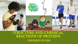
  • 1. STRUCTURE AND CHEMICAL REACTIONS OF PROTEINS PREPARED BY RGB
  • 2. PROTEIN COMPOSITION Proteins are precisely defined sequences of amino acids (residues) linked through peptide bonds to form polymers (hundreds or thousands of molecules combined to produce a single macromolecule. A peptide is a molecule formed by linking together a small number of amino acids (from two to a dozen) through peptide bonds; both peptides and proteins usually have a free amine group on one end of a molecule (the N – terminal residue) and a free carboxyl group on the other end of the molecule (the C – terminal residue). The structure, function, and activity of a protein is determined to a large extent by the number, sequence, and chemical properties of its constituent amino acids.
  • 3. PROTEIN COMPOSITION All proteins are composed of varying sequence of 20 amino acids called common amino acids. a. Other amino acids that occur in proteins are modifications of the common amino acids b. Modifications include addition of functional groups and bond formation between certain groups within the amino acid. c. Amino acids containing modifications are labeled derived or modified amino acids; modifications occur after the amino acids are incorporated into the protein. The names of 20 common amino acids are usually written as three-letter abbreviation with the first letter capitalized or as a single capital letter designation.
  • 5. STRUCTURE OF AMINO ACIDS Amino acids contain a central carbon (C), called the α carbon, to which four substituents are bound: an amine group (-NH2), a carboxylic acid group (-COOH), a hydrogen atom (H), and a group unique to each amino acid called the side chain or R group. a. Three of the four substituents on the α carbon (-NH2, - COOH, and –H) are common to all amino acids. b. The fourth substituent, the side chain (indicated by R), varies and confers distinctive chemical properties to each amino acid. c. In standard nomenclature for amino acids, the carbons of the R group are labeled with Greek letters, starting with β for the first carbon attached to the α carbon.
  • 6. STRUCTURE OF AMINO ACIDS For example, in glutamic acid (three linear carbons in the R group), the carbons are labeled β, γ, and δ, with the δ carbon being he final carbonyl carbon in this R group. All amino acids found in proteins are called α amino acids because the amine group is bonded to the α carbon; however, amino acids with an amine group attached to other carbon atoms have important biochemical functions. a. β-alanine, β-amino acid, is a building block of pantothenic acid, which is one of the structural units of coenzyme A. b. γ-aminobutyric acid (GABA), a γ-amino acid, is involved in the transmission of nerve impulses.
  • 7. STRUCTURE OF AMINO ACIDS Because four different substituents are bonded to the α carbon of an amino acid, the α carbon is the chiral center and the amino acids (except glycine) are optically active. a. Amino acids can be either D- or L-stereoisomers, depending on the orientation of substituents around the α carbon. b. All amino acids found in proteins are L-amino acids; D- amino acids are found in certain bacterial cell walls and in some peptide antibiotics. The structure and function of an amino acid is determined to a large extent by the characteristics of its R group.
  • 8. STRUCTURE OF AMINO ACIDS One simple way to remember amino acids is to classify them as having one of three different types of side chains (R groups); amino acids with nonpolar side chains, amino acids with charged polar side chains, and amino acids with uncharged polar side chains. a. The nine amino acids described below have a nonpolar side chain and various structures and sizes. 1. Glycine (Gly, G), the simplest amino acid, has two identical substituents (two hydrogen atoms) bonded to the α carbon; because the α carbon is achiral, glycine is the only amino acid that is not optically active. 2. Alanine (Ala, A) has a methyl group; valine (Val, V) has an isopropyl group; leucine (LEU, L) and isoleucine (Ile, I) contain isomers of butane.
  • 9. STRUCTURE OF AMINO ACIDS 3. Phenylalanine (Phe, F) is so named because of its R group can be described as a phenyl group attached to the methyl group of alanine. 4. Proline (Pro, P) is uniquely in that its α-amino group, as well as its α carbon, are incorporated into a single cyclic structure; proline is more correctly classified as an imino acid rather than an amino acid, because its α-NH2 group contains a secondary nitrogen (one that is bonded to two other alkyl (R) groups). 5. The R group of tryptophan (Trp, W) contains indole (a five- member, nitrogen-containing ring fused to a benzene ring). 6. Methionine (Met, M) contains sulfur in a thioether (R-S-R) linkage.
  • 10. STRUCTURE OF AMINO ACIDS b. The five amino acids described below here have charged polar side chains. (1) Aspartate and glutamate are the only two common amino acids that bear a negative charge on the R group at neutral pH. - Aspartate (Asp, D) contains a carboxyl group (-COOH) separated from the α carbon by a methylene (-CH2-) group. - Glutamate (Glu, E) contains a carboxyl group (-COOH) separated from the α carbon by two methylene groups. (2) Lysine, arginine, and histidine are the only three common amino acids that bear a positive charge on the R group at neutral pH.
  • 11. STRUCTURE OF AMINO ACIDS - Lysine (Lys, K) contains a butylamine side chain. - Arginine (Arg, R) contains a guanidinium group separated from the α carbon by three methylene groups. - Histidine (His, H) contains a five –member heterocyclic ring, known as a imidazole group. c. The seven amino acids described below have uncharged polar side chains. (1) Serine (Ser, S) contains a hydroxymethyl (- CH2OH) group attached to the α carbon; threonine (Thr, T) contains ethanol attached to the α carbon; tyrosine (Tyr, Y) contains phenol (a benzene ring bearing a hydroxyl group); cysteine (Cys, C) contains a sulfhydryl (-SH) group.
  • 12. STRUCTURE OF AMINO ACIDS (2) Asparagine (Asn, N) and glutamine (Gln, Q) are structurally similar to aspartate and glutamate, respectively, except that an amide group replaces the carboxyl group in the side chain of each; the designation Asx and Glx represent the sum of aspartate and asparagine, or glutamate and glutamine, in a protein respectively.
  • 13. CHEMICAL PROPERTIES OF AMINO ACIDS The chemical properties of the R group determine the chemical properties of each amino acid. The number of ionizable groups in an amino acid determine the extent to which it will be ionized at a given pH in the body. Amino acids may be categorized according to the functional groups and chemical properties of their side chains; some amino acids groups fit more than one classification. a. The amino acids with aliphatic (non-ring) side chains are glycine, alanine, valine, leucine and isoleucine. - Glycine and alanine have the smallest aliphatic side chains; they play an important structural role in proteins by fitting into spaces too small for a larger amino acid. - Valine, leucine, and isoleucine have longer aliphatic side chains that are hydrophobic; they tend to congregate in structures that enable them to avoid water.
  • 14. CHEMICAL PROPERTIES OF AMINO ACIDS b. The aromatic (benzene-related) amino acids, phenylalanine, tyrosine, and tryptophan, all have the aromatic group attached to the α carbon via a methylene (- CH2-) group. - Phenylalanine and tryptophan are hydrophobic and tend to form structures that enable them to avoid water. - The hydroxyl group on tyrosine makes it more hydrophilic and a more chemically reactive acid than phenylalanine. - Although histidine and proline contain ring structures, they are usually classified differently. c. Cysteine and methionine, the two sulfur – containing amino acids, are hydrophobic. - Two cysteine residues can bond via their thiol (-SH) groups to form the amino acid cystine; the thiol group of cysteine is chemically reactive and easily forms a disulfide (-S-S-) through oxidation.
  • 15. CHEMICAL PROPERTIES OF AMINO ACIDS - The formation of disulfide bonds is important in protein structures; disulfide bonds commonly join two different peptide chains together or link two different cysteine residues within the same peptide. d. Serine and threonine have a side chain hydroxyl group that makes them both hydrophilic and chemically reactive. e. In proline, the incorporation of nitrogen from the α-amine group into the side chain constrains the rotation of the molecule around the C – N bond; this bond constraint affect the structure of a protein causing it to bend at the position of proline residue. f. The acidic amino acids aspartate and glutamate are dicarboxylic, monobasic amino acids because they have two carboxyl groups (one bound to the α carbon and one in the side chain) but only one amine group (bound to the α carbon); the carboxyl groups of both amino acids are negatively charged at physiologic pH (7.4).
  • 16. CHEMICAL PROPERTIES OF AMINO ACIDS g. Amino acids with basic side chains are lysine, arginine, and histidine. - The side chain amine group makes these amino acids extremely polar and hydrophilic; at physiologic pH, both lysine and arginine have positively charged amine group. - At physiologic pH, the imidazole ring in histidine exists primarily in the unchanged form because the imidazole ring has pK’a of approximately 6. h. Asparagine and glutamine, the two amino acids that contain an amide group in the side chain, are not charged at physiologic pH.
  • 17. IONIZATION OF AMINO ACIDS All free amino acids contain at least two ionizable groups, the α-carboxyl group and the α-amine group; the charge on the amino acid R groups within a protein affects the three- dimensional structure and reactivity of the protein. Except for the first and last amino acids in a protein, all of the α-carboxyl and the α-amine groups within a protein participate in peptide bonds and thus do not contribute to the protein’s overall charge. The overall charge of a protein is determined by the charge on the amino acid side chains (the charge on the R groups) and the charge on the protein’s N- terminal and C-terminal residues.
  • 18. IONIZATION OF AMINO ACIDS a. The pK’a of the α COOH is 2.35; at physiologic pH, the proton is removed and the carboxyl group is in the ionized form with a-1 charge. b. The pK’a of the α NH2 is 9.69; at physiologic pH, the NH2 is protonated to the ionized form (-NH3 —1) with a +1 charge. All amino acids contain both an acidic group (the α-carboxyl group) and a basic group (the α-amine group) in the same molecule. a. An amino acid without an ionizable side chain will have one +1 charge (from the α-amine group) and one –1 charge (from the α-carboxyl group) at physiologic pH; its net charge is zero. b. A charged compound with a net charge of zero is called a zwitterion.
  • 19. IONIZATION OF AMINO ACIDS The pH at which a compound exists as zwitterion is the isoelectric point (pl). The pH of the environment determines the degree of ionization of an amino acid; this is illustrated in the titration of the two ionizable groups of alanine. a. At pH <2.35, the two ionizable groups (the α COOH and the α NH2) are completely protonated; the carboxyl group is un-ionized and esists as – COOH; the amine group has a +1 charge and exists as – NH3 +1. b. At pH = 2.35, which is the pK’a of the carboxyl group, equal amounts of the ionized (-COO—1) and unionized (- COOH) carboxyl group are present.
  • 20. IONIZATION OF AMINO ACIDS c. As the pH increases beyond 2.35, the carboxyl group on most of the molecules remains unprotonated and thus is negatively charged; the amine group is still protonated because the pH has not reached the amine pK’a of 9.69, and hence the amine group on most molecules is still charged. d. At the pH midway between the two pK’a values, the negative charge from the carboxyl group equals the positive charge from the amine group; this pH ([2.35+9.69]2 = 6.02) is the isoelectric point of alanine, the pH at which alanine exists as zwitterion. e. At pH=9.69, which is the pK’a of the amine group, equal amounts of un-ionized (-NH2) and ionized (-NH3 +1) amine groups are present; the carboxyl group, well beyond its pK’a, remains ionized (-COO—1)
  • 21. IONIZATION OF AMINO ACIDS f. At pH>9.69, none of the ionizable groups of alanine are protonated; the carboxyl group has a negative charge (-COO— 1) and the amine group is un-ionized (-NH2). An ionizable group in the side chain of an amino acid has its own pK’a; in these amino acids, the side chain pK’a also affects the isoelectric point.
  • 22. LEVELS OF PROTEIN STRUCTURE All proteins have at least three levels of structures: primary, secondary, and tertiary. Proteins composed of more than one polypeptide chain also have a quaternary structure. Monomers are proteins composed of only one polypeptide chain; proteins composed of two or more polypeptide chains are called oligomers and are classified by number of polypeptide chains; dimers have two chains; trimers, three chains, tetramers, four chains, and so on. The primary, secondary, tertiary, and quaternary structure of a protein controls its three-dimensional shape, which partly determines its activity.
  • 23. PRIMARY STRUCTURE OF PROTEIN Amino acids are linked by peptide bonds. a. A peptide bond forms as result of a condensation reaction between the carbonyl carbon from the α COOH of one amino acid and the amine nitrogen from the α NH2 of the next amino acid; the reaction eliminates a molecule of water. b. The peptide bond is rigid and planar; it has a bond length intermediate between that of a single bond and a double bond. c. The partial double-bond character of the peptide bond restricts its ability to rotate, making it the backbone of the protein. d. Other bonds in amino acids are single bonds and therefore have more freedom of rotation than peptide bonds. Insulin
  • 24. PRIMARY STRUCTURE OF PROTEIN The primary structure of a protein is the sequence and number of amino acids linked by peptide bonds. a. The convention for writing the primary structure of an amino acid is to name its sequence of amino acid from left to right. b. The first amino acid (the N-terminal amino acid) has a free α-amine group; the carboxyl group of this first amino acid participates in the formation of the first peptide bond. c. Subsequent amino acids are written to the right of the N-terminal amino acid; all of these residues are linked by peptide bonds at their α-amine and α-carboxyl groups. d. The last amino acid (the C –terminal amino acid) has a free α-carboxyl group. Insulin
  • 25. SECONDARY STRUCTURE OF PROTEIN Intramolecular and intermolecular hydrogen bonding in the primary structure give the protein its secondary structure. The α helix, the β pleated sheet, and the collagen triple helix are three different types of secondary structure. The α helix occurs in many different fibrous and globular proteins; it is formed by intramolecular hydrogen bonds between the carboxyl group oxygen of one amino acid and the amine hydrogen of the fourth amino acid in front of it. a. Every carboxyl oxygen and amine hydrogen participates in the intramolecular bonding; the combined forces of all these hydrogen bonds give the protein an overall rodlike structure.
  • 26. SECONDARY STRUCTURE OF PROTEIN b. Formation of the α helix occurs spontaneously in a pattern of 3.6 amino acids per turn of the α helix. c. Amino acids that are three to four residues apart in the primary structure are spatially close to each other in the α helix; R groups that are adjacent to each other in the primary structure are spatially distant in the α helix. d. The extent of α-helical structure in a protein depends on its constituent amino acids; some amino acids (Leu, Ala, Glu, Met) tend to favor α helix formation, whereas others (Val, Ser, Asp, Asn) tend to destabilize it. The β pleated sheet is the secondary structure of silk, a fibrous protein produced by spiders and insects; it is formed by intermolecular hydrogen bonds between a carboxyl group oxygen in one amino acid and an amine group hydrogen in an adjacent polypeptide.
  • 27. SECONDARY STRUCTURE OF PROTEIN a. A parallel β pleated sheet is formed when adjacent polypeptids are oriented in the same direction (from N- terminal to C-terminal or vice versa). b. An antiparallel β pleated sheet is formed when adjacent polypeptides are oriented opposite directions (one chain oriented from the N- to C- terminal and adjacent chain oriented from C- to N- terminal). c. Proteins that have β pleated sheets assume an extended, rather than a rod-shaped or coiled, structure. Collagen, the most abundant structural protein found in vertebrates, is a major component of connective tissues, such as bone, teeth, cartilage, and tendon; the secondary structure of collagen is a triple helix composed of three entwined polypeptide chains and bound by intermolecular hydrogen bonds.
  • 28. SECONDARY STRUCTURE OF PROTEIN a. The collagen triple helix has a high concentration of proline and glycine; often every third residue in each of the three chains is a glycine molecule. b. Two derived amino acids, hydroxyproline and hydroxylysine, occur frequently; the sequence – Gly – Pro – hydroxyproline – occurs frequently. c. There are 3.3 amino acids per turn of the triple helix, with approximately every third residue projecting into the helix interior. d. The high concentration of glycine (the smallest amino acid) prevents crowding in the interior of the helix.
  • 29. TERTIARY STRUCTURE OF PROTEIN A protein’s tertiary structure results from interactions between amino acid side chains of each of the residues; these interactions between R groups cause a polypeptide to assume its normal three-dimensional configuration. Many types of noncovalent interactions are involved, such as van der Waals forces, ionic bonding, hydrophobic bonding, and hydrogen bonding. a. Residues with hydrophilic side groups usually are located on the exterior of the molecule, oriented to interact with the aqueous environment. b. Residues with hydrophobic side groups usually are located on the interior o the molecule, oriented so the hydrophobic groups can interact with each other and exclude water. One type of covalent bond that influences tertiary structures is the disulfide bond of the derived amino acid.
  • 30. QUATERNARY STRUCTURE OF PROTEIN Most proteins are composed of more than one polypeptide chain; these polypeptide subunits associate with each other in a defined geometric arrangement called the quaternary structure. Formation of the quaternary structure involves the same noncovalent interactions as in the formation of a tertiary structure, but the interactions occur between polypeptide chains (interchain) rather than within a polypeptide chain (intrachain). Oligomeric proteins composed of identical subunits are called homogeneous; those composed of different subunits are called heterogeneous.
  • 31. IDENTIFICATION OF PROTEIN The amino acid composition of protein can be discerned by cleaving all the peptide bonds, then separating and identifying the constituent amino acids. The amino acid sequence of a protein can be discerned by using various methods to cleave only selected peptide bonds, then assimilating all the information to deduce the amino acid sequence; this process is similar to putting together the pieces of the puzzle. Proteins are also identified and characterized by spectroscopy, ultracentrifugation, chromatography, and electrophoresis. More sophisticated aspects of protein characterization are accomplished by X-ray diffraction, circular dichroism spectroscopy, nuclear magnetic resonance spectroscopy, and mass spectroscopy.
  • 32. DETERMINING AMINO ACID COMPOSITION AND SEQUENCE In the first stage of determining the amino acid composition of a protein, all the peptide bonds are hydrolyzed by an acid or a base. a. In acid hydrolysis, the protein solution is usually treated with 6 N hydrochloric acid at 110OC for 24 hours. b. In base hydrolysis, the protein solution is usually treated with 2 to 4 N sodium hydroxide at 100 OC for 4 to 8 hours. c. Under acid hydrolysis, aspartate cannot be distinguished from asparagine and glutamate cannot be distinguished from glutamine; the totals for both types of molecules are identified as Asx and Glx, respectively. d. Tryptophan is acid-labile and requires base hydrolysis for identification.
  • 33. DETERMINING AMINO ACID COMPOSITION AND SEQUENCE In the second stage of determining the amino acid composition of a protein, the hydrolyzed amino acids are separated, identified, and quantitated. a. Amino acids can be separated by thin layer chromatography or liquid chromatography, which is the separation of molecules on a solid support according to their physical or chemical properties. b. Amino acids can be identified and quantitated by reacting them with reagents that produce a colored or fluorescent product, the intensity of which is proportional to the concentration of the amino acid present.
  • 34. DETERMINING AMINO ACID COMPOSITION AND SEQUENCE (1)Ninhydrin, a reagent that turns blue when reacted with amino acids and yellow when reacted with imino acids (proline), is sensitive to microgram (10—6 g) quantities of amino acids. (2)Fluorescamine, a reagent that reacts with the α-amine group to produce a fluorescent derivative, is more sensitive than ninhydrin; fluorescamine can detect nanogram (10—9 g) quantities of amino acids. After determining the amino acid composition of the protein, the determination of the structure includes identifying the N-terminal residue and C-terminal residue.
  • 35. DETERMINING AMINO ACID COMPOSITION AND SEQUENCE a. The N-terminal amino acid is identified by the reaction of its free amine group in the intact protein. (1)The reagent fluoronitrobenzene (FDNB) reacts with the free amine group of a protein to form a covalent dinitrophenyl derivative. (2)The protein is subjected to acid hydrolysis and chromatographic separation as previously described. (3)The dinitrophenyl derivative has different chromatographic properties than the other amino acids and can thus be identified. b. The free amine group in the side chain of lysine also reacts with FDNB and must be considered when interpreting results.
  • 36. DETERMINING AMINO ACID COMPOSITION AND SEQUENCE c. An N-terminal amino acid that is modified on the amine group will not have a free amine group to react with FDNB. d. Other reagents, such as dabsyl chloride and dansyl chloride, frequently replace FDNB; they permit detection of smaller quantities of the amino acid. e. The Edman degradation is a method of identifying an entire sequence of amino acids in a protein by successively releasing individual N-terminal residues that can be separated from the intact protein and identified.
  • 37. DETERMINING AMINO ACID COMPOSITION AND SEQUENCE (1)The reagent phenyl isothiocyanate (PITC, also called Edman’s reagent) reacts with the N-terminal amino acid to form a thiazolinone derivative, which is then extracted with organic solvent and converted to a more stable molecule called a phenylthiohydantoin (PTH) amino acid. (2)The technique used in the Edman degradation separates the N-terminal residue only and leaves the rest of the polypeptide intact. (3)The separated PTH amino acid is then identified using standard identification techniques, such as chromatography and electrophoresis. (4)The next amino acid is the new N-terminal amino acid and is sequentially labeled and removed without altering the remaining protein. (5)The cycles are repeated, with a new N-terminal amino acid generated during each cycle, until all amino acids have been separated and identified in sequence.
  • 38. DETERMINING AMINO ACID COMPOSITION AND SEQUENCE f. The identification of the C-terminal residue in a protein is accomplished by treating the intact protein with a carboxypeptidase, a class of enzymes that selectively catalyze the removal of the C-terminal amino acid from the peptide. (1)Other enzymes that cleave specific peptide bonds on their carboxyl side include trypsin, which cleaves the residues that contain positively charged R groups (Lys and Arg), and chymotrypsin, which cleaves at residues with aromatic R groups (Phe, Tyr, and Trp) and bulky aliphatic R groups (Ile and Val). (2)Cyanogen bromide is a chemical that cleaves at the carboxyl side of the peptide bond of methionine.
  • 39. DISSOCIATION OF PROTEIN SUBUNITS Proteins with multiple subunits must be broken down to individual subunits to characterize their amino acid composition and sequence. Denaturation is the process of treating a protein with heat or chemical reagents to break it down into its constituent subunits. a. Denaturation causes loss of a protein’s secondary, tertiary, and quaternary structure, but its primary structure may be maintained. b. In vivo, changes in pH, ionic strength, and temperature denature proteins; in vitro, detergents, urea, organic solvents, acids or bases denature proteins. c. Denatured proteins lose their normal biochemical activity and are nonfunctional. d. Disulfide binds in proteins are disrupted when reducing agents, such as β-mercaptoethanol or dithiothreitol, are added.
  • 40. IDENTIFICATION, SEPARATION AND CHARACTERIZATION OF PROTEINS The presence of a protein and its integrity can be detected by spectroscopy, a technique for identifying and quantitating unknown molecules by virtue of their interaction with light. a. Absorption spectroscopy identifies peptide bonds and certain functional groups by measuring the absorbance of different wavelengths UV and Visible light. 1. Peptide bonds absorb UV light in the range of 180 to 230 nanometers (nm) 2. The aromatic rings in phenylalanine, tryptophan and tyrosine absorb UV in 260 to 300 nm. 3. Denaturation changes in secondary and tertiary structure appear as a difference in light absorbance between 180 to 230 nm.
  • 41. IDENTIFICATION, SEPARATION AND CHARACTERIZATION OF PROTEINS Fluorescence spectroscopy, a more sensitive measurement than UV or visible spectroscopy, detects the presence and quantity of a protein. 1. Fluorescence occurs when a molecule in an excited state, produced from absorption of short wavelength (high energy) light, re-emits some of its excess energy not emitted as light dissipates as heat. 2. Only proteins containing amino acids with aromatic side chains (Phe, Tyr, Trp) will naturally fluoresce. 3. A protein that does not naturally fluoresce can be made to fluoresce by covalently bonding a fluorescent group to the protein.
  • 42. IDENTIFICATION, SEPARATION AND CHARACTERIZATION OF PROTEINS Ultracentrifugation separates proteins according to size. a. The sedimentation coefficient of a protein, subjected to a centrifugal (outward) force, depends on he protein’s mass, density, and shape and on the density of the suspending solution. b. The Svedberg (S) unit, a measure of the sedimentation coefficient of a particle, is equal to 10—13 second. c. The magnitude of the sedimentation coefficient gives a relative value that can characterize the molecular weight of protein.
  • 43. IDENTIFICATION, SEPARATION AND CHARACTERIZATION OF PROTEINS Gel filtration chromatography (also called size exclusion chromatography) separates proteins according to size. a. Inert porous beads are packed into a column; the pore size is selected to correspond to the molecular weight of molecules small enough to enter the pores. b. A buffered protein solution is then applied to the top of the packed column. c. High molecular weight proteins are too large to enter the pores in the beads and thus go directly to the column. d. Low molecular weight proteins enter the pores and thus have a longer path to traverse on their way through the column; some midsize proteins can enter the pores while others cannot; they travel through the column at rates between high and low molecular weight proteins. e. The elution time of each protein is related to its molecular size; large proteins elute first, midsize protein next and small proteins elute last.
  • 44. IDENTIFICATION, SEPARATION AND CHARACTERIZATION OF PROTEINS Ion exchange chromatography separates proteins according to their charge. a. Inert beads coated with either negatively or positively charged groups are packed into a column, and a buffered protein solution is then applied to the top of the packed column. b. Proteins with a net positive charge will bind, via ionic bonds, to beads coated with negatively charged groups (cation exchange); conversely, proteins with net negative charge will bind to beads coated with positively charged groups (anion exchange). c. The addition of buffer solution containing a higher concentration of the same type of ions as in the original protein solution displaces, or exchanges, the proteins ionic bonding to the charged groups on the beads; this allows the researcher to control when the protein of interest elutes from the column.
  • 45. IDENTIFICATION, SEPARATION AND CHARACTERIZATION OF PROTEINS Affinity chromatography separates proteins that bind specifically with certain chemical groups. a. Inert beads coated with specific chemical groups are packed into a column; a buffered protein solution is then applied to the top of the packed column. b. A protein with an affinity for the specific chemical group will bond to that group and consequently will be retained on the column; proteins that lack affinity will not bind and will flow directly through the column. c. Addition of higher concentration of a soluble form of the specific chemical group displaces the protein from the binding sites on the beads; the protein then elutes separately.
  • 46. IDENTIFICATION, SEPARATION AND CHARACTERIZATION OF PROTEINS Electrophoresis, the separation of charged molecules by their movement on a solid support while exposed to an electric field, also aids in the separation of different proteins. a. Electrophoresis separates proteins on the basis of their mass if they can be made to have the same charge. b. The anionic detergent Sodium dodecyl sulfate (SDS) is added to a protein solution; SDS binds strongly to most proteins, causing denaturation and imparting a large net negative charge to each protein.
  • 47. IDENTIFICATION, SEPARATION AND CHARACTERIZATION OF PROTEINS c. The large negative charge assumed by each protein masks its intrinsic charge; each protein in the SDS solution tends to have the same charge-to-mass ratio and a similar shape. d. The SDS-treated proteins are placed on the solid support, along with several marker proteins of known molecular weight, and are exposed to an electric field; the proteins vary in the distance they move on the support in direct relation to their molecular weight.
  • 48. IDENTIFICATION, SEPARATION AND CHARACTERIZATION OF PROTEINS Isoelectric focusing separates proteins on the basis of their isoelectric points. a. The support medium is a gel in which a pH gradient has been established. b. The protein mixture is applied to the gel; proteins migrate through the gel until they reach the pH at which they have no net charge (their pl); with no net charge, proteins cease to migrate. c. Because each protein has its own pl, separation of a mixture of proteins can be achieved.
  • 49. DETERMINATION OF HIGHER LEVELS OF PROTEIN STRUCTURE The three-dimensional structure of a protein (its secondary, tertiary and quaternary structure) may be determined by X-ray diffraction. The amount and type of secondary structure in a protein is determined by circular dichroism (CD) spectroscopy. Nuclear magnetic resonance spectroscopy (NMR) provides structural and functional information about the environment of a particular atom of a protein. Mass Spectroscopy (MS) can determine the structure of very small quantities of protein.
  • 50. DIVERSITY OF PROTEIN FUNCTIONS The different functions of PROTEINS include… ▪ …are involved in some way in all the biochemical reactions that make life possible; each species possesses proteins that are distinct from those of all other species. ▪ …transport substances; for example, the protein hemoglobin transports both oxygen and carbon dioxide in the blood. ▪ …catalyze biochemical reactions; most enzymes are proteins. ▪ …help maintain structure; for example, the protein collagen constitutes part of fibrous connective tissues in skin, bone, tendon, cartilage, blood vessels, and teeth. ▪ …facilitate movement; for example, the proteins actin and myosin mediate muscle contraction. ▪ …serve as storage compounds; for example, the protein ferritin stores iron in the body. ▪ … protect organisms against foreign invaders, such as viruses, bacteria and other toxins they produce; for example, immunoglobulins (antibodies) are proteins that react with foreign compounds (antigen). ▪ Serve as hormones, the messengers and regulators of metabolism; for example, the protein hormone insulin regulates glucose levels in the blood.
  • 51. TRANSPORT ROLE OF PROTEINS: HEMOGLOBIN Hemoglobin (Hb), an oligomeric protein found in erythrocytes (red blood cells), is composed of four polypeptide chains (subunits), two each of two identical subunits. Hemoglobin is a globular protein with considerable α- helical secondary structure; different hemoglobin types vary in subunit composition. a. Hb A, the major type of adult hemoglobin of two α and two β polypeptide chains. b. Hb A2, composing approximately 3% of adult hemoglobin, is composed of two α and two δ polypeptide chains. c. Hb F (fetal hemoglobin), found in fetuses and newborns, is composed of two α and two γ polypeptide chains.
  • 52. TRANSPORT ROLE OF PROTEINS: HEMOGLOBIN Each of the four polypeptide chains in hemoglobin is tightly bound to its own molecule of heme, which functions as a prosthetic group (a nonprotein portion of a protein molecule that is necessary for its activity). a. Heme is a heterocyclic molecule that contains an iron atom in its center; the heme in the hemoglobin is called Fe (II) heme, iron protoporphyrin IX, or ferroprotoporphyrin IX. b. Protophyrin IX , the nonmetal portion of the heme molecule, consists of four pyrrole rings linked by methane bridges to form a tetrapyrrole structure.
  • 53. TRANSPORT ROLE OF PROTEINS: HEMOGLOBIN c. The iron atom bound in the center of the protoporphyrin ring is the actual binding site of O2 in the hemoglobin molecule; each iron atom binds one O2 molecule. d. Hemoglobin binds O2 when iron is in the reduced state (abbr. Fe+2, Iron (II), ferrous); hemoglobin does not bind when iron is in the oxidized – Fe+3, Iron (III), ferric – state. e. Because each of the four polypeptide chains in hemoglobin has its own heme group, one hemoglobin binds four O2 molecules (the iron in each heme binds one O2 molecule). f. The four heme groups are located in nonpolar crevices near the surface of the hemoglobin molecule, but far apart from each other.
  • 54. TRANSPORT ROLE OF PROTEINS: HEMOGLOBIN Hemoglobin binds O2 for transport to cells that require oxygen and then releases O2 at those cells; oxyhemoglobin is hemoglobin bound to O2; deoxyhemoglobin is hemoglobin without bound O2. a. O2 binding to hemoglobin is cooperative; binding of the first O2 molecule to the first heme group facilitates binding of subsequent O2 molecules to the other three heme groups. b. The cooperative binding of O2 to hemoglobin demonstrates an allosteric effect, an effect in which the binding of one molecule at a specific site of protein facilitates the binding of subsequent molecules at a distant sites on a protein; in hemoglobin, the binding of the first O2 molecule facilitates the binding of subsequent O2 molecules, even though the binding sites are distant from each other.
  • 55. TRANSPORT ROLE OF PROTEINS: HEMOGLOBIN c. O2 binding to deoxyhemoglobin breaks 8 ionic bonds and causes partial disruption of its normal quaternary structure as it becomes oxyhemoglobin. d. The molecule 2,3-bisphosphoglycerate (BPG) causes hemoglobin to release O2 molecules at body cells needing oxygen. (1)One molecule of BPG binds in the central cavity of deoxyhemoglobin molecule, stabilizing its quaternary structure and thereby favoring the deoxygenated form of hemoglobin. (2)Upon reoxygenation, the conformation of hemoglobin changes, making the central activity of the protein too small for BPG; without BPG, hemoglobin can take up O2 and form oxyhemoglobin.
  • 56. TRANSPORT ROLE OF PROTEINS: HEMOGLOBIN e. An oxygen dissociation curve is a graphic representation of the saturation of binding sites (heme groups) as a function of the amount of O2 present; O2 is measured as the partial pressures of gaseous O2 (PO2) in units of torr. 1. The PO2 at which 50% of binding sites are saturated with O2 is termed the P50. 2. The P50 for different O2 binding proteins is unique and predicts the affinity of the protein for O2. 3. A partial pressure of 26 torr is required to saturate half the binding sites in hemoglobin, but only 1 torr saturates half the binding sites in myoglobin, indicating that myoglobin has a higher affinity for O2 than hemoglobin. 4. The sigmoidal (S-shaped) oxygen dissociation curve for hemoglobin reflects the cooperative binding of O2 to hemoglobin; in contrast, the hyperbolic oxygen dissociation curve for myoglobin suggests no cooperative binding.
  • 57. TRANSPORT ROLE OF PROTEINS: HEMOGLOBIN The Bohr effect describes the relationships between the binding of O2, CO2, and H+1 to hemoglobin. a. Metabolically active tissues require O2 and produce CO2 and H+1 as by – products; hemoglobin releases O2 to these tissues and picks up CO2 and H+1. b. Accumulation of CO2 and H+1 in the hemoglobin molecule decreases its affinity for O2. c. CO2 generates H+1 in erythrocytes by the reaction CO2 + H2O⇄ H+1 + HCO3 +1, catalyzed by carbonic anhydrase. d. Hemoglobin transports CO2 to the lungs, which eliminate CO2 in expired air; H+1 is handled in two ways: 1. hemoglobin’s four N-terminal amine groups and the imidazole groups of its histidine residues take up H+1; these groups are responsible for hemoglobin’s blood-buffering capacity. 2. Deoxyhemoglobin can take up H+1 without a resultant change in pH, if the extent to which H+1 is generated from CO2 is just sufficient to meet the ability of deoxyhemoglobin to accept H+1; this is referred to as the isohydric carriage of CO2 and is independent of hemoglobin’s buffering ability.
  • 58. CATALYTIC ROLE OF PROTEINS: ENZYMES Many proteins function as biological catalysts, catalysts in living organisms are called enzymes. a. Enzymes accelerate biochemical reaction rates by reducing the energy of activation needed to reach the transition state between reactant and product. b. In enzyme-catalyzed biochemical reactions, the reactant molecule that binds first to the enzyme is called the substrate. c. The active site of an enzyme are those amino acids that come into direct contact with the substrate and bind it. d. Two different models describe how substrates bind to enzymes; the lock-and-key model assumes that the substrate and enzyme have complementary shapes that fit perfectly; the induced fit model assumes that enzyme’s active site changes its shape to fit the substrate’s molecular configuration as the substrate binds.
  • 59. CATALYTIC ROLE OF PROTEINS: ENZYMES e. Like all true catalysts, enzymes are not consumed by reactions they catalyze; after participating in a reaction, they recycle and catalyze additional reactions. f. Enzymes are selective in the reactions they catalyze; many enzymes catalyze only one specific biological reaction. g. Some enzymes require nonprotein groups called cofactors to function optimally; a cofactor may be a metal ion such as Zn+2, or an organic molecule, such as NAD+1 (nicotinamide adenine dinucleotide); organic molecules that functions as cofactors are called coenzymes; a cofactor bound to an enzyme is known as a prosthetic group. h. An enzyme without its cofactor is called an apoenzyme; an enzyme associated with its cofactor is called a holoenzyme. i. Enzymes requiring metal ions as cofactors are known as metalloenzymes.
  • 60. CATALYTIC ROLE OF PROTEINS: ENZYMES j. Many organisms are unable to synthesize certain essential coenzymes and thus must consume these coenzyme in the diet; in humans, most the water-soluble vitamins are dietary precursors of coenzymes. k. Isoenzymes are either structurally different forms of the same enzyme or oligomeric proteins with various combinations of different subunits; isoenzymes usually occur in different tissues of the same organism, use different substrates, but catalyze similar types of reactions. l. Enzymes are classified as allosteric or nonallosteric; the activity of an allosteric enzyme is controlled by the binding of a second molecule, called an effector or modulator; the binding of the modulator may either decrease or increase the enzyme’s activity; nonallosteric enzymes show no modulation through binding of other molecules.
  • 61. CATALYTIC ROLE OF PROTEINS: ENZYMES Enzymes require optimal conditions for optimal catalytic activity. a. Because enzymes must bind precisely with their substrates, anything that changes the enzyme’s three – dimensional shape changes its ability to catalyze reactions; enzymes that have been altered through physical or chemical means so they are no longer active are called denatured. b. Enzymes require an optimal temperature for optimal activity; enzymes are heat-labile and denature at temperatures exceeding normal physiologic temperature (37OC). c. Enzymes require optimal pH and ionic strength of the surrounding medium affect the changes on specific amino acid residues; the charges on the amino acid residues affect the molecule’s configuration and therefore its activity. d. Enzyme-catalyzed reactions require optimal concentrations of enzyme, substrate, and cofactors for the maximal reaction rate to be attained.
  • 62. CATALYTIC ROLE OF PROTEINS: ENZYMES Enzymes are named by attaching the suffix “ase” to the type of reaction they catalyze. a. Oxidoreductases are class 1 enzymes; they catalyze oxidation- reduction reactions; for example, alcohol dehydrogenase catalyzes the oxidation of an alcohol to an aldehyde. b. Transferases are class 2 enzymes; they catalyze the transfer of a group from one molecule to another; for example, phosphotransferase catalyzes the transfer of phosphoryl group from one molecule to another. c. Hydrolases are class 3 enzymes; they catalyze the breaking of covalent bonds using water; for example, peptidase hydrolyzes a peptide bond.
  • 63. CATALYTIC ROLE OF PROTEINS: ENZYMES d. Lyases are class 4 enzymes; they either remove a group by splitting a bond and forming a double bond or add a group to a double bond and form a single bond; for example, decarboxylase removes a carboxyl group to form carbon dioxide. e. Isomerases are class 5 enzymes; they catalyze internal atom rearrangements in a molecule; for example, racemase catalyzes the rearrangement (isomerization) of substituents on an alpha carbon atom. f. Ligases are class 6 enzymes; they catalyze the formation of covalent bond and therefore require energy, usually supplied by the hydrolysis of high-energy phosphate bonds; for example, pyruvate carboxylase catalyzes the formation of carbon-to- carbon bond from pyruvate and carbon dioxide to form the compound oxaloacetate.
  • 64. CATALYTIC ROLE OF PROTEINS: ENZYMES Kinetics, the study of the rate (velocity) of reactions, uses Michaelis- Menten equation to describe the rate of enzyme-catalyzed reactions; the Michaelis-Menten equation generally predicts the reaction rate for single substrate reactions involving nonallosteric enzymes only. a. In first-order reactions involving a single substrate, the initial reaction rate (Vi) depends only on the concentration of the substrate [S]; in zero-order reactions involving a single substrate, the rate of reaction remains the same regardless of the concentration of the substrate because all the active sites on the enzyme are already saturated with substrate. b. For single-substrate reactions at a constant enzyme concentration, nonallosteric enzymes display first-order kinetics as the concentration of substrate increases; when the concentration of the substrate is high enough to saturate all available enzyme active sites, the reaction displays zero-order kinetics.
  • 65. CATALYTIC ROLE OF PROTEINS: ENZYMES c. The Michaelis-Menten equation describes enzyme- catalyzed reactions that follow the hyperbolic curve shown. d. The concentration of a substrate required to achieve one- half of an enzyme’s maximal velocity is a constant termed Km; each enzyme has a unique, characteristic Km; the lower the Km, the higher the affinity of the enzyme for its substrate. e. The Lineweaver-Burk plot is a plot of a reciprocal of the Michaelis-Menten equation; the reciprocal of the Michaelis-Menten equation is equivalent to a straight-line equation of the form y=ax+b, where a is the slope of the line and b is the y-intercept of the line.
  • 66. CATALYTIC ROLE OF PROTEINS: ENZYMES f. A Lineweaver-Burk provides a way to find the Km and Vmax for a particular reaction because its x- axis (1/[S]) intercept is – 1/Km and its y-axis (1/Vi) intercept is 1/Vmax. g. The Lineweaver-Burk plot also is called a double- reciprocal plot because 1/[S] and 1/Vi are plotted on the x-axis and y-axis respectively. h. The slope of a Lineweaver-Burke plot is equal to Km/Vmax.
  • 67. CATALYTIC ROLE OF PROTEINS: ENZYMES The rate of enzyme-catalyzed reactions may be affected by the presence of inhibitors or by allosteric effects. a. Inhibitors are substances that decrease the rate of an enzyme-catalyzed reaction. b. Competitive inhibitors are substances with a molecular structure similar to the substrate; they compete with the substrate for binding at the enzyme’s active site, but do not change the enzymes affinity for the substrate; competitive inhibition may be overcome by increasing the substrate concentration.
  • 68. CATALYTIC ROLE OF PROTEINS: ENZYMES c. Noncompetitive inhibitors are substances bearing no structural similarity to the substrate; they bind to the free enzyme or enzyme-substrate complex at a site other than the active site, thus reducing the enzyme’s affinity for the substrate; noncompetitive inhibition cannot be overcome by increasing the substrate concentration. d. Irreversible inhibitors form a covalent bond at the enzyme’s active site, thus rendering the enzyme inactive. e. A Lineweaver-Burk plot of reaction rates in the presence of an inhibitor can distinguish the type of the inhibitor.
  • 69. CATALYTIC ROLE OF PROTEINS: ENZYMES (1)A competitive inhibitor alters the Km but not the Vmax; because the competitive inhibitor resembles and competes with the substrate for binding at the active site, more than the usual concentration of substrate (Km) is needed to achieve one-half maximal velocity. (2)A noncompetitive inhibitor decreases the Vmax, does not alter the Km; a noncompetitive inhibitor decreases the overall maximum reaction velocity (decreased Vmax), but it does not change the concentration of the substrate required to achieve one-half the lowered maximal velocity (Km) because it does not bind at the active site.
  • 70. CATALYTIC ROLE OF PROTEINS: ENZYMES f. Allosteric effects are the changes in an enzyme’s activity caused by conformational changes in the protein at sites distant from the active site. g. Allosteric activators increase the reaction rate; allosteric inhibitors decrease the reaction rate. h. Allosteric effects are important in ensuring that the body’s metabolic processes are coordinated to meet the body’s metabolic needs under different circumstances. The turnover number (kcat) of an enzyme is a measure of an enzyme’s activity; it is defined as the number of moles of substrate transformed per minute per mole of enzyme under optimal conditions.
  • 71. STRUCTURAL ROLE OF PROTEINS: COLLAGEN Collagen is an extracellular fibrous protein capable of providing great structural support; it is water – insoluble and the most abundant protein in vertebrates. The secondary structure of collagen is a triple helix, a rigid structure that gives the protein a high tensile strength. Collagen is composed of three individual polypeptide chains joined by interchain cross-links into a triple- helix; collagen’s secondary and tertiary structures include many intramolecular and intermolecular covalent cross-links. Humans have at least 5 types of collagen, each with a different amino acid composition.
  • 72. STRUCTURAL ROLE OF PROTEINS: COLLAGEN The body makes mature collagen from simpler, less mature forms of the protein called procollagen and tropocollagen. a. Procollagen, an immature and inactive form of collagen, is a propeptide (a peptide that must be modified before it is functional). (1) In the rough endoplasmic reticulum, procollagen is modified by hydroxylation of certain amino acid residues and addition of carbohydrate units; procollagen polypeptides containing these modifications form a triple-helical structure. (2) The procollagen still contains extra amino acids, which must be removed. b. Hydrolysis of the peptide bonds linking the extra amino acids to procollagen yields tropocollagen, the next level of collagen maturity. c. Collagen, the mature protein, is produced when cross-linking occurs within and among the three helical chains to produce a triple helix. (1) Intramolecular cross-links form between lysine residues. (2) Intermolecular cross-links form between hydroxylysine and lysine residues.
  • 73. MOVEMENT ROLE OF PROTEINS: ACTIN AND MYOSIN Actin and myosin, two proteins found in muscle cells, mediate muscle contraction. Muscle cell cytoplasm (called sarcoplasm) contains fibers called myofibrils, which are composed of repeating functional units called sarcomeres. Two types of protein filaments are found in the sarcomere: thick filaments composed primarily of myosin, and thin filaments composed primarily of actin but also containing the proteins tropomyosin and troponin. Electron microscopy preparations of skeletal muscle reveal that a sarcomere contains alternating regions of dark A band and a light I band.
  • 74. MOVEMENT ROLE OF PROTEINS: ACTIN AND MYOSIN a. The A band contains both thick and thin filaments. b. The central, less dense, region of the A band, called the H zone, contains only thick filaments (myosin filaments). c. The H zone is bisected by a dark M line, which results from the overlap of thick filaments in this region. d. The I band, which contains only thin filaments (actin filaments), is bisected by a dense Z line, an area rich in proteins other than actin. Sequential binding and release between the proteins of the thick and thin filaments is the biochemical event that brings about muscle movement.
  • 75. MOVEMENT ROLE OF PROTEINS: ACTIN AND MYOSIN a. Myosin, the major structural protein of the thick filaments, is a large, oligomeric, fibrous protein with globular portions that appear as projections or heads from the thick filament. b. Myosin is also an enzyme that catalyzes the hydrolysis of adenosine triphosphate (ATP) to adenosine diphosphate (ADP); ATP hydrolysis provides the energy for muscle contraction. c. Myosin is a binding protein; the binding and subsequent release of myosin and actin causes muscle movement. d. The small, monomeric protein, actin, is the primary component of thin filaments; the proteins tropomyosin and troponin are bound to actin; tropomyosin is a double- stranded α helix; troponin is composed of three subunits, one of which binds calcium ions (Ca+2).
  • 76. MOVEMENT ROLE OF PROTEINS: ACTIN AND MYOSIN e. Calcium ions are the physiologic regulators of muscle contraction; Ca+2 is sequestered in the sarcoplasmic reticulum (analogous to the endoplasmic reticulum of nonmuscle cells) in resting skeletal muscle. (1) A nerve impulse causes release of Ca+2from the sarcoplasmic reticulum into the sarcoplasm. (2) Released Ca+2 binds to troponin and induces a conformational change in both troponin and tropomyosin. f. The sliding filament model explains how contraction occurs after muscles have been stimulated by Ca+2 movement into the sarcoplasm. (1) The length of thick and thin filaments does not change; rather, the sarcomere shortens on contraction because the thick and thin filaments slide past from each other. (2) As calcium binds to troponin, the myosin globular head (called a cross bridge) binds to actin. (3) The subsequent calcium-mediated conformational change in tropomyosin induces a tilting and sliding of the myosin globular head; sliding continues until the myosin globule detaches from actin and becomes available to bind to actin again at a new site.
  • 77. STORAGE ROLE OF PROTEINS: FERRITIN Ferritin, a major protein involved in the storage of iron in body tissues, is an apoprotein (a protein without its prosthetic group) to which iron can reversibly bind. The structure of ferritin is well suited to its role. a. Ferritin is a very large protein, containing 24 subunits, with a large internal cavity for iron storage. b. Each molecule of ferritin can store as many as 4, 500 iron ions, although in most individuals less than 3, 000 are stored. c. Channels from ferritin’s surface to its cavity allow the uptake and release of iron. Ferritin facilitates the conversion of Iron (II) or Fe+2 – to the Iron (III) or Fe+3 – form of iron; iron is stored in its ferric form.
  • 78. PROTECTIVE ROLE OF PROTEINS: IMMUNOGLOBULINS Immunoglobulins (Ig), also called antibodies (Ab), are specific proteins produced in response to an antigen (Ag) (a specific foreign compound in the body). a. Immunoglobulins play a key role in a part of the body’s immune system called the humoral immune system. b. The humoral immune system relies on immunoglobulins to mark antigens for destruction by phagocytes (leukocytes, or specialized white blood cells that ingest and break down antigens as well as worn-out, diseased, or foreign cells).
  • 79. PROTECTIVE ROLE OF PROTEINS: IMMUNOGLOBULINS c. Immunoglobulins occur on the cell membrane of B- lymphocytes, where they act as binding sites for antigens. d. Antigens also induce clonal selection, a proliferation of B cells and helper T cells (leukocytes that recognize and bind to the antigen). e. Binding of antigen to immunoglobulin at the surface of B- lymphocytes induces helper T-lymphocytes to bind to B- lymphocytes, which in turn causes B-lymphocytes to rapidly divide and multiply. f. Immature B-lymphocytes (called plasma cells) resulting from this rapid cell proliferation secrete large amounts of free immunoglobulin, which circulates the blood. g. This free immunoglobulin binds to free antigen and “marks” them for destruction by phagocytes.
  • 80. PROTECTIVE ROLE OF PROTEINS: IMMUNOGLOBULINS Immunoglobulins are glycoproteins (compounds in which protein is linked to carbohydrate), with protein constituting the major portion of the molecule. a. The protein portion of a basic immunoglobulin molecule is a four-chain polypeptide, with each chain having both intra- and inter- chain disulfide bonds. b. There are two identical light (L) chains and two identical heavy (H) chains, each with a variable and constant region. (1)The N-terminal portion of both heavy and light chains is the variable region because its amino acid sequence differs in each immunoglobulin. (2)The C-terminal portions of the light chains and the remainders of the heavy chains are called constant regions because their amino acid sequence is the same in each immunoglobulin. c. The N-terminal region of each chain binds to antigen; the C-terminal region binds the chain to its host cell.
  • 81. PROTECTIVE ROLE OF PROTEINS: IMMUNOGLOBULINS Humans have five classes of immunoglobulins, each with a characteristic type of heavy chain (labeled α, δ, ϵ, γ, or μ); there are two different types of light chains, but these occur in all five classes of hemoglobin. a. Immunoglobulin G (IgG), the most abundant immunoglobulin, accounts for approximately 70% of immunoglobulins produced; IgG is a monomer and is so named because it contains γ heavy chains. b. IgM, usually the first immunoglobulin produced in response to an antigen, is a pentamer containing μ heavy chains. c. IgA, the primary immunoglobulin found in such secretions as saliva, accounts for 15% to 20% of immunoglobulins; it is a monomer in humans and is so named because it contains α heavy chains.
  • 82. PROTECTIVE ROLE OF PROTEINS: IMMUNOGLOBULINS d. IgD, a monomer present in only trace amounts, is of unknown function and is named for its δ heavy chains. e. IgE is a monomer named for its ϵ heavy chains; it is associated with allergic reactions, such as hay fever, and is found most frequently in histamine-containing cells, such as mast cells and basophils. An immunoglobulin binds an antigen at the variable regions of its heavy and light chains. a. The specific antigen-binding site is called the hypervariable region because its amino acid sequence differs in every immunoglobulin; the hypervariable region forms a cleft in which the antigen binds via many noncovalent bonds and interactions.
  • 83. PROTECTIVE ROLE OF PROTEINS: IMMUNOGLOBULINS b. An antigen may be multivalent (contains more than one group that the body will recognize as foreign) and hence may elicit the production of more than one immunoglobulin. c. Because each antigen induces production of a unique immunoglobulin, the immune system requires a very large number of immunoglobulins (1 million to 1 billion) d. The production of this quantity of proteins is accomplished by rearrangements of DNA that encodes immunoglobulins; this biochemical feat allows great diversity in immunoglobulin production with minimal amounts of cell-stored DNA.
  • 84. PROTECTIVE ROLE OF PROTEINS: IMMUNOGLOBULINS Organisms exhibit two main classes of immune responses to invasion by foreign compounds. a. In the primary response to antigen exposure, immunoglobulins (mostly IgM and IgG) appear in the blood approximately 7 days after invasion of an antigen; the blood concentration of immunoglobulins rises, reaches a plateau, then declines in approximately another 7 to 10 days. b. Special B-lymphocytes called memory cells are already “primed” to react quickly against the antigen after the primary response occurs; they are part of secondary response to an antigen. c. Exposure to the same antigen for a second time provokes the memory cells to produce immunoglobulins more quickly, at higher concentrations, and for a longer duration than in the primary response.
  • 85. ROLE OF PROTEINS AS HORMONES: INSULIN A hormone is a potent compound produced at one site in the body that travels to another target site to exert its effects. Hormones are chemically classified as either polypeptides, steroids, or amines; polypeptide hormones include growth hormone, adrenocorticotrophic hormone (ACTH), thyroid- stimulating hormone, oxytocin and insulin. The hormone, insulin, is synthesized by the β cells of the pancreas and travels in the blood to its target sites (for example, to liver cells or adipose cells). Insulin causes its target cells to take up glucose from the blood; the net effect is that the target cells have glucose to oxidize for energy and the blood glucose concentration drops.
  • 86. ROLE OF PROTEINS AS HORMONES: INSULIN Two processing steps are required to produce insulin: a. A compound called preproinsulin is synthesized on ribosomes in the pancreas; this compound contains an N- terminal signal sequence that targets preproinsulin to the membrane of the endoplasmic reticulum. b. The signal sequence is removed is removed to yield proinsulin, which is targeted to the Golgi apparatus; in the Golgi apparatus, additional internal residues are removed to yield functional insulin. Mature insulin consists of two polypeptide chains covalently linked by the disulfide bonds of cystine residues.
  • 87. FUNCTIONS OF CONJUGATED PROTEINS Many important biochemical proteins, called conjugated proteins, consists partly of protein and partly of some other molecule class. Proteins containing one or more covalently bound carbohydrate residue as prosthetic groups are called glycoproteins. Proteins complexed with lipids in noncovalent interactions are lipoproteins. Proteins tightly bound to nucleic acids are nucleoproteins.
  • 88. FUNCTIONS OF GLYCOPROTEINS In glycoproteins, the carbohydrate, typically N- acetylglucosamine or N-acetylgalactosamine, is bonded to a serine, threonine, or asparagine residue in the process of glycosylation. a. Serine and threonine residues are linked to carbohydrates via the side-chain hydroxyl group oxygen; this process therefore is called O- glycosylation. b. Asparagine residues are linked to carbohydrates via side-chain amine group nitrogen; this process therefore is called N-glycosylation. Carbohydrates are attached to proteins only after protein synthesis is completed; this process is called post-translational modification.
  • 89. FUNCTIONS OF GLYCOPROTEINS The amount of carbohydrates bonded to proteins varies from less than 5% to more than 80% carbohydrate. Glycoproteins are typically found in the cell membranes, where the carbohydrate portion is always located on the membrane’s external surface. a. The carbohydrates aid in orienting the protein into the correct position in the membrane’s lipid bilayer. b. Glycoproteins aid in transporting substances into and out of the cell. c. Glycoproteins act as recognition and binding sites (receptors) for substances to be taken up by the cell. Glycoproteins in erythrocyte membranes are antigens that determine whether the individual has type A (presence of N-acetylgalactosamine), B (presence of D-galactose), AB (presence of both N-acetylgalactosamine and D-galactose), or O (absence of both N-acetylgalactosamine and D-galactose) blood. Glycoproteins are components of the mucus secreted by the epithelial cells; mucus lubricates and protects tissues lined by these cells. Many proteins found in blood plasma are glycoproteins, including immunoglobulins (previously described), clotting proteins, and several polypeptide hormones.
  • 90. FUNCTIONS OF LIPOPROTEINS The lipid component of a lipoprotein may be a phospholipid, cholesterol, or triglyceride; the amount of each lipid varies among the lipoproteins. Lipoproteins are synthesized in the intestine and liver and then released into the blood. Lipoproteins in the blood function both in transport and metabolism of lipids; quantitative changes in blood lipoproteins are predictive of atherosclerosis. The five major classes of lipoproteins are named based on their density, determined by ultracentrifugation; the higher the protein content, the denser the lipoprotein. Each type of lipoprotein has a distinct protein composition.
  • 91. FUNCTIONS OF LIPOPROTEINS a. Chylomicrons, the least dense lipoproteins, appear in the blood only after eating; they are composed of approximately 2% protein and 98% lipid, the major portion of which are triglycerides. b. Very-low-density lipoproteins (VLDLs) transport triglycerides from the liver to other tissues; VLDLs are 5% to 10% protein and 90% to 95% lipid; triglycerides comprise about half the lipid content, with the other half divided between cholesterol and phospholipids. c. Intermediate-density lipoproteins (IDLs) are 15% to 20% protein and 80% to 85% lipid; the lipid portion consists approximately equal amounts of phospholipids, cholesterol or triglycerides.
  • 92. FUNCTIONS OF LIPOPROTEINS c. Low-density lipoproteins (LDLs) transport cholesterol from the liver; its site of synthesis to other tissues; LDLs are 20% to 25% protein and 75% to 80% lipid; the lipid portion consists mostly of cholesterol, with smaller amounts of phospholipids and some triglycerides. d. High-density lipoproteins (HDLs) transport cholesterol from tissues back to the liver for removal from the body; HDLs are 45% protein and 55% lipid; the lipid portion is mainly phospholipid. The protein portion of a lipoprotein, termed an apolipoprotein, is composed of a hydrophobic interior that associates with nonpolar lipids, and a hydrophilic exterior that associates with both polar lipids and aqueous environment.
  • 93. FUNCTIONS OF LIPOPROTEINS a. The apolipoprotein has a high degree of α-helical structure. b. Every third or fourth amino acid residue has an ionic side chain, forming a polar edge along the helix; this polar region associates with the polar “head” portion of the phospholipid molecule and the aqueous extracellular fluid. c. The amino acid residues containing hydrophobic side chains interact with the nonpolar phospholipid “tail” and other nonpolar lipids.
  • 94. FUNCTIONS OF NUCLEOPROTEINS Nucleoproteins package the two nucleic acids, deoxyribonucleic acid (DNA) and ribonucleic acid (RNA). In eukaryotic cells, DNA complexes with small basic proteins called histones to form chromosomes; half the chromosomal mass is DNA. Ribosomal RNA associates with proteins to form ribonucleoprotein complexes or ribosomes of the cell; approximately two- thirds of the ribosomal mass is RNA.
  • 95. PROTEIN METABOLISM Proteins are composed of amino acids joined through peptide bonds; proteins ingested as part of the diet are broken down in the gut to their constituent amino acids, which are then absorbed from the small intestine directly into the blood. Cells within the body continually synthesize proteins (anabolism) and break them down (catabolism) to their component amino acids; this cyclic synthesis and breakdown of proteins helps cells regulate metabolism. To maintain the proper balance between protein degradation and synthesis, cells cleave proteins into their constituent amino acids as well as reuse, degrade, and synthesize amino acids as needed.
  • 96. PROTEIN METABOLISM Proteases are enzymes that cleave peptide bonds between specific amino acids, resulting in free amino acids; excess free amino acids (those not needed for protein synthesis) are neither stored nor excreted but are catabolized in the liver. a. During amino acid catabolism, the α-amine group is removed and either recycled in the formation of new amino acids or excreted as urea. b. Carbon backbones of amino acids are metabolized to pyruvate, to various intermediates of the tricarboxylic acid (TCA) cycle, or to acetyl coenzyme A (CoA) and thus become resources for gluconeogenesis or fatty acid synthesis.
  • 97. DEAMINATION AND TRANSAMINATION OF AMINO ACIDS The removal of the α-amine group from an amino acid is termed as deamination; deamination occurs through two primary mechanisms: transamination and oxidative deamination (a process of deaminating glutamate only). Deamination of amino acids results in the formation of glutamate, aspartate, or ammonium ions (NH4 +1); high levels of NH4 +1 are toxic o humans. a. Aquatic organisms can excrete NH4 +1 directly; birds and land- dwelling reptiles convert NH4 +1 to uric acid, which is excreted. b. Land-dwelling vertebrate organisms, including humans, convert NH4 +1 to urea, which is secreted as a component of urine.
  • 98. DEAMINATION AND TRANSAMINATION OF AMINO ACIDS Transamination is the transfer of an α-amine group from an amino acid to an α-keto acid to yield a new amino acid and the α- keto acid of the original amino acid: Amino acida + α-keto acidb ⟶ α-keto acida + Amino acidb a. Most transamination reactions use α-ketoglutarate or oxaloacetate as the amine group acceptor; the most common transamination reaction in human metabolism is the transfer of amine group to α-ketoglutarate to form the amino acid glutamate. Amine group from an amino acid + α-ketoglutarate ⟶ α-keto acid + glutamate
  • 99. DEAMINATION AND TRANSAMINATION OF AMINO ACIDS b. Aminotransferase (previously called transaminases) are enzymes with specificity for transferring the amine group from particular amino acids to α-keto acids; for example, aspartate aminotransferase (AST) transfers the amine group from alanine to α-ketoglutarate, forming glutamate and pyruvate. c. Pyridoxal phosphate (pyridoxal P), derived from pyridoxine (vitamin B6), is a prosthetic group of aminotransferases. (1)Pyridoxal P forms a covalent bond with the side chain amine group of a specific lysine residue at the active site of the aminotransferase; this bond is called Schiff’s base. (2)Upon binding of the substrate displaces to the aminotransferase, the α-amine group of the amino group from the enzyme.
  • 100. DEAMINATION AND TRANSAMINATION OF AMINO ACIDS The other primary deamination reaction, oxidative deamination of glutamate is catalyzed by glutamate dehydrogenase, to produce NH4 +1 and α-ketoglutarate. a. Unlike other dehydrogenases, glutamate dehydrogenase uses either NAD+1 or nicotinamide adenine dinucleotide phosphate (NADP++1+); glutamate dehydrogenase is the only known enzyme that can use both NAD+1 or NADP+1. b. Guanosine triphosphate (GTP) and ATP inhibit glutamate dehydrogenase, while guanosine diphosphate (GDP) and ADP activate it; a decrease in the energy charge accelerates the oxidation of amino acids for greater energy production.
  • 101. THE UREA CYCLE Excretion of NH4 +1 as urea occurs in a series of reactions called the urea cycle; urea is synthesized in the liver, transported in the blood, and removed from the blood by the kidneys. The urea cycle consumes four high – energy bonds in three molecules of ATP for every two amine groups eliminated through urea formation; the first two steps of the cycle take place in the mitochondrion; the remainder of the cycle takes place in the cytosol; the overall reaction for the production of urea through the urea cycle are as follows: NH3 + HCO3 +1 + Aspartate + 3ATP ⟶ Urea + Fumarate + 2ADP + 2Pi + AMP +PPi
  • 102. THE UREA CYCLE a. In Step 1, carbamoyl phosphate is synthesized from NH4 +1, CO2, H2O, and two ATP molecules (catalyzed by carbamoyl phosphate synthetase); each ATP molecule is hydrolyzed to ATP, consuming two high – energy bonds. b. In Step 2, carbamoyl phosphate combines with ornithine to form citrulline; the enzyme ornithine transcarbamoylase catalyzes the reaction; citrulline diffuses from the mitochondria and into the cytosol; ornithine is similar to oxaloacetate of the TCA cycle in that it is regenerated with each turn of the cycle.
  • 103. THE UREA CYCLE c. In Step 3, the α-amine group of aspartate provides the second amine group that ultimately becomes urea; the enzyme argininosuccinate synthetase catalyzes the formation of argininosuccinate; this reaction is driven by the hydrolysis of the last molecule of ATP in this cycle to AMP, thus consuming two more high – energy bonds. d. In Step 4, the enzyme argininosuccinase catalyzes the cleavage of argininosuccinse into arginine and fumarate. e. In Step 5, the enzyme arginase cleaves urea from arginine and ornithine is regenerated for another turn of the urea cycle.
  • 104. CATABOLISM OF AMINO ACID CARBON The carbon skeletons of deaminated amino acids are converted to compounds that can produce glucose, be oxidized in the TCA cycle, or generate ketone bodies, fatty acids, or hormones; the results depend on the structure of the initial amino acid. All carbon skeletons from the twenty amino acids can be converted into the following seven molecules: acetyl CoA, acetoacetyl CoA, pyruvate, or the TCA cycle intermediates α-ketoglutarate, succinyl CoA, fumarate, or oxaloacetate. a. Amino acids that are catabolized to acetyl CoA or acetoacetyl CoA are called ketogenic amino acids because excess acetyl CoA or acetoacetyl CoA leads to the formation of ketone bodies.
  • 105. CATABOLISM OF AMINO ACID CARBON b. Amino acids that are catabolized to pyruvate, α-ketoglutarate, succinyl CoA, fumarate, or oxaloacetate and are called glucogenic; they can be converted to phosphoenolpyruvate and then to glucose via glucogenesis; glucose cannot be synthesized from acetyl CoA or acetoacetyl CoA. c. Some amino acids are both ketogenic and glucogenic because their catabolism proceeds through steps that yield both ketogenic and glucogenic end products.
  • 106. AMINO ACID ANABOLISM After reduction by certain bacteria that dwell on the roots on the roots of leguminous plants, nitrogen (N2) from the atmosphere is incorporated from the atmosphere into the amino acids as NH4 +1; this process is called nitrogen fixation. In eukaryotes inorganic nitrogen, as NH4 +1, is incorporated into the amino acids as glutamate and glutamine. a. NH4 +1 reacts with α-ketoglutarate (produced in the TCA cycle), forming glutamate; glutamate dehydrogenase catalyzes this reaction. b. Glutamate reacts with NH4 +1, to form glutamine and H+1; glutamine synthetase catalyzes this reaction.
  • 107. AMINO ACID ANABOLISM c. In contrast, prokaryotes use glutamine as the nitrogen donor to produce glutamate by the reductive amination of α-ketoglutarate; glutamate synthetase catalyzes this reaction. The eleven amino acids that humans can synthesize are called nonessential; the term means that these amino acids can be synthesized by the body and therefore do not have to be obtained in the diet; it does not mean that they are unnecessary for life. Humans must obtain nine amino acids from the diet because they lack the enzymes necessary to synthesize these molecules; these amino acids are called essential.
  • 108. AMINO ACID ANABOLISM The carbon skeletons of all the nonessential amino acids are derived from the intermediates of glycolysis, the pentose phosphate pathway, the TCA cycle, and other amino acids. a. Alanine is produced from pyruvate and glutamate, catalyzed by alanine aminotransferase. b. Aspartate is produced from oxaloacetate and glutamate, catalyzed by aspartate aminotransferase. c. Asparagine is synthesized from aspartate and glutamine; this reaction requires the hydrolysis of ATP to AMP and pyrophosphate. d. Tyrosine is produced by the hydroxylation of phenylalanine (an essential amino acid) in a reaction catalyzed by phenylalanine hydroxylase and requiring tetrahydrobiopterin as a cofactor.
  • 109. AMINO ACID ANABOLISM e. Glutamate is synthesized from NH4 +1 and α- ketoglutarate; the reaction, catalyzed by glutamate dehydrogenase, requires either NADPH or NADH as cofactor. f. Glutamine, proline, and arginine are produced from glutamate. g. Serine is synthesized from 3-phosphoglycerate, an intermediate of glycolysis. h. Glycine is synthesized from serine. i. Cysteine is synthesized from serine and methionine (an essential amino acid); S- adenosylmethionine (SAM), an “active” form of methionine, is a methyl group donor.