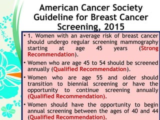
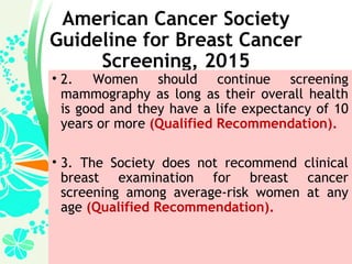
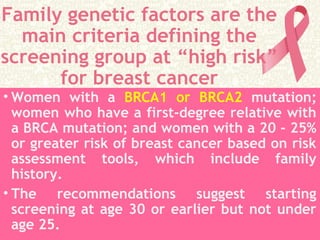
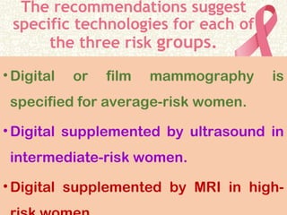
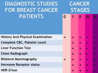
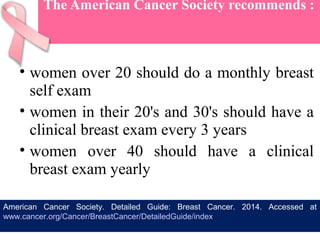
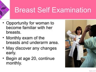
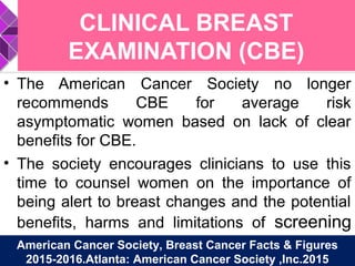
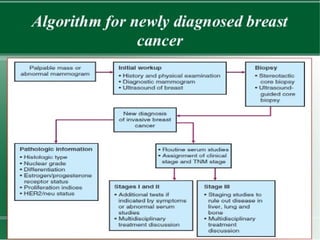
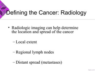
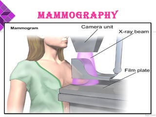
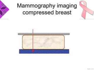
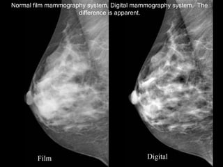
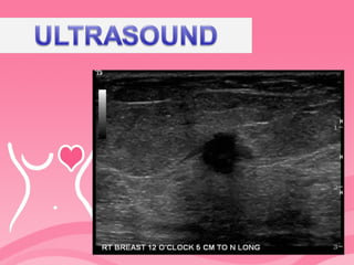
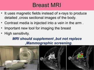
Presented by:-
Jaspreet Kaur Sodhi
Associate Professor
Institute of Nursing Education
GTBS© Hospital,Ludhiana
DIAGNOSTDIAGNOST
IC TESTSIC TESTS
ININ
BREASTBREAST
CANCERCANCER
SkIN REDNESS &
ASymmETRy
ENLARGEMENT
DImplING
NIpplE RETRACTION
NIpplE DISCHARGE
EARly DETECTIONS
REDuCES
THE RISk Of DyING
fROm BREAST
CANCER.
American Cancer Society
Guideline for Breast Cancer
Screening, 2015
• 1. Women with an average risk of breast cancer
should undergo regular screening mammography
starting at age 45 years (Strong
Recommendation).
• Women who are age 45 to 54 should be screened
annually (Qualified Recommendation).
• Women who are age 55 and older should
transition to biennial screening or have the
opportunity to continue screening annually
(Qualified Recommendation).
• Women should have the opportunity to begin
annual screening between the ages of 40 and 44
(Qualified Recommendation).
American Cancer Society
Guideline for Breast Cancer
Screening, 2015
• 2. Women should continue screening
mammography as long as their overall health
is good and they have a life expectancy of 10
years or more (Qualified Recommendation).
• 3. The Society does not recommend clinical
breast examination for breast cancer
screening among average-risk women at any
age (Qualified Recommendation).
Average Risk of Breast
Cancer
•Women without a personal history
of breast cancer.
•A suspected or confirmed genetic
mutations e.g. BRCA
•History of previous radiotherapy to
chest at a young age.
Family genetic factors are the
main criteria defining the
screening group at “high risk”
for breast cancer
• Women with a BRCA1 or BRCA2 mutation;
women who have a first-degree relative with
a BRCA mutation; and women with a 20 - 25%
or greater risk of breast cancer based on risk
assessment tools, which include family
history.
• The recommendations suggest starting
screening at age 30 or earlier but not under
age 25.
The recommendations suggest
specific technologies for each of
the three risk groups.
•Digital or film mammography is
specified for average-risk women.
•Digital supplemented by ultrasound in
intermediate-risk women.
•Digital supplemented by MRI in high-
DIAGNOSTIC STUDIES
FOR BREAST CANCER
PATIENTS
CANCER
STAGES
0 1 II III IV
History And Physical Examination + + + + +
Complete CBC, Platelet count + + + +
Liver Function Test + + + +
Chest Radiograph + + + +
Bilateral Mammography + + + + +
Hormone Receptor status + + + +
HER-2/nue + + + +
The American Cancer Society recommends :
• women over 20 should do a monthly breast
self exam
• women in their 20's and 30's should have a
clinical breast exam every 3 years
• women over 40 should have a clinical
breast exam yearly
American Cancer Society. Detailed Guide: Breast Cancer. 2014. Accessed at
www.cancer.org/Cancer/BreastCancer/DetailedGuide/index
American Cancer Society,
• No longer recommends that all women
perform monthly breast self
examination(BSE).
• But, All Women should become familiar
with both the appearance and feel of
their breast and report any changes
promptly to their physician.
American Cancer Society, Breast Cancer Facts & Figures
2015-2016.Atlanta: American Cancer Society ,Inc.2015
Breast Self Examination
• Opportunity for woman to
become familiar with her
breasts.
• Monthly exam of the
breasts and underarm area.
• May discover any changes
early.
• Begin at age 20, continue
monthly.
When to do BSE?
• Menstruating women- 5 to 7 days
after the beginning of their period.
• Menopausal women - same date
each month.
• Pregnant women – same date each
month.
• Takes about 20 minutes.
• Perform BSE at least once a month.
• Examine all breast tissue.
•Stand in front of a
mirror and look at
each breast
separately.
• Note the size, shape,
colour, contour and
direction of your
breasts and nipples.
Raise your
arms over
your head
and look at
your breasts,
as you turn
slowly from
side to side.
Press your
hands on
your hips and
push your
shoulders
forward. Look
at each breast
separately.
Stand in
front of a
mirror and
start BSE
just below
the collar
bone.
•Use the 3 middle
fingertips of your left
hand for the right breast.
•Apply firm pressure and
make small circles as
you go back or forth in a
pattern covering all the
breast area including the
nipple.
•Extend the examination
to the breast tissue in
the underarm.
•Change your hand and
repeat BSE on the
opposite breast.
•Lie down and raise one arm
above your head.
•Examine your breasts as before,
omitting the underarm.
•Change your arm and repeat BSE
on the opposite breast.
Month .......................
Record:
normal ridges,
lumpy areas, freckles
or moles.
Remember, what you
are looking for are
unusual changes
from your normal.
Record your observations
CLINICAL BREAST
EXAMINATION (CBE)
• The American Cancer Society no longer
recommends CBE for average risk
asymptomatic women based on lack of clear
benefits for CBE.
• The society encourages clinicians to use this
time to counsel women on the importance of
being alert to breast changes and the potential
benefits, harms and limitations of screening
mammography.American Cancer Society, Breast Cancer Facts & Figures
2015-2016.Atlanta: American Cancer Society ,Inc.2015
Defining the Cancer: Radiology
• Radiologic imaging can help determine
the location and spread of the cancer
– Local extent
– Regional lymph nodes
– Distant spread (metastases)
MAMMOGRAPHY
Mammography Technique
Mammography can be
viewed in 2 axis:-
• Cranio-caudal(CC)
• Medio-Lateral
Mammography imaging
compressed breast
a- The breast are almost
entirely fatty. Mammography is
highly sensitive in this setting.
b- There are scattered areas of
fibro-glandular density. The
term density describes the
degree of x-ray attenuation of
breast tissue but not discrete
mammographic findings.
c- The breasts are
heterogeneously dense, which
may obscure small masses.
Some areas in the breasts are
sufficiently dense to obscure
small masses.
d - The breasts are extremely
dense, which lowers the
sensitivity of mammography.
a- The breast are almost
entirely fatty. Mammography
is highly sensitive in this
setting.
b-There are scattered areas of
fibro-glandular density. The
term density describes the
degree of x-ray attenuation of
breast tissue but not discrete
mammographic findings.
c-The breasts are
heterogeneously dense, which
may obscure small masses.
Some areas in the breasts are
sufficiently dense to obscure
small masses.
d-The breasts are extremely
dense, which lowers the
sensitivity of mammography.
•The density of a mass is related to the expected
attenuation of an equal volume of fibro-glandular
tissue.
•High density is associated with malignancy.
•It is extremely rare for breast cancer to be low
density.
BI-RADS
BReAst IMAGInG
RePORtInG And
dAtA sYsteM
Mammography Sensitivity in
Younger Women
Screening mammograms
miss up to 25% of
breast cancers in
women in their 40s,
compared to 10% of
cancers for older
women
After the initial breast
screening,
a follow up
or
Diagnostic Mammographic
is often recommended
when the
BI-RADS category was 3 or
higher.
Digital Mammography
• In digital mammography, the film used to record the
mammogram is replaced by a sensitive digital
detector. The detector provides
• An electrical signal that is digitized, stored in a
computer and displayed on a monitor.
• Unlike film, where the image characteristics are
fixed, in digital mammography, the displayed image
can be adjusted during viewing to enhance visibility
of anatomical information.
• Digital mammography was developed with the
intention of improving performance of mammography
in dense breasts.
Film Digital
Normal film mammography system, Digital mammography system. The
difference is apparent.
Advantages of Mammography
• Early detection of breast cancer by
mammography leads to greater range of
treatment options, including:
less-extensive surgery (eg: breast-conserving
surgery like lumpectomy versus mastectomy)
Use of chemotherapy with fewer side effects
Limitations of Mammography
• Not all the breast cancer will be detected by
mammogram and some breast cancers that are
screen-detected still have poor prognosis.
• False Positive testing.
Radiation exposure
• Many people are concerned about
radiation exposure, the dose required for
a mammogram is very small and the risk
of harm is minimal.
ULtRAsOUn
d• It is particularly useful for investigating
mass lesions and distinguishing
whether they are cystic, solid or
suspicious of malignancy
• However, ultrasound has limited value
in the fatty breast due to lack of
contrast
ULtRAsOUnd
• Unlike MRI, breast ultrasound does not require
the injection of a contrast agent.
• Ultrasound is well tolerated by patients and
does not expose them to ionizing radiation.
• As well, ultrasound is a reasonable alternative
to MRI for women who would otherwise be
eligible for MRI examination, but who either
cannot or are not willing to have an MRI
examination..
Breast MRIBreast MRI
• It uses magnetic fields instead of x-rays to produce
detailed ,cross sectional images of the body.
• Contrast media is injected into a vein in the arm .
• Important new tool for imaging the breast
• High sensitivity.
MRI should supplement ,but not replace
,Mammographic screening.
Principle behind Breast MRI
• As breast cancers grow, they ensure their
blood supply by sending out signals that
recruit the development of new blood
vessels, a process referred to as tumour
angiogenesis.
• These vessels are poorly formed and are
leaky. If an intravenous injection of a
chelated Gd contrast agent is performed, the
agent that leaks from these vessels will pool
in the extra-vascular space and then wash
out.
• Breast MRI produces three-dimensional
images that allow the pattern of leakage
and washout to be monitored.
• The amount of uptake of the contrast agent,
the shape of the enhancing areas and the
kinetics of uptake provide information that
allows very high sensitivity in detecting
breast cancers and distinguishing them
from non-cancerous structures in the
breast.
Advantages of
MRI
• There is no ionizing
radiation, no breast
compression, it’s not
affected by breast
density and it
provides greater than
90% sensitivity.
• MRI is also useful to
guide biopsies and to
show the response of
cancers to neo-
adjuvant
chemotherapy.
Disadvantages of
MRI
• cost of the equipment
• Need for an
intravenous injection
of a contrast medium
and the longer time
required for imaging.
Breast Computed Tomography
imaging a “pendant” breast
Triple Diagnosis
It refers to the concurrent
use of Physical Examination,
Mammography, and skilled
FNAC for diagnosing
palpable breast lumps.
FNAC
If a lump is detected on clinical
examination, but if not picked up by
mammogram or a USG , the lump has
to be further probed and ideal test is
FINE NEEDLE
ASPIRATION
CYTOLOGY.
FNAC
The National Cancer Institute
recommendation for the diagnosis
of breast aspiration cytology:
Advantages of FNAC
• Well tolerated
outpatient procedure
performed by
palpation or under
ultrasound.
Disadvantages of
FNAC
• Low yield of cells
without normal
surrounding structure.
• Need experienced
cytopathologist.
Breast Cancer Diagnosis
• Any breast change or lump
needs to be evaluated.
• Breast cancer needs to be
diagnosed by biopsy
– Fine needle aspiration
– Core needle biopsy
– Surgical biopsy
BIOPsY
Ultrasound-Guided Breast
Biopsy
• Doctors use ultrasound to
guide the needle during the
biopsy.
•This method is used when
lumps are hard to feel on a
breast exam or see on a
mammogram.
Stereotactic core needle
biopsy localization.
•A three-dimensional (3-D) x-ray
guides a biopsy needle to a lump or
other change that cannot be felt on a
breast exam.
•With one type of equipment, patient
lie face down on an exam table with a
hole in it.
• The hole allows patient’s breast to
hang below the table, where the x-ray
machine and needle are.
• Or, special equipment can be
attached to a standard mammogram
machine to x-ray your breast from two
Breast Biopsy
A core needle biopsy of the area is recommendedA core needle biopsy of the area is recommended
Non-
Surgical
Surgical
Needle Biopsies
• Fine Needle – A thin, hollow needle is used to
remove a sample of tissue. The procedure is quick
and can be done in a doctor’s office.
• Core Needle – A larger needle is inserted through a
small incision in the skin, and a small core of tissue
is removed. This type of needle biopsy is done
with the assistance of mammography or ultrasound
imaging using stereotactic techniques with the aid
of the computer, which calculates the precise
location of the lump.
CORE NEEDLE BIOPSY
CORE NEEDLE BIOPSY
• A large –bore needle is placed into the
area of suspicion and a core of tissue is
removed.
• Usually several core biopsies are taken
of the area to ensure adequate sampling
of the abnormality .
• This may be done with palpation, under
stereotactic mammography ,or
ultrasound guidance.
Advantages of Core
Needle Biopsy
• High yield of tissue so
less chance of false
readings.
• Tissue is removed with
normal structure and
architecture intact,
which enables more
accurate interpretation.
Disadvantages of
Core Needle Biopsy
• Greater risk of bleeding,
bruising ,or infection.
• Slightly more
uncomfortable
INCISIONAL BIOPSY
Incisional Biopsy
• A surgical procedure where only a
small amount of the abnormality is
removed through an open incision.
• Generally reserved for lumps that are
larger
• Performed under local anesthesia in a
hospital or outpatient clinic
Advantages of
Incisional
Biopsy.
• High yield of tissue
makes diagnosis
easier.
• Does not remove
large amounts of
tissue for benign
lesions.
Disadvantages of
MRI
• Does not remove all of
the cancer and,
therefore ,another
procedure is
necessary.
Excisional Biopsy
Excisional Biopsy
• A surgical procedure that removes the
entire suspected area plus some
surrounding normal tissue.
• Standard procedure for lumps that are
smaller than an inch or so in diameter
• Similar to a lumpectomy
• Performed under local anesthetic or
general anesthesia in a hospital or
outpatient clinic
Advantages of
Excisional Biopsy
• Removes entire lump.
• May be the only surgical
treatment needed.
• Larger samples provide
information for treatment
plan
Disadvantages of
Excisional Biopsy
• May remove large
amounts of tissue that are
benign.
• Removing tissue can
change the look and feel
of the breast.
• Surgical procedure
• Expensive
• Side effects such as
infection or blood
collection under the skin
FNAC
•The sensitivity of FNAC in diagnosis
of breast lesion is 90-95 %.
•Definitive treatment is often based
on cytological diagnosis without
histological confirmation, unless
there is disagreement between
cytology and clinical and/or
mammographic assessment .
Karin Lindholm-Breast. Orell S.R., Sterrett G.F., Whitaker D. — Fine needle
aspiration cytology. 4th edition. Reed Elsevier India Private Limited, New Delhi:
Chapter7, pg.167; 2005.
• FNAC is a valuable method, although moderately less
sensitive than CB.
• CB is the preferred method for preoperative diagnosis
when sampling FNAC provides scarce material and
suspicion of a fibrotic and collagenous lesion such as
lobular carcinoma and radial scar arises.
• FNAC is most accurate when experienced cytologists
are available and when immediate assessment by
professionals is performed for evaluation of material
adequacy, so that additional aspirations can be done
when needed.
Berner A, Davidson B, Sigstad E,Risberg .Fine-needle aspiration cytology vs. core
biopsy in the diagnosis of breast lesions. Diagn Cytopathol. 2003 Dec;29(6):344-8.
Cancer is diagnosed
in 1 of 5
breast biopsies
Staging
The Stages of Breast Cancer
Breast Cancer is diagnosed according to stages (stages 0
through IV) under the TNM classification.
Factors used in staging of Breast Cancer:
• Tumor Size
Size of primary tumor
• Nodal status
Indicates presence or absence of cancer cells in lymph
nodes
• Metastasis
Indicates if cancer cells have spread from the affected
breast to other areas of the body (i.e. skin, liver, lungs,
Source: National Cancer Institute
Breast cancer
Spread to lymph nodes
Supraclavicular
Subclavicular
Distal (upper)
axillary
Central (middle)
axillary
Proximal (lower)
axillary
Mediastinal
Internal mammary
Interpectoral
(Rotter’s)
Breast Cancer: Stage I
T1a: TT1a: T ≤≤ 0.5 cm0.5 cm
T1b: 0.5 cm < TT1b: 0.5 cm < T ≤≤ 1 cm1 cm
T1c: 1 cm < TT1c: 1 cm < T ≤≤ 2 cm2 cm
T1 N0 M0T1 N0 M0
TT ≤≤ 2 cm2 cm
T1T1
N0 = no regional lymph node metastasis
M0 = no distant metastasis
Breast Cancer: Stage IIA
T2 N0 M0T2 N0 M0
N1 = metastasis to movable ipsilateral axillary lymph node(s)
M0 = no distant metastasis
2 cm < T2 cm < T << 5 cm5 cm
No evidenceNo evidence
of tumorof tumor
T0T0
T0T0
T1T1
N1 M0N1 M0}
T2T2
Breast Cancer: Stage IIB
T3 N0 M0T3 N0 M0
N1 = metastasis to movable ipsilateral axillary lymph node(s) (p) N1a, N1b
M0 = no distant metastasis
T > 5 cmT > 5 cm
T2 N1 M0T2 N1 M0
T3T3
Breast Cancer: Stage IIIA
Metastasis to ipsilateral axillary lymph node(s)
N1 = movable
N2 = fixed to one another or to other structures
M0 = no distant metastasis
T3 N1 M0T3 N1 M0 T0T0
T1T1
T2T2
T3T3
N2 M0N2 M0
Breast Cancer: Stage IIIB
Any T N3 M0Any T N3 M0
N3 = metastasis to ipsilateral internal mammary lymph node(s)
M0 = no distant metastasis
Tumor of any size
with direct extension
to chest wall or skin
T4d = inflammatory
carcinoma
T4 any N M0T4 any N M0
T4T4
Breast Cancer: Stage IV
M1 = distant metastasis (including metastases to cervical, or contralateral
internal mammary lymph nodes)
Any T any N M1
FDG PET scanFDG PET scanBone ScanBone Scan
Staging for Distant Disease:Staging for Distant Disease:
Breast CancerBreast Cancer
Tumor in the breast, butTumor in the breast, but
not elsewherenot elsewhere
Multiple boneMultiple bone
metastasesmetastases
Patient APatient A Patient BPatient B
The most common sites of distant disease in breast cancer are the bones,The most common sites of distant disease in breast cancer are the bones,
liver and lungsliver and lungs
Progress on Biomarkers of
Cancer Diagnosis and
Prognosis
According to Dr. Norman
Boyd,
• Hormone levels vary with the proportion
of dense and non-dense tissue in the
breast.
• Excess estrogen in dense breasts is
believed to accelerate tumour growth.
• Breast density risks apply mostly to
premenopausal women, which is what
makes the 40 - 49 age group, even up to
age 55, so important.
Estrogen receptors(ER)
&
Progesterone receptors(PR)
• Normal breast cells and some breast cancer
cells contains receptors that attaches to
estrogen and progesterone.
• These 2 hormones often fuel the growth of
breast cancer cells.
• Cancer removed during biopsy or surgery is
checked if it has ER or PR.
HORMONE RECEPTOR
POSITIVE
• Breast cancer that have estrogen receptors
are referred to as ER- Positive cancers.
• Breast cancer that have progesterone
receptors are referred to as PR- Positive
cancers.
Tamoxifen
Blocks estrogen from entering into the cell,
blocking estrogen-dependent growth
Nucleus
Tumor cell
Estrogen
biosynthesis
Estrogen
biosynthesis
from muscle & fat
DeVita, et al. Cancer Principles and Practice of Oncology. 6th
ed 2001
Aromatase
Inhibitors Aramatase
Ki-67.
•Ki-67 is a protein in cells that
increases as they prepare to divide
into new cells.
• A staining process can measure the
percentage of tumor cells that are
positive for Ki-67.
• The more positive cells there are,
the more quickly they are dividing
and forming new cells.
What is a biomarker?
Biomarkers are anatomical,
physiological, biochemical or
molecular parameters associated
with the presence and severity of
specific disease states detectable
and measurable by a variety of
methods including physical
examination, laboratory assays and
medical imaging.
Potential uses for
Biomarkers in Oncology
Molecular biomarkers• Gene expression
 microarray
 quantitative reverse transcription-polymerase chain
reaction (RT- PCR)
• Gene amplification
 (FISH/CISH)
• Gene sequence
 DNA sequencing
• Protein expression
 Immunohistochemistry (IHC)
 Enzyme-linked immunosorbent assay (ELISA)
IHC, FISH and ELISA are widely established in clinical
pathology laboratories
HER2 (Human Epidermal Growth
Factor 2)Testing In Breast Cancer
HER2 receptors: Proteins made by the HER2 gene
that receive signals that stimulate cells to grow
and multiply.
HER2 POSITIVE
• HER2-positive breast cancer
is a breast cancer that tests
positive for a protein called
human epidermal growth
factor receptor 2 (HER2),
which promotes the growth of
cancer cells.
IHC
(Immuno-HistoChemistry):
• The IHC test shows whether there is too much
HER2-receptor protein in the cancer cells.
• The results of the IHC test can be 0
(negative), 1+ (also negative), 2+ (borderline),
or 3+ (positive; the HER2 protein is
overexpressed).
FISH
Fluorescent in situ hybridization.
• FISH is a technique
that measures gene amplification using fluorescently
labeled DNA (probe).
• FISH is a fast and highly sensitive technique that provides
an objective scoring system.
• The FISH test shows whether there are too many copies
of the HER2 gene in the cancer cells.
• The results of the FISH test can be positive (extra HER2
gene copies— amplified) or negative (normal number of
HER2 gene copies—not amplified).
PROCESS OF FISH
The principles of fluorescence in situ hybridization.
© The Author 2010. Published by Oxford University Press.
Luminal A (HR+/HER2-).
• Most (74%) breast cancers express the
estrogen receptor (ER+) and/or the
progesterone receptor (PR+) but not HER2
(HER2-).
• These cancers tend to be slow-growing and
less aggressive than other subtypes.
• Luminal A tumors are associated with the most
favorable prognosis, particularly in the short
term, in part because expression of hormone
receptors is predictive of a favorable response
to hormonal therapy
Triple Negative Breast
Cancer
• Overall, about 12% of breast cancers are triple
negative, so called because they are ER-, PR-,
and HER2-
• They are also more common in premenopausal
women and those with a BRCA1 gene mutation.
• The majority (about 75%) of triple negative
breast cancers fall in to the basal-like subtype.
• Triple negative breast cancers have a poorer
short-term prognosis than other breast cancer
types, in part because there are currently no
targeted therapies for these tumors
Triple Negative Breast Cancer
• Triple Negative Breast Cancer
– Estrogen Receptor (ER) Negative
– Progesterone Receptor (PR) Negative
– HER2 Receptor Negative
• Considered to have a poorer prognosis than many
other types of breast cancer
• Many existing targeted therapies do not have a
place in TN Breast Cancer therapy (e.g. Herceptin,
Tamoxifen)
Luminal B (HR+/HER2+).
• Luminal B breast cancers are ER+
and/or PR+ and are further defined by
being highly positive for Ki67 (indicator
of a large proportion of actively
dividing cells) or HER2.
• About 10% of breast cancers are ER+
and/or PR+ and HER2+.
• Luminal B breast cancers tend to be
higher grade and more aggressive than
luminal A breast cancers.
HER2-enriched (HR-/HER2+).
• About 4% of breast cancers produce excess
HER2 and do not express hormone receptors.
• These cancers tend to grow and spread more
aggressively than other breast cancers and
are associated with poorer short-term
prognosis compared to ER+ breast cancers.
• However, the recent widespread use of
targeted therapies for HER2+ cancers has
reversed much of the adverse prognostic
impact of HER2 overexpression.
PROGNOSIS OF HER2nue
• In about 1 of every 5 breast cancers, the
cancer cells have a gene mutation that
makes an excess of the HER2 protein.
• HER2-positive breast cancers tend to be
more aggressive than other types of
breast cancer.
• They're less likely to be sensitive to
hormone therapy, though many people
with HER2-positive breast cancer can still
benefit from hormone therapy.
• BRCA1: An abnormal gene, known as
BReast CAncer gene 1, associated with a
higher risk of developing breast cancer.
• BRCA2: An abnormal gene, known as
BReast CAncer gene 2, associated with a
higher risk of developing breast cancer.
• ER-negative: A cancer that does not
have estrogen receptors.
• ER-positive: A cancer that has estrogen
receptors
• IHC (ImmunoHistoChemistry) test: A test
used to measure proteins, including the HER2
protein.
• FISH (Fluorescence In Situ Hybridization)
test: A test for multiple genes, including the
HER2 gene.
CanadIan BREaST CanCER FOundaTIOn – OnTaRIO
REgIOn
EaRlIER dETECTIOn and dIagnOSIS OF BREaST CanCER:a
REPORT FROm IT’S aBOuT TImE! a COnSEnSuS COnFEREnCE
amEndEd OCTOBER 13, 2010
amERICan CanCER SOCIETy. dETaIlEd guIdE: BREaST
CanCER. 2014. aCCESSEd aT
www.CanCER.ORg/CanCER/BREaSTCanCER/dETaIlEdguI
dE/IndEx On SEPTEmBER 3, 2015.
amERICan CanCER SOCIETy. BREaST CanCER FaCTS &
FIguRES 2015-2016. aTlanTa: amERICan CanCER
SOCIETy, InC. 2015.
Thank You

Breast cancer

  • 1.
    Presented by:- Jaspreet KaurSodhi Associate Professor Institute of Nursing Education GTBS© Hospital,Ludhiana DIAGNOSTDIAGNOST IC TESTSIC TESTS ININ BREASTBREAST CANCERCANCER
  • 7.
  • 8.
  • 9.
  • 10.
  • 11.
  • 12.
    EARly DETECTIONS REDuCES THE RISkOf DyING fROm BREAST CANCER.
  • 14.
    American Cancer Society Guidelinefor Breast Cancer Screening, 2015 • 1. Women with an average risk of breast cancer should undergo regular screening mammography starting at age 45 years (Strong Recommendation). • Women who are age 45 to 54 should be screened annually (Qualified Recommendation). • Women who are age 55 and older should transition to biennial screening or have the opportunity to continue screening annually (Qualified Recommendation). • Women should have the opportunity to begin annual screening between the ages of 40 and 44 (Qualified Recommendation).
  • 15.
    American Cancer Society Guidelinefor Breast Cancer Screening, 2015 • 2. Women should continue screening mammography as long as their overall health is good and they have a life expectancy of 10 years or more (Qualified Recommendation). • 3. The Society does not recommend clinical breast examination for breast cancer screening among average-risk women at any age (Qualified Recommendation).
  • 16.
    Average Risk ofBreast Cancer •Women without a personal history of breast cancer. •A suspected or confirmed genetic mutations e.g. BRCA •History of previous radiotherapy to chest at a young age.
  • 17.
    Family genetic factorsare the main criteria defining the screening group at “high risk” for breast cancer • Women with a BRCA1 or BRCA2 mutation; women who have a first-degree relative with a BRCA mutation; and women with a 20 - 25% or greater risk of breast cancer based on risk assessment tools, which include family history. • The recommendations suggest starting screening at age 30 or earlier but not under age 25.
  • 18.
    The recommendations suggest specifictechnologies for each of the three risk groups. •Digital or film mammography is specified for average-risk women. •Digital supplemented by ultrasound in intermediate-risk women. •Digital supplemented by MRI in high-
  • 19.
    DIAGNOSTIC STUDIES FOR BREASTCANCER PATIENTS CANCER STAGES 0 1 II III IV History And Physical Examination + + + + + Complete CBC, Platelet count + + + + Liver Function Test + + + + Chest Radiograph + + + + Bilateral Mammography + + + + + Hormone Receptor status + + + + HER-2/nue + + + +
  • 20.
    The American CancerSociety recommends : • women over 20 should do a monthly breast self exam • women in their 20's and 30's should have a clinical breast exam every 3 years • women over 40 should have a clinical breast exam yearly American Cancer Society. Detailed Guide: Breast Cancer. 2014. Accessed at www.cancer.org/Cancer/BreastCancer/DetailedGuide/index
  • 21.
    American Cancer Society, •No longer recommends that all women perform monthly breast self examination(BSE). • But, All Women should become familiar with both the appearance and feel of their breast and report any changes promptly to their physician. American Cancer Society, Breast Cancer Facts & Figures 2015-2016.Atlanta: American Cancer Society ,Inc.2015
  • 22.
    Breast Self Examination •Opportunity for woman to become familiar with her breasts. • Monthly exam of the breasts and underarm area. • May discover any changes early. • Begin at age 20, continue monthly.
  • 23.
    When to doBSE? • Menstruating women- 5 to 7 days after the beginning of their period. • Menopausal women - same date each month. • Pregnant women – same date each month. • Takes about 20 minutes. • Perform BSE at least once a month. • Examine all breast tissue.
  • 24.
    •Stand in frontof a mirror and look at each breast separately. • Note the size, shape, colour, contour and direction of your breasts and nipples.
  • 25.
    Raise your arms over yourhead and look at your breasts, as you turn slowly from side to side.
  • 26.
    Press your hands on yourhips and push your shoulders forward. Look at each breast separately.
  • 27.
    Stand in front ofa mirror and start BSE just below the collar bone.
  • 28.
    •Use the 3middle fingertips of your left hand for the right breast. •Apply firm pressure and make small circles as you go back or forth in a pattern covering all the breast area including the nipple. •Extend the examination to the breast tissue in the underarm. •Change your hand and repeat BSE on the opposite breast.
  • 29.
    •Lie down andraise one arm above your head. •Examine your breasts as before, omitting the underarm. •Change your arm and repeat BSE on the opposite breast.
  • 30.
    Month ....................... Record: normal ridges, lumpyareas, freckles or moles. Remember, what you are looking for are unusual changes from your normal. Record your observations
  • 31.
    CLINICAL BREAST EXAMINATION (CBE) •The American Cancer Society no longer recommends CBE for average risk asymptomatic women based on lack of clear benefits for CBE. • The society encourages clinicians to use this time to counsel women on the importance of being alert to breast changes and the potential benefits, harms and limitations of screening mammography.American Cancer Society, Breast Cancer Facts & Figures 2015-2016.Atlanta: American Cancer Society ,Inc.2015
  • 33.
    Defining the Cancer:Radiology • Radiologic imaging can help determine the location and spread of the cancer – Local extent – Regional lymph nodes – Distant spread (metastases)
  • 34.
  • 35.
    Mammography Technique Mammography canbe viewed in 2 axis:- • Cranio-caudal(CC) • Medio-Lateral
  • 36.
  • 38.
    a- The breastare almost entirely fatty. Mammography is highly sensitive in this setting. b- There are scattered areas of fibro-glandular density. The term density describes the degree of x-ray attenuation of breast tissue but not discrete mammographic findings. c- The breasts are heterogeneously dense, which may obscure small masses. Some areas in the breasts are sufficiently dense to obscure small masses. d - The breasts are extremely dense, which lowers the sensitivity of mammography. a- The breast are almost entirely fatty. Mammography is highly sensitive in this setting. b-There are scattered areas of fibro-glandular density. The term density describes the degree of x-ray attenuation of breast tissue but not discrete mammographic findings. c-The breasts are heterogeneously dense, which may obscure small masses. Some areas in the breasts are sufficiently dense to obscure small masses. d-The breasts are extremely dense, which lowers the sensitivity of mammography.
  • 39.
    •The density ofa mass is related to the expected attenuation of an equal volume of fibro-glandular tissue. •High density is associated with malignancy. •It is extremely rare for breast cancer to be low density.
  • 40.
  • 42.
    Mammography Sensitivity in YoungerWomen Screening mammograms miss up to 25% of breast cancers in women in their 40s, compared to 10% of cancers for older women
  • 43.
    After the initialbreast screening, a follow up or Diagnostic Mammographic is often recommended when the BI-RADS category was 3 or higher.
  • 44.
    Digital Mammography • Indigital mammography, the film used to record the mammogram is replaced by a sensitive digital detector. The detector provides • An electrical signal that is digitized, stored in a computer and displayed on a monitor. • Unlike film, where the image characteristics are fixed, in digital mammography, the displayed image can be adjusted during viewing to enhance visibility of anatomical information. • Digital mammography was developed with the intention of improving performance of mammography in dense breasts.
  • 45.
    Film Digital Normal filmmammography system, Digital mammography system. The difference is apparent.
  • 46.
    Advantages of Mammography •Early detection of breast cancer by mammography leads to greater range of treatment options, including: less-extensive surgery (eg: breast-conserving surgery like lumpectomy versus mastectomy) Use of chemotherapy with fewer side effects
  • 47.
    Limitations of Mammography •Not all the breast cancer will be detected by mammogram and some breast cancers that are screen-detected still have poor prognosis. • False Positive testing.
  • 48.
    Radiation exposure • Manypeople are concerned about radiation exposure, the dose required for a mammogram is very small and the risk of harm is minimal.
  • 50.
    ULtRAsOUn d• It isparticularly useful for investigating mass lesions and distinguishing whether they are cystic, solid or suspicious of malignancy • However, ultrasound has limited value in the fatty breast due to lack of contrast
  • 51.
    ULtRAsOUnd • Unlike MRI,breast ultrasound does not require the injection of a contrast agent. • Ultrasound is well tolerated by patients and does not expose them to ionizing radiation. • As well, ultrasound is a reasonable alternative to MRI for women who would otherwise be eligible for MRI examination, but who either cannot or are not willing to have an MRI examination..
  • 54.
    Breast MRIBreast MRI •It uses magnetic fields instead of x-rays to produce detailed ,cross sectional images of the body. • Contrast media is injected into a vein in the arm . • Important new tool for imaging the breast • High sensitivity. MRI should supplement ,but not replace ,Mammographic screening.
  • 55.
    Principle behind BreastMRI • As breast cancers grow, they ensure their blood supply by sending out signals that recruit the development of new blood vessels, a process referred to as tumour angiogenesis. • These vessels are poorly formed and are leaky. If an intravenous injection of a chelated Gd contrast agent is performed, the agent that leaks from these vessels will pool in the extra-vascular space and then wash out.
  • 56.
    • Breast MRIproduces three-dimensional images that allow the pattern of leakage and washout to be monitored. • The amount of uptake of the contrast agent, the shape of the enhancing areas and the kinetics of uptake provide information that allows very high sensitivity in detecting breast cancers and distinguishing them from non-cancerous structures in the breast.
  • 57.
    Advantages of MRI • Thereis no ionizing radiation, no breast compression, it’s not affected by breast density and it provides greater than 90% sensitivity. • MRI is also useful to guide biopsies and to show the response of cancers to neo- adjuvant chemotherapy. Disadvantages of MRI • cost of the equipment • Need for an intravenous injection of a contrast medium and the longer time required for imaging.
  • 58.
    Breast Computed Tomography imaginga “pendant” breast
  • 59.
    Triple Diagnosis It refersto the concurrent use of Physical Examination, Mammography, and skilled FNAC for diagnosing palpable breast lumps.
  • 60.
    FNAC If a lumpis detected on clinical examination, but if not picked up by mammogram or a USG , the lump has to be further probed and ideal test is FINE NEEDLE ASPIRATION CYTOLOGY.
  • 61.
  • 62.
    The National CancerInstitute recommendation for the diagnosis of breast aspiration cytology:
  • 63.
    Advantages of FNAC •Well tolerated outpatient procedure performed by palpation or under ultrasound. Disadvantages of FNAC • Low yield of cells without normal surrounding structure. • Need experienced cytopathologist.
  • 64.
    Breast Cancer Diagnosis •Any breast change or lump needs to be evaluated. • Breast cancer needs to be diagnosed by biopsy – Fine needle aspiration – Core needle biopsy – Surgical biopsy
  • 66.
  • 67.
    Ultrasound-Guided Breast Biopsy • Doctorsuse ultrasound to guide the needle during the biopsy. •This method is used when lumps are hard to feel on a breast exam or see on a mammogram.
  • 68.
    Stereotactic core needle biopsylocalization. •A three-dimensional (3-D) x-ray guides a biopsy needle to a lump or other change that cannot be felt on a breast exam. •With one type of equipment, patient lie face down on an exam table with a hole in it. • The hole allows patient’s breast to hang below the table, where the x-ray machine and needle are. • Or, special equipment can be attached to a standard mammogram machine to x-ray your breast from two
  • 69.
    Breast Biopsy A coreneedle biopsy of the area is recommendedA core needle biopsy of the area is recommended Non- Surgical Surgical
  • 70.
    Needle Biopsies • FineNeedle – A thin, hollow needle is used to remove a sample of tissue. The procedure is quick and can be done in a doctor’s office. • Core Needle – A larger needle is inserted through a small incision in the skin, and a small core of tissue is removed. This type of needle biopsy is done with the assistance of mammography or ultrasound imaging using stereotactic techniques with the aid of the computer, which calculates the precise location of the lump.
  • 71.
  • 72.
    CORE NEEDLE BIOPSY •A large –bore needle is placed into the area of suspicion and a core of tissue is removed. • Usually several core biopsies are taken of the area to ensure adequate sampling of the abnormality . • This may be done with palpation, under stereotactic mammography ,or ultrasound guidance.
  • 73.
    Advantages of Core NeedleBiopsy • High yield of tissue so less chance of false readings. • Tissue is removed with normal structure and architecture intact, which enables more accurate interpretation. Disadvantages of Core Needle Biopsy • Greater risk of bleeding, bruising ,or infection. • Slightly more uncomfortable
  • 74.
  • 75.
    Incisional Biopsy • Asurgical procedure where only a small amount of the abnormality is removed through an open incision. • Generally reserved for lumps that are larger • Performed under local anesthesia in a hospital or outpatient clinic
  • 76.
    Advantages of Incisional Biopsy. • Highyield of tissue makes diagnosis easier. • Does not remove large amounts of tissue for benign lesions. Disadvantages of MRI • Does not remove all of the cancer and, therefore ,another procedure is necessary.
  • 77.
  • 78.
    Excisional Biopsy • Asurgical procedure that removes the entire suspected area plus some surrounding normal tissue. • Standard procedure for lumps that are smaller than an inch or so in diameter • Similar to a lumpectomy • Performed under local anesthetic or general anesthesia in a hospital or outpatient clinic
  • 79.
    Advantages of Excisional Biopsy •Removes entire lump. • May be the only surgical treatment needed. • Larger samples provide information for treatment plan Disadvantages of Excisional Biopsy • May remove large amounts of tissue that are benign. • Removing tissue can change the look and feel of the breast. • Surgical procedure • Expensive • Side effects such as infection or blood collection under the skin
  • 80.
    FNAC •The sensitivity ofFNAC in diagnosis of breast lesion is 90-95 %. •Definitive treatment is often based on cytological diagnosis without histological confirmation, unless there is disagreement between cytology and clinical and/or mammographic assessment . Karin Lindholm-Breast. Orell S.R., Sterrett G.F., Whitaker D. — Fine needle aspiration cytology. 4th edition. Reed Elsevier India Private Limited, New Delhi: Chapter7, pg.167; 2005.
  • 81.
    • FNAC isa valuable method, although moderately less sensitive than CB. • CB is the preferred method for preoperative diagnosis when sampling FNAC provides scarce material and suspicion of a fibrotic and collagenous lesion such as lobular carcinoma and radial scar arises. • FNAC is most accurate when experienced cytologists are available and when immediate assessment by professionals is performed for evaluation of material adequacy, so that additional aspirations can be done when needed. Berner A, Davidson B, Sigstad E,Risberg .Fine-needle aspiration cytology vs. core biopsy in the diagnosis of breast lesions. Diagn Cytopathol. 2003 Dec;29(6):344-8.
  • 83.
    Cancer is diagnosed in1 of 5 breast biopsies
  • 84.
  • 86.
    The Stages ofBreast Cancer Breast Cancer is diagnosed according to stages (stages 0 through IV) under the TNM classification. Factors used in staging of Breast Cancer: • Tumor Size Size of primary tumor • Nodal status Indicates presence or absence of cancer cells in lymph nodes • Metastasis Indicates if cancer cells have spread from the affected breast to other areas of the body (i.e. skin, liver, lungs, Source: National Cancer Institute
  • 87.
    Breast cancer Spread tolymph nodes Supraclavicular Subclavicular Distal (upper) axillary Central (middle) axillary Proximal (lower) axillary Mediastinal Internal mammary Interpectoral (Rotter’s)
  • 88.
    Breast Cancer: StageI T1a: TT1a: T ≤≤ 0.5 cm0.5 cm T1b: 0.5 cm < TT1b: 0.5 cm < T ≤≤ 1 cm1 cm T1c: 1 cm < TT1c: 1 cm < T ≤≤ 2 cm2 cm T1 N0 M0T1 N0 M0 TT ≤≤ 2 cm2 cm T1T1 N0 = no regional lymph node metastasis M0 = no distant metastasis
  • 89.
    Breast Cancer: StageIIA T2 N0 M0T2 N0 M0 N1 = metastasis to movable ipsilateral axillary lymph node(s) M0 = no distant metastasis 2 cm < T2 cm < T << 5 cm5 cm No evidenceNo evidence of tumorof tumor T0T0 T0T0 T1T1 N1 M0N1 M0} T2T2
  • 90.
    Breast Cancer: StageIIB T3 N0 M0T3 N0 M0 N1 = metastasis to movable ipsilateral axillary lymph node(s) (p) N1a, N1b M0 = no distant metastasis T > 5 cmT > 5 cm T2 N1 M0T2 N1 M0 T3T3
  • 91.
    Breast Cancer: StageIIIA Metastasis to ipsilateral axillary lymph node(s) N1 = movable N2 = fixed to one another or to other structures M0 = no distant metastasis T3 N1 M0T3 N1 M0 T0T0 T1T1 T2T2 T3T3 N2 M0N2 M0
  • 92.
    Breast Cancer: StageIIIB Any T N3 M0Any T N3 M0 N3 = metastasis to ipsilateral internal mammary lymph node(s) M0 = no distant metastasis Tumor of any size with direct extension to chest wall or skin T4d = inflammatory carcinoma T4 any N M0T4 any N M0 T4T4
  • 93.
    Breast Cancer: StageIV M1 = distant metastasis (including metastases to cervical, or contralateral internal mammary lymph nodes) Any T any N M1
  • 94.
    FDG PET scanFDGPET scanBone ScanBone Scan Staging for Distant Disease:Staging for Distant Disease: Breast CancerBreast Cancer Tumor in the breast, butTumor in the breast, but not elsewherenot elsewhere Multiple boneMultiple bone metastasesmetastases Patient APatient A Patient BPatient B The most common sites of distant disease in breast cancer are the bones,The most common sites of distant disease in breast cancer are the bones, liver and lungsliver and lungs
  • 95.
    Progress on Biomarkersof Cancer Diagnosis and Prognosis
  • 96.
    According to Dr.Norman Boyd, • Hormone levels vary with the proportion of dense and non-dense tissue in the breast. • Excess estrogen in dense breasts is believed to accelerate tumour growth. • Breast density risks apply mostly to premenopausal women, which is what makes the 40 - 49 age group, even up to age 55, so important.
  • 97.
    Estrogen receptors(ER) & Progesterone receptors(PR) •Normal breast cells and some breast cancer cells contains receptors that attaches to estrogen and progesterone. • These 2 hormones often fuel the growth of breast cancer cells. • Cancer removed during biopsy or surgery is checked if it has ER or PR.
  • 98.
    HORMONE RECEPTOR POSITIVE • Breastcancer that have estrogen receptors are referred to as ER- Positive cancers. • Breast cancer that have progesterone receptors are referred to as PR- Positive cancers.
  • 99.
    Tamoxifen Blocks estrogen fromentering into the cell, blocking estrogen-dependent growth Nucleus Tumor cell Estrogen biosynthesis Estrogen biosynthesis from muscle & fat DeVita, et al. Cancer Principles and Practice of Oncology. 6th ed 2001 Aromatase Inhibitors Aramatase
  • 100.
    Ki-67. •Ki-67 is aprotein in cells that increases as they prepare to divide into new cells. • A staining process can measure the percentage of tumor cells that are positive for Ki-67. • The more positive cells there are, the more quickly they are dividing and forming new cells.
  • 101.
    What is abiomarker? Biomarkers are anatomical, physiological, biochemical or molecular parameters associated with the presence and severity of specific disease states detectable and measurable by a variety of methods including physical examination, laboratory assays and medical imaging.
  • 102.
  • 103.
    Molecular biomarkers• Geneexpression  microarray  quantitative reverse transcription-polymerase chain reaction (RT- PCR) • Gene amplification  (FISH/CISH) • Gene sequence  DNA sequencing • Protein expression  Immunohistochemistry (IHC)  Enzyme-linked immunosorbent assay (ELISA) IHC, FISH and ELISA are widely established in clinical pathology laboratories
  • 104.
    HER2 (Human EpidermalGrowth Factor 2)Testing In Breast Cancer
  • 105.
    HER2 receptors: Proteinsmade by the HER2 gene that receive signals that stimulate cells to grow and multiply.
  • 107.
    HER2 POSITIVE • HER2-positivebreast cancer is a breast cancer that tests positive for a protein called human epidermal growth factor receptor 2 (HER2), which promotes the growth of cancer cells.
  • 108.
    IHC (Immuno-HistoChemistry): • The IHCtest shows whether there is too much HER2-receptor protein in the cancer cells. • The results of the IHC test can be 0 (negative), 1+ (also negative), 2+ (borderline), or 3+ (positive; the HER2 protein is overexpressed).
  • 109.
    FISH Fluorescent in situhybridization. • FISH is a technique that measures gene amplification using fluorescently labeled DNA (probe). • FISH is a fast and highly sensitive technique that provides an objective scoring system. • The FISH test shows whether there are too many copies of the HER2 gene in the cancer cells. • The results of the FISH test can be positive (extra HER2 gene copies— amplified) or negative (normal number of HER2 gene copies—not amplified).
  • 110.
  • 112.
    The principles offluorescence in situ hybridization. © The Author 2010. Published by Oxford University Press.
  • 113.
    Luminal A (HR+/HER2-). •Most (74%) breast cancers express the estrogen receptor (ER+) and/or the progesterone receptor (PR+) but not HER2 (HER2-). • These cancers tend to be slow-growing and less aggressive than other subtypes. • Luminal A tumors are associated with the most favorable prognosis, particularly in the short term, in part because expression of hormone receptors is predictive of a favorable response to hormonal therapy
  • 115.
    Triple Negative Breast Cancer •Overall, about 12% of breast cancers are triple negative, so called because they are ER-, PR-, and HER2- • They are also more common in premenopausal women and those with a BRCA1 gene mutation. • The majority (about 75%) of triple negative breast cancers fall in to the basal-like subtype. • Triple negative breast cancers have a poorer short-term prognosis than other breast cancer types, in part because there are currently no targeted therapies for these tumors
  • 116.
    Triple Negative BreastCancer • Triple Negative Breast Cancer – Estrogen Receptor (ER) Negative – Progesterone Receptor (PR) Negative – HER2 Receptor Negative • Considered to have a poorer prognosis than many other types of breast cancer • Many existing targeted therapies do not have a place in TN Breast Cancer therapy (e.g. Herceptin, Tamoxifen)
  • 117.
    Luminal B (HR+/HER2+). •Luminal B breast cancers are ER+ and/or PR+ and are further defined by being highly positive for Ki67 (indicator of a large proportion of actively dividing cells) or HER2. • About 10% of breast cancers are ER+ and/or PR+ and HER2+. • Luminal B breast cancers tend to be higher grade and more aggressive than luminal A breast cancers.
  • 118.
    HER2-enriched (HR-/HER2+). • About4% of breast cancers produce excess HER2 and do not express hormone receptors. • These cancers tend to grow and spread more aggressively than other breast cancers and are associated with poorer short-term prognosis compared to ER+ breast cancers. • However, the recent widespread use of targeted therapies for HER2+ cancers has reversed much of the adverse prognostic impact of HER2 overexpression.
  • 119.
    PROGNOSIS OF HER2nue •In about 1 of every 5 breast cancers, the cancer cells have a gene mutation that makes an excess of the HER2 protein. • HER2-positive breast cancers tend to be more aggressive than other types of breast cancer. • They're less likely to be sensitive to hormone therapy, though many people with HER2-positive breast cancer can still benefit from hormone therapy.
  • 121.
    • BRCA1: Anabnormal gene, known as BReast CAncer gene 1, associated with a higher risk of developing breast cancer. • BRCA2: An abnormal gene, known as BReast CAncer gene 2, associated with a higher risk of developing breast cancer.
  • 122.
    • ER-negative: Acancer that does not have estrogen receptors. • ER-positive: A cancer that has estrogen receptors
  • 123.
    • IHC (ImmunoHistoChemistry)test: A test used to measure proteins, including the HER2 protein. • FISH (Fluorescence In Situ Hybridization) test: A test for multiple genes, including the HER2 gene.
  • 131.
    CanadIan BREaST CanCERFOundaTIOn – OnTaRIO REgIOn EaRlIER dETECTIOn and dIagnOSIS OF BREaST CanCER:a REPORT FROm IT’S aBOuT TImE! a COnSEnSuS COnFEREnCE amEndEd OCTOBER 13, 2010 amERICan CanCER SOCIETy. dETaIlEd guIdE: BREaST CanCER. 2014. aCCESSEd aT www.CanCER.ORg/CanCER/BREaSTCanCER/dETaIlEdguI dE/IndEx On SEPTEmBER 3, 2015. amERICan CanCER SOCIETy. BREaST CanCER FaCTS & FIguRES 2015-2016. aTlanTa: amERICan CanCER SOCIETy, InC. 2015.
  • 132.

Editor's Notes

  • #5 Before getting into symptoms of breast cancer and screening for breast cancer, I think it is appropriate to talk just a little bit about the normal physiology and anatomy of the breast. The breast is a sebaceous gland that is composed primarily of fatty tissue. For purposes of description, the external structure of the breast can be divided into 4 quadrants: the Upper inner quadrant, the lower inner quadrant, the lower outer quadrant and the upper outer quadrant. The upper-outer quadrant of the breast is thicker than the remainder of the breast. This quadrant contains a greater bulk of mammary tissue than the other quadrants and both benign and malignant tumors occur most frequently there. The breast borders are probably more extensive than you realize. The upper border of breast tissue begins at the collarbone. The lower border is at the base of a properly fitted bra. The inner border is the edge of the sternum and the outer border is the anterior axillary line which is the underarm or arm pit. Some women have tails or axillary projections of breast tissue that extend further than the anterior axillary lines into the armpit. It is important this this area be included in the breast self-examination.
  • #9 18. Breast Cancer This patient has breast cancer. The “peau d’orange” or dimpling effect of the skin is apparent.
  • #13 New slide
  • #23 Monthly breast self-exam or BSE is monthly examination of the breasts and underarm area by the patient where she is looking and feeling for changes. It is an opportunity for the patient to become familiar with her breasts so if there is a change, she’ll be able to detect it more quickly. Breast self-exam should begin at the age of 20 and continue monthly. In, November 2002, the United States Preventative Services Task Force found that evidence is insufficient to recommend teaching BSE. They found fair evidence that BSE caused false-positive results which increased costs to the health care system. Because there have not been any good studies done to decide the benefit/risk ratio of BSE, this group dropped BSE from its recommendations. Many doctors still teach BSE during clinical breast exams and feel there is benefit to a woman being familiar her own body. More studies need to be done.
  • #24 Despite the controversy, because some women will still want to utilize BSE, the guidelines are included in this presentation. Premenopausal women should perform BSE five to seven days following the start of menstruation. Postmenopausal women and pregnant women should mark their calendar and perform BSE the same day each month. BSE usually takes around 20 minutes and it is important that all breast tissue be examined during the self-exam. Although there have been no studies, which demonstrate a survival advantage to performing monthly BSE, it remains a cornerstone of the recommendation for early breast cancer detection by the ACS and NCI.
  • #41 New slide
  • #46 On the screen, we see the difference between the same patient that has had a mammogram on a normal film mammography system, and also on a new digital mammography system. The difference is apparent. Prevention of cancer should be the main goal in oncology However, that won’t happen at a rapid enough rate to eliminate the need to treat active cancers. Thus the secondary goal is to create more selective therapies that can kill cancer cells more efficiently with less side effects. We must support research to these ends.
  • #50 Right breast 12 o’clock invasive ductal carcinoma 11 mm on mammo and US, has US bx. Multiple MRI masses and non-mass-like enhancement at 12 o’clock spanning 40 mm. Invasive ductal carcinoma and ductal carcinoma in situ by MRI guided core needle biopsy, breast conservation candidate due to breast size.
  • #65 Change to Fine needle aspiration Remove “Stereotactic” Change to surgical BIOPSY
  • #69 Image guided biopsy developed that can be done in radiation suite- abnormalities on mammogram- such as these calcs shown on left can be imaged and bx. Exceeding impt in era of mammo screening with more lesions detected before any lump can be detected.
  • #84 Diagnosis
  • #88 31. Breast Cancer: Spread to Lymph Nodes Axillary lymph nodes are most commonly involved in breast cancer. Internal mammary nodes can be involved if the tumor affects the internal part of the breast. Supraclavicular metastasis is rare unless axillary or internal mammary metastases are present.
  • #89 35. Breast Cancer: Stage I Stage I disease T1 N0 M0 is common. T1 tumors are 2 cm in dimension. T1a tumors are 0.5 cm. T1b tumors are &amp;gt;0.5 cm, but 1 cm in greatest dimension. T1c tumors are 1 cm, but no more than 2 cm in greatest dimension. This early breast cancer is often diagnosed on screening mammograms.
  • #90 36. Breast Cancer: Stage IIA Stage IIA is also common. It is T0 N1 M0, T1 N1 M0, or T2 N0 M0. T2 lesions are over 2 cm and up to 5 cm.
  • #91 37. Breast Cancer: Stage IIB Stage IIB lesions are either T2 N1 M0 or T3 N0 M0 tumors. T3 tumors are &amp;gt;5 cm. The prognosis of these cancers is intermediate. Pathologic confirmation of the diagnosis is mandatory (N1a is micrometastasis 0.2 cm; N1b is metastasis to lymph nodes &amp;gt;0.2 cm).
  • #92 38. Breast Cancer: Stage IIIA Stage IIIA tumors are T0-3 N2 M0 or T3 N1 M0. N2 lesions are fixed to one another or to other structures. The prognosis for this stage is not good. Chemotherapy is usually used for treatment.
  • #93 39. Breast Cancer: Stage IIIB Stage IIIB disease is either T4 any N M0 or any T N3 M0. T4 lesions are tumors of any size with extension to chest wall or skin. T4a involves only extension to the chest wall, while T4b presents as edema, skin ulceration on the breast, or satellite skin nodules to the same breast. T4c involves both T4a and T4b.
  • #94 40. Breast Cancer: Stage IV Stage IV, or metastatic, breast cancer is a lethal disease. The most common sites of metastases are soft tissue (skin or draining lymph nodes), bone, and viscera (eg, liver, lung).
  • #96 William Cho
  • #100 Tamoxifen works at the level of the cell to block estrogen from entering into the cell.