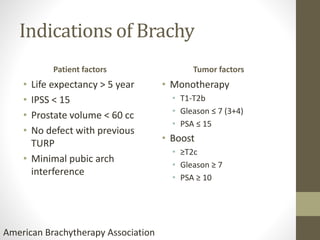
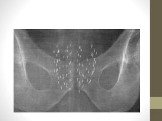
Prostate Cancer
Brachytherapy
Dr Ali Azher, MD (Radiation Oncology)
The Gujarat Cancer & Research Institute, BJ Medical College,
Ahmedabad, Gujarat
aliazhermuhammed@gmail.com
Brachytherapy
• Excellent long term tumor control
• Outcomes comparable to RP and RT
• Radium to Iridium
• 2D to IMRT
• Dying art or missing opportunity
• Poor Man’s IMRT
Brief History
• 1910 - Young used urethral radium for treatment of prostate
cancer
• 1917 –Barringer inserted radium needles transperineally in
the prostate
• 1930 - Flocks first injected radioactive gold liquid into prostate
• 1972 –Willet Whitmore described an open implant technique
using Iodine 125
• 1970 -Whitmer et al. at MSKCC in New York first performed
prostate seed implants using iodine seeds
• 1983 - Holm performed the first closed implant,using needles
and ultrasound guidance
Types
• Interstitial - radioactive sources are inside the tumor
• Breast, prostate
• Contact or plesiobrachytherapy – close to the tumor
• Intracavitary, intraluminal, endovascular, surface
• Cervix, esophagus
• ULDR – 0.01 to 0.3 Gy/hr
• LDR – 0.4 to 2.0 Gy/hr
• MDR – 2-12 Gy/hr
• HDR – greater than 12 Gy/hr
• PDR
• Combine physical advantages of HDR & radiobiological
advantages of LDR
• 3 Gy/hr
• Short duration
• Short pulses 10-30 min
TYPES
• LDR(0.4-2Gy/hr)
• LDR brachytherapy or ‘seed’ implant involves the insertion of
permanent radioactive sources directly into the prostate.
• HDR(>12Gy/hr)
• HDR brachytherapy also involves radioactive material being
placed directly into the prostate but, unlike LDR seeds, the
placement of the material is temporary and for shorter periods –
usually for a day or two at a time
Key concepts
• Highly conformal dose at high dose rate
Small
volume
High dose
Short
treatment
limits
toxicity to
surrounding
tissues
Radiobiology in brief
• α (alpha) – initial slope; lethal damage, intrinsic
radiosensitivity, linearly dependent
• β (beta) – curviness – sublethal damage proportional to the
square of dose
• The α/β ratio thus determines sensitivity of a cell to
alterations in fraction size.
• Rapidly proliferating cells are not very sensitive to fraction size
(high α/β).
• Slowly proliferating cells are very sensitive to fraction size (low
α/β)
• Tpot – potential doubling time
• Prostate 42 days
• Prostate tumors contain small fractions of cycling cells
• α/β is low 1.5Gy
• Similar to late responding normal tissues (3Gy)
• If α/β < normal tissue
• Hypofraction has an advantage
• If α/β > normal tissue
• Hypofraction has a disadvantage
Quick review of risk groups
Risk group Features
Very low T1C + GS ≤6 + PSA <10 + fewer than 3 bx positive ≤50% cancer
cells in core, PSA density <0.15
Low T1 T2a + GS ≤6 + PSA <10
Intermediate
favorable
T2b T2c/GS 7/PSA 10-20 + biopsy cores <50%
Intermediate
unfavorable
T2b T2c/GS 7/PSA 10-20
High T3a/ GS 8/PSA >20
Very high T3b T4/Primary GS 5/>cores with GS 8-10
Regional Any T, N1, M0
Metastatic Any T, Any N, M1
• Very low
• Expected patient survival ≥20yrs
• Low
• ≥10yrs
• Favorable intermediate
• Unfavorable intermediate
• EBRT+BT±ADT
• High or very high
• >5yrs EBRT+BT+ADT
Indications of Brachy
Patient factors
• Life expectancy > 5 year
• IPSS < 15
• Prostate volume < 60 cc
• No defect with previous
TURP
• Minimal pubic arch
interference
Tumor factors
• Monotherapy
• T1-T2b
• Gleason ≤ 7 (3+4)
• PSA ≤ 15
• Boost
• ≥T2c
• Gleason ≥ 7
• PSA ≥ 10
American Brachytherapy Association
• As monotherapy : for low risk patients
• As Boost : for intermediate and high risk pts
• As salvage therapy : for recurrent cases
Contraindications
Absolute
• Limited life expectancy
• Unacceptable operative
risk
• Distant metastases
• Absence of a rectum or
rectal fistula
• Large TURP defect
• Ataxia telangiectasia
Relative
• High IPSS (>20)
• History of prior pelvic RT
• TURP defect
• Large median lobe
• Gland size > 60 cc
• Inflammatory bowel
disease
History
Urologic history
• Prior TURP/Urethral surgery/BPH
• Medication for urinary obstructive symptoms
• Erectile function
• Prior diagnosis of cancer, especially rectal or bladder cancer
• Prior pelvic RT, surgery, or fracture
• Inflammatory bowel disease/ Connective tissue disorder
• Documentation of the IPSS
• Documentation of erectile function, International Index of
Erectile function score
Work-up
• Prostate biopsy within the last 12 months
• PSA
• DRE
• Prostate volume
• Can tolerate extended dorsal lithotomy position
• Suitability for anesthesia
• MR imaging T2W
Planning – volume study
• Prostate size and length
• Pubic arch interference
• any other reasons patient cannot tolerate brachytherapy
Pubic Arch Interference
• Gland should be <60cc, optimally <50cc
• In larger glands
• pubic arch may interfere with needle placement
• Inadequate dose coverage
• More seeds
• Increase in central urethral dose – potential risk of urinary
morbidity
• Combined Androgen Blockade (CAB) – 30% reduction in volume
after 3 months
Seed implantation
• Loose
• Stranded
• Dosimetricaly similar
• Seed migration is less with stranded seeds (lung & perinueum)
• Stranded I 125 seeds higher D90, V100 & V150 & higher
urethral dosimetry (avoid in the region of apex)
• Perineal, Retropubic and suprapubic approaches
• Perineal approach
• Needles are introduced above the anus and are guided into the
prostate with the index finger in the rectum.
• Suprapubic Cystotomy Approach
• The needles are placed directly in the prostate through the open
bladder with a finger in rectum guiding the placement.
• Retropubic approach
• This approach is used most extensively, though it’s a more
difficult procedure.
Radio isotopes
• ULDR
• Iodine 125
• Palladium 103
• Cesium 131
Isotope Half life (d) Energy Kev Dose rate
Iodine 125 59.6 28 7cGY/h
Palladium 103 17 22 19cGy
Cesium 9.6 29
Pd 103 more intense radiation prostatits in first month
but recovered soon
TYPES OF PERMANENT
IMPLANTS
Classic LDR Ultra LDR
222Rn seeds, 198Au seeds 103Pd, 125I and 131Cs
Half-lives of a few days Uses longer half life
High-energy γ-rays emitted by Low-energy photon emitters
Monoenergetic Cascade of energy
The patient must be confined to
the hospital until the source
strength decays to a safe level
(two to three half-lives or about
10 days)
No
Thin lead foils (0.2mm) – almost
complete shielding
• HDR BT
• Cs 137
• Co 60
• Ir192 optimal choice
• T1/2 =73.8 days
• γ ray energies (0.136 to 0.613 MeV)
• Effective γ rays energy approx 0.380 MeV
• Effectively shielded
LDR procedure
• Patient is placed under spinal or general anesthetia
• Supine in lithotomy position & catheterisation
• Contrast is placed in the bladder
• Applicator template is secured to TRUS apparatus
• Reference plane 0.0 (base of the prostate)
• Needles are inserted
• USG axial & sagittal views will guide the placement of the
needle
• Seeds are dropped within the prostate
• Lineal alignment & spacing
• Plain X ray to evaluate symmetry & spacing
• Bladder irrigation & cystoscopy to evacuate migrated seeds &
clots
• Catheter removed & remain in recovery until urinate
• Minimum peripheral dose
• Maximum dose that covers 100% of target volume
• Dependent on the position of the seeds
• Dose may vary upto 25%
• 90% of target will get dose
Post Plan
• Post op day 1
• Wait for 4 weeks for resolution of inflammation & edema
• MR imaging to visualise prostatic tissue better
• Chest Xray to look for seed migration
HDR Procedure
• Patient is placed under spinal or general anesthetia
• Supine in lithotomy position & catheterisation
• Contrast is placed in the bladder
• Urethra can be identified using foley.
• Needles are placed in the prostate at regular intervals
• For CT-based planning, images should be contiguous and no
more than 3 mm thick in axial plane. Should extend at least 9
mm above and below the target volume
• Patients are discharged same day after regaining urinary
function
• Simulation
• Contouring
• OARs
• Prostate
Prescription doses
• Permanent LDR BT
Isotope Monotherapy Boost
Iodine 125 145 Gy 110 Gy
Palladium 103 125 Gy 100 Gy
Cesium 131 115 Gy 85 Gy
HDR Monotherapy
• 8.5Gy x 4 in one implant (75.6 Gy)
• 7 Gy x 6 in two implants (76 Gy)
Boost dose
• HDR BT
• Flexibility in source positioning
• Accurate source positioning
• Immobilised target
• Stable geometry
• Target volume dose optimisation
• Effective cell kill
• No exposure
• XRT 45 Gy
• LDR – 110Gy for Iodine 125, 100Gy for Palladium 103
• HDR – 21Gy/2 fraction or 15Gy single fraction
• D90 – dose to 90% of prostate
• D90 >90% better PSA relapse free outcomes
• V100 – volume of prostate receiving 100% of Dose
Organ Volume Constraint
CTV V100 >95%
CTV V125 50-60%
CTV V150 <50%
Rectum D2cc <70%
Rectum V75 <1 cc
Bladder V75 <1 cc
Urethra V115 <1cc
OARs
• Rectum
• Urethra
• Penile bulb
• Neurovascular bundles
Toxicities
• Acute
• Urinary retention 1.5% - 22%
• Transient urinary morbidity – urinary frequency, urgency &
dysuria (peak 1-3 months post implantation)
• Rectal – diarrhea, proctitis 2-5%
Late toxicities
• Chronic cystitis (3-7 %)
• Incontinence (1% for non-TURP, 25-42% for TURP)
• Rectal ulceration (< 1 %)
• Urethral necrosis (< 1 %)
• Erectile dysfunction 20-25%
ASCENDE-RT TRIAL
• Androgen Suppression Combined with Elective Nodal and
Dose Escalated Radiation Therapy
• 9 year PSA Free survival 83% Vs 62%
• No survival differences
Intermediate &
high risk
patients
RT+BT+ADT
(12m)
RT+ADT (12m)
Salvage therapy
• Improvements in imaging & dosimetry – reduces the risk of
treatment related complications
• Recurrent diseases documented histologically
• Preferred candidates
• No clinical/radiological e/o distant mets
• Adequate urinary function
• > 5yrs of life expectancy
• > 2yrs of Disease free interval from primary RT
• Long PSA DT > 6-9months at the time of recurrence
Doses
• Permanent LDR seed implants
• Pd 103 D90 100Gy
• 5-10 yr PSA RFS in 10-53%
• HDR BT
• 36Gy in 6# weekly

Prostate Cancer Brachytherapy

  • 1.
    Prostate Cancer Brachytherapy Dr AliAzher, MD (Radiation Oncology) The Gujarat Cancer & Research Institute, BJ Medical College, Ahmedabad, Gujarat aliazhermuhammed@gmail.com
  • 2.
    Brachytherapy • Excellent longterm tumor control • Outcomes comparable to RP and RT • Radium to Iridium • 2D to IMRT • Dying art or missing opportunity • Poor Man’s IMRT
  • 3.
    Brief History • 1910- Young used urethral radium for treatment of prostate cancer • 1917 –Barringer inserted radium needles transperineally in the prostate • 1930 - Flocks first injected radioactive gold liquid into prostate • 1972 –Willet Whitmore described an open implant technique using Iodine 125 • 1970 -Whitmer et al. at MSKCC in New York first performed prostate seed implants using iodine seeds • 1983 - Holm performed the first closed implant,using needles and ultrasound guidance
  • 4.
    Types • Interstitial -radioactive sources are inside the tumor • Breast, prostate • Contact or plesiobrachytherapy – close to the tumor • Intracavitary, intraluminal, endovascular, surface • Cervix, esophagus
  • 6.
    • ULDR –0.01 to 0.3 Gy/hr • LDR – 0.4 to 2.0 Gy/hr • MDR – 2-12 Gy/hr • HDR – greater than 12 Gy/hr • PDR • Combine physical advantages of HDR & radiobiological advantages of LDR • 3 Gy/hr • Short duration • Short pulses 10-30 min
  • 7.
    TYPES • LDR(0.4-2Gy/hr) • LDRbrachytherapy or ‘seed’ implant involves the insertion of permanent radioactive sources directly into the prostate. • HDR(>12Gy/hr) • HDR brachytherapy also involves radioactive material being placed directly into the prostate but, unlike LDR seeds, the placement of the material is temporary and for shorter periods – usually for a day or two at a time
  • 8.
    Key concepts • Highlyconformal dose at high dose rate Small volume High dose Short treatment limits toxicity to surrounding tissues
  • 9.
    Radiobiology in brief •α (alpha) – initial slope; lethal damage, intrinsic radiosensitivity, linearly dependent • β (beta) – curviness – sublethal damage proportional to the square of dose • The α/β ratio thus determines sensitivity of a cell to alterations in fraction size. • Rapidly proliferating cells are not very sensitive to fraction size (high α/β). • Slowly proliferating cells are very sensitive to fraction size (low α/β)
  • 11.
    • Tpot –potential doubling time • Prostate 42 days • Prostate tumors contain small fractions of cycling cells • α/β is low 1.5Gy • Similar to late responding normal tissues (3Gy) • If α/β < normal tissue • Hypofraction has an advantage • If α/β > normal tissue • Hypofraction has a disadvantage
  • 12.
    Quick review ofrisk groups Risk group Features Very low T1C + GS ≤6 + PSA <10 + fewer than 3 bx positive ≤50% cancer cells in core, PSA density <0.15 Low T1 T2a + GS ≤6 + PSA <10 Intermediate favorable T2b T2c/GS 7/PSA 10-20 + biopsy cores <50% Intermediate unfavorable T2b T2c/GS 7/PSA 10-20 High T3a/ GS 8/PSA >20 Very high T3b T4/Primary GS 5/>cores with GS 8-10 Regional Any T, N1, M0 Metastatic Any T, Any N, M1
  • 13.
    • Very low •Expected patient survival ≥20yrs • Low • ≥10yrs • Favorable intermediate • Unfavorable intermediate • EBRT+BT±ADT • High or very high • >5yrs EBRT+BT+ADT
  • 14.
    Indications of Brachy Patientfactors • Life expectancy > 5 year • IPSS < 15 • Prostate volume < 60 cc • No defect with previous TURP • Minimal pubic arch interference Tumor factors • Monotherapy • T1-T2b • Gleason ≤ 7 (3+4) • PSA ≤ 15 • Boost • ≥T2c • Gleason ≥ 7 • PSA ≥ 10 American Brachytherapy Association
  • 15.
    • As monotherapy: for low risk patients • As Boost : for intermediate and high risk pts • As salvage therapy : for recurrent cases
  • 16.
    Contraindications Absolute • Limited lifeexpectancy • Unacceptable operative risk • Distant metastases • Absence of a rectum or rectal fistula • Large TURP defect • Ataxia telangiectasia Relative • High IPSS (>20) • History of prior pelvic RT • TURP defect • Large median lobe • Gland size > 60 cc • Inflammatory bowel disease
  • 17.
    History Urologic history • PriorTURP/Urethral surgery/BPH • Medication for urinary obstructive symptoms • Erectile function • Prior diagnosis of cancer, especially rectal or bladder cancer • Prior pelvic RT, surgery, or fracture • Inflammatory bowel disease/ Connective tissue disorder • Documentation of the IPSS • Documentation of erectile function, International Index of Erectile function score
  • 18.
    Work-up • Prostate biopsywithin the last 12 months • PSA • DRE • Prostate volume • Can tolerate extended dorsal lithotomy position • Suitability for anesthesia • MR imaging T2W
  • 19.
    Planning – volumestudy • Prostate size and length • Pubic arch interference • any other reasons patient cannot tolerate brachytherapy
  • 20.
    Pubic Arch Interference •Gland should be <60cc, optimally <50cc • In larger glands • pubic arch may interfere with needle placement • Inadequate dose coverage • More seeds • Increase in central urethral dose – potential risk of urinary morbidity • Combined Androgen Blockade (CAB) – 30% reduction in volume after 3 months
  • 21.
    Seed implantation • Loose •Stranded • Dosimetricaly similar • Seed migration is less with stranded seeds (lung & perinueum) • Stranded I 125 seeds higher D90, V100 & V150 & higher urethral dosimetry (avoid in the region of apex)
  • 23.
    • Perineal, Retropubicand suprapubic approaches
  • 24.
    • Perineal approach •Needles are introduced above the anus and are guided into the prostate with the index finger in the rectum. • Suprapubic Cystotomy Approach • The needles are placed directly in the prostate through the open bladder with a finger in rectum guiding the placement. • Retropubic approach • This approach is used most extensively, though it’s a more difficult procedure.
  • 25.
    Radio isotopes • ULDR •Iodine 125 • Palladium 103 • Cesium 131 Isotope Half life (d) Energy Kev Dose rate Iodine 125 59.6 28 7cGY/h Palladium 103 17 22 19cGy Cesium 9.6 29 Pd 103 more intense radiation prostatits in first month but recovered soon
  • 26.
    TYPES OF PERMANENT IMPLANTS ClassicLDR Ultra LDR 222Rn seeds, 198Au seeds 103Pd, 125I and 131Cs Half-lives of a few days Uses longer half life High-energy γ-rays emitted by Low-energy photon emitters Monoenergetic Cascade of energy The patient must be confined to the hospital until the source strength decays to a safe level (two to three half-lives or about 10 days) No Thin lead foils (0.2mm) – almost complete shielding
  • 27.
    • HDR BT •Cs 137 • Co 60 • Ir192 optimal choice • T1/2 =73.8 days • γ ray energies (0.136 to 0.613 MeV) • Effective γ rays energy approx 0.380 MeV • Effectively shielded
  • 28.
    LDR procedure • Patientis placed under spinal or general anesthetia • Supine in lithotomy position & catheterisation • Contrast is placed in the bladder • Applicator template is secured to TRUS apparatus • Reference plane 0.0 (base of the prostate) • Needles are inserted • USG axial & sagittal views will guide the placement of the needle • Seeds are dropped within the prostate • Lineal alignment & spacing • Plain X ray to evaluate symmetry & spacing • Bladder irrigation & cystoscopy to evacuate migrated seeds & clots
  • 29.
    • Catheter removed& remain in recovery until urinate
  • 32.
    • Minimum peripheraldose • Maximum dose that covers 100% of target volume • Dependent on the position of the seeds • Dose may vary upto 25% • 90% of target will get dose
  • 33.
    Post Plan • Postop day 1 • Wait for 4 weeks for resolution of inflammation & edema • MR imaging to visualise prostatic tissue better • Chest Xray to look for seed migration
  • 34.
    HDR Procedure • Patientis placed under spinal or general anesthetia • Supine in lithotomy position & catheterisation • Contrast is placed in the bladder • Urethra can be identified using foley. • Needles are placed in the prostate at regular intervals • For CT-based planning, images should be contiguous and no more than 3 mm thick in axial plane. Should extend at least 9 mm above and below the target volume • Patients are discharged same day after regaining urinary function
  • 36.
  • 37.
    Prescription doses • PermanentLDR BT Isotope Monotherapy Boost Iodine 125 145 Gy 110 Gy Palladium 103 125 Gy 100 Gy Cesium 131 115 Gy 85 Gy
  • 38.
    HDR Monotherapy • 8.5Gyx 4 in one implant (75.6 Gy) • 7 Gy x 6 in two implants (76 Gy)
  • 39.
    Boost dose • HDRBT • Flexibility in source positioning • Accurate source positioning • Immobilised target • Stable geometry • Target volume dose optimisation • Effective cell kill • No exposure • XRT 45 Gy • LDR – 110Gy for Iodine 125, 100Gy for Palladium 103 • HDR – 21Gy/2 fraction or 15Gy single fraction
  • 40.
    • D90 –dose to 90% of prostate • D90 >90% better PSA relapse free outcomes • V100 – volume of prostate receiving 100% of Dose Organ Volume Constraint CTV V100 >95% CTV V125 50-60% CTV V150 <50% Rectum D2cc <70% Rectum V75 <1 cc Bladder V75 <1 cc Urethra V115 <1cc
  • 41.
    OARs • Rectum • Urethra •Penile bulb • Neurovascular bundles
  • 42.
    Toxicities • Acute • Urinaryretention 1.5% - 22% • Transient urinary morbidity – urinary frequency, urgency & dysuria (peak 1-3 months post implantation) • Rectal – diarrhea, proctitis 2-5%
  • 43.
    Late toxicities • Chroniccystitis (3-7 %) • Incontinence (1% for non-TURP, 25-42% for TURP) • Rectal ulceration (< 1 %) • Urethral necrosis (< 1 %) • Erectile dysfunction 20-25%
  • 44.
    ASCENDE-RT TRIAL • AndrogenSuppression Combined with Elective Nodal and Dose Escalated Radiation Therapy • 9 year PSA Free survival 83% Vs 62% • No survival differences Intermediate & high risk patients RT+BT+ADT (12m) RT+ADT (12m)
  • 45.
    Salvage therapy • Improvementsin imaging & dosimetry – reduces the risk of treatment related complications • Recurrent diseases documented histologically • Preferred candidates • No clinical/radiological e/o distant mets • Adequate urinary function • > 5yrs of life expectancy • > 2yrs of Disease free interval from primary RT • Long PSA DT > 6-9months at the time of recurrence
  • 46.
    Doses • Permanent LDRseed implants • Pd 103 D90 100Gy • 5-10 yr PSA RFS in 10-53% • HDR BT • 36Gy in 6# weekly